Parasitology 2nd lecture
advertisement
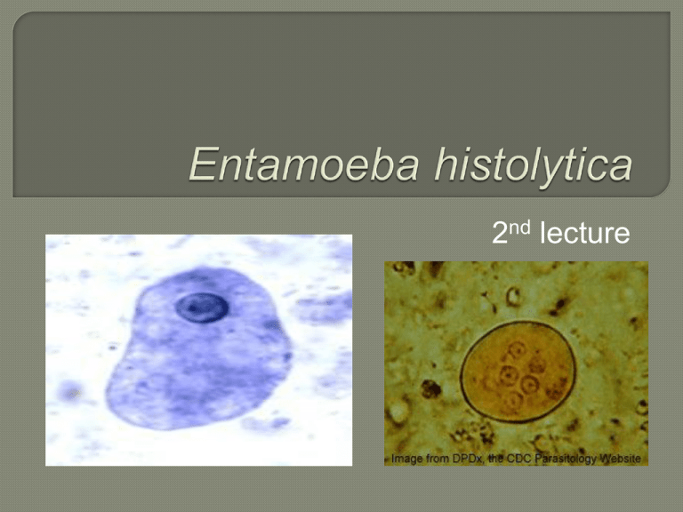
2nd lecture Intestinal and luminal protozoa significant to human health include: Entamoeba histolytica (Amebae). Balantidium coli (Ciliates) The ciliates are a group of protozoans characterized by the presence of hair-like organelles called cilia, which are identical in structure to flagella but typically shorter and present in much larger numbers Giardia lamblia and Trichomonas vaginalis (Flagellates). AMEBIASIS (amebic dysentery, amebic hepatitis) Amebas are unicellular organisms common in the environment: many are parasites of vertebrates and invertebrates. Relatively few species inhabit the human intestine and only Entamoeba histolytica is identified as a human intestinal pathogen. Entamoeba histolytica is an anaerobic parasitic protozoan, part of the genus Entamoeba. E. histolytica is estimated to infect about 50 million people worldwide. Many older textbooks state that 10% of the world population is infected. E histolytica has a relatively simple life cycle that alternates between trophozoite and cyst stages. The trophozoite is the actively metabolizing, mobile stage, and the cyst is dormant and environmentally resistant. Diagnostic concern centers on both stages. Trophozoites vary remarkably in size-from 10 to 60 μm or more in diameter, and when they are alive they may be actively motile. Amebas are anaerobic organisms and do not have mitochondria. The finely granular endoplasm contains the nucleus and food vacuoles, which in turn may contain bacteria or red blood cells. The parasite is sheathed by a clear outer ectoplasm. Nuclear morphology is best seen in permanent stained preparations. The cyst is a spherical structure, 10-20 μm in diameter, with a thin transparent wall. Fully mature cysts contain four nuclei with the characteristic amebic morphology. Rod-like structures (chromatoidal bars) are present variably, but are more common in immature cysts. Inclusions in the form of glycogen masses also may be present. A number of non-pathogenic amebae can parasitize the human gastrointestinal tract and may cause diagnostic confusion. The fecal-oral transmission of the ameba usually involves contaminated food or water. The parasite can also be transmitted directly by sexual contact. The active (trophozoite) stage exists only in the host and in fresh loose feces; cysts survive outside the host in water, soils and on foods, especially under moist onditions. The cysts are readily killed by heat and by freezing temperatures, and survive for only a few months outside of the host. When cysts are swallowed they cause infections by excysting (releasing the trophozoite stage) in the digestive tract. The trophozoite stage is readily killed in the environment and cannot survive passage through the acidic stomach to cause infection. E. histolytica, as its name suggests (histo–lytic = tissue destroying), is pathogenic; infection can lead to amoebic dysentery or amoebic liver abscess. Symptoms can include fulminating dysentery, bloody diarrhea, weight loss, fatigue, abdominal pain, and amoeboma. The amoeba can actually 'bore' into the intestinal wall, causing lesions and intestinal symptoms, and it may reach the blood stream. From there, itcan reach different vital organs of the human body, usually the liver, but sometimes the lungs, brain, spleen, etc. A common outcome of this invasion of tissues is a liver abscess, which can be fatal if untreated. It can be diagnosed by stool samples but it is important to note that certain other species are impossible to distinguish by microscopy alone. Trophozoites may be seen in a fresh fecal smear and cysts in an ordinary stool sample. ELISA (Enzymelinked immunosorbent assay) or RIA (Radioimmunoassay) can also be used. Multiplication and Life Cycle Amebas multiply in the host by simple binary fission. Most multiplication occurs in the host, and survival outside the host depends on the desiccation-resistant cyst form. Encystment occurs apparently in response to desiccation as the ameba is carried through the colon. After encystment, the nucleus divides twice to produce a quadrinucleate mature cyst. Excystment occurs after ingestion and is followed by rapid cell division to produce four amebas which undergo a second division. Each cyst thus yields eight tiny amebas. 0.5 - 50% of the population world wide harbors E. histolytica parasites with the higher rates of infection being in underdeveloped countries. 1 - 3% of the population of the USA is infected. Infection is associated with poor hygiene. Humans are the principal host, although dogs, cats and rodents may be infected. Fecal-oral transmission occurs when food preparation is not sanitary or when drinking water is contaminated. Contamination may come directly from infected food handlers or indirectly from faulty sewage disposal. The prevalence of amebiasis in underdeveloped countries reflects the lack of adequate sanitary systems. Amebas are found in all climates, arctic to tropical. Good Sanitation. Proper sewage system. Avoidance of contamination of food & H2O. Health Education Personal Hygiene. THANK YOU
