Test 2 Study Guide.doc
advertisement
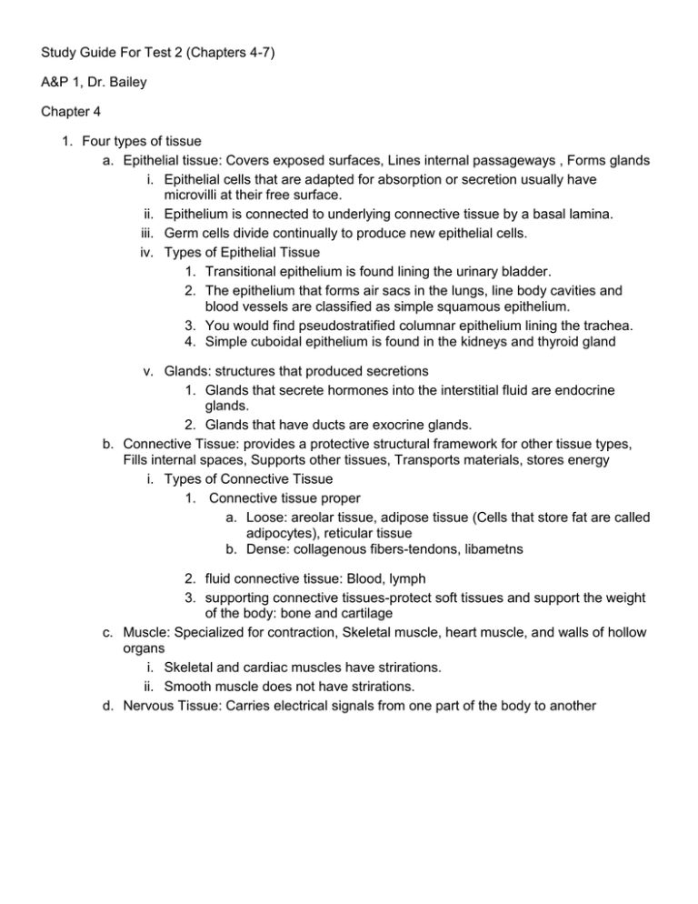
Study Guide For Test 2 (Chapters 4-7) A&P 1, Dr. Bailey Chapter 4 1. Four types of tissue a. Epithelial tissue: Covers exposed surfaces, Lines internal passageways , Forms glands i. Epithelial cells that are adapted for absorption or secretion usually have microvilli at their free surface. ii. Epithelium is connected to underlying connective tissue by a basal lamina. iii. Germ cells divide continually to produce new epithelial cells. iv. Types of Epithelial Tissue 1. Transitional epithelium is found lining the urinary bladder. 2. The epithelium that forms air sacs in the lungs, line body cavities and blood vessels are classified as simple squamous epithelium. 3. You would find pseudostratified columnar epithelium lining the trachea. 4. Simple cuboidal epithelium is found in the kidneys and thyroid gland v. Glands: structures that produced secretions 1. Glands that secrete hormones into the interstitial fluid are endocrine glands. 2. Glands that have ducts are exocrine glands. b. Connective Tissue: provides a protective structural framework for other tissue types, Fills internal spaces, Supports other tissues, Transports materials, stores energy i. Types of Connective Tissue 1. Connective tissue proper a. Loose: areolar tissue, adipose tissue (Cells that store fat are called adipocytes), reticular tissue b. Dense: collagenous fibers-tendons, libametns 2. fluid connective tissue: Blood, lymph 3. supporting connective tissues-protect soft tissues and support the weight of the body: bone and cartilage c. Muscle: Specialized for contraction, Skeletal muscle, heart muscle, and walls of hollow organs i. Skeletal and cardiac muscles have strirations. ii. Smooth muscle does not have strirations. d. Nervous Tissue: Carries electrical signals from one part of the body to another Chapter 5: Integumentary System i. ii. Layers of the Epidermis: provides a barrier against bacteria as well as chemical and mechanical injuries a. stratum corneum: The tough superficial layer of the epidermis with large amounts of keratin. Loacated in thick and thin skin. b. stratum lucidum: located in the thick skin of the palms and soles c. stratum granulosum. d. stratum spinosum. e. Stratum basale (germinativum): innermost epidermal layer, has layers of stem cells that constantly divide to renew the epidermis. Melanocytes are cells located in the stratum basale, they make melanin. The differences in skin pigmentation among individuals do not reflect different numbers of melanocytes, but different layers of synthetic activity (there for people with darker skin make more melanin than people with lighter skin, but have the same number of melanocytes as people with lighter skin). Albino people have a normal amount of melanocytes, but they cells cannot produce melanin. Chapter 6: Osseous Tissue and Bone Structure I. Five Primary Functions of the Skeletal System a. Support b. Storage of Minerals (calcium) and Lipids (yellow marrow) c. Blood Cell Production (red marrow) d. Protection e. Leverage (force of motion) II. Bone Shapes a. Sutural Bones: Small, irregular bones, Found between the flat bones of the skull b. Irregular Bones: Have complex shapes, Examples: spinal vertebrae, pelvic bones c. Short Bones: Small and thick, Examples: ankle and wrist bones d. Flat Bones: Thin with parallel surfaces, Found in the skull, sternum, ribs, and scapulae e. Long Bones: Long and thin, Found in arms, legs, hands, feet, fingers, and toes f. Sesamoid Bones: Small and flat, Develop inside tendons near joints of knees, hands, and feet III. Structure of a Long Bone a. Diaphysis: The shaft , A heavy wall of compact bone, or dense bone, A central space called medullary (marrow) cavity b. Epiphysis : Wide part at each end, Articulation with other bones, Mostly spongy (cancellous) bone, Covered with compact bone (cortex) c. Metaphysis: Where diaphysis and epiphysis meet IV. Periosteum : Covers outer surfaces of bones, Consists of outer fibrous and inner cellular layers V. Bone Cells a. Osteocytes : Mature bone cells that maintain the bone matrix, Live in lacunae , Are between layers (lamellae) of matrix, Connect by cytoplasmic extensions through canaliculi in lamellae, Do not divide; Two major functions of osteocytes: To maintain protein and mineral content of matrix, To help repair damaged bone b. Osteoblasts : Immature bone cells that secrete matrix compounds (osteogenesis), Osteoid — matrix produced by osteoblasts, but not yet calcified to form bone, Osteoblasts surrounded by bone become osteocytes, c. Osteoprogenitor Cells: Mesenchymal stem cells that divide to produce osteoblasts, Located in endosteum, the inner cellular layer of periosteum, Assist in fracture repair d. Osteoclasts: Secrete acids and protein-digesting enzymes, Giant, multinucleate cells, Dissolve bone matrix and release stored minerals (osteolysis), Derived from stem cells that produce macrophages VI. The structure of compact bone: The Osteon is the basic unit, Osteocytes are arranged in concentric lamellae., Around a central canal containing blood vessels VII. The Structure of Spongy Bone: Does not have osteons: The matrix forms an open network of trabeculae; Trabeculae have no blood vessels; The space between trabeculae is filled with red bone marrow Chapter 7: The Axial Skelton I. 80 bones make up the axial skeleton II. Skull a. The skull contains 22 bones: 8 cranial bones, 14 facial bones b. Foramina: Openings for nerves and blood vessels c. The membranous areas between the cranial bones of the fetal skull are fontanels III. Functions of the axial skeleton? provides an attachment for muscles that move the appendicular skeleton; provides an attachment for muscles that move the head, neck, and trunk; provides an attachment for muscles involved in respiration ; provides protection for the brain and spinal cord IV. Regions of the Vertebral Column • Cervical (C)- 7 cervical vertebra • Thoracic (T)-12 throacic vertebra • Lumbar (L)-5 lumbar vertebra (also have the widest intervertebral disks) • Sacral (S)-1 fused vertebra • Coccygeal (Co)-1 fused vertebra V. Ribs a. True ribs are ribs 1-7 (Connected to the sternum by costal cartilages) b. False ribs are Ribs 8–12 (Do not attach directly to the sternum); 11 and 12 are floating ribs c. Ribs articulate with the thoracic vertebra VI. The Sternum: A flat bone, In the midline of the thoracic wall a. Three parts of the sternum i. The manubrium ii. The sternal body iii. The xiphoid process Images to Know for the Lab Exam


