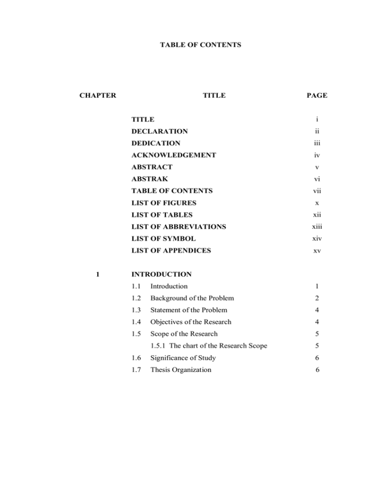
TABLE OF CONTENTS CHAPTER TITLE PAGE
advertisement

TABLE OF CONTENTS CHAPTER 1 TITLE PAGE TITLE i DECLARATION ii DEDICATION iii ACKNOWLEDGEMENT iv ABSTRACT v ABSTRAK vi TABLE OF CONTENTS vii LIST OF FIGURES x LIST OF TABLES xii LIST OF ABBREVIATIONS xiii LIST OF SYMBOL xiv LIST OF APPENDICES xv INTRODUCTION 1.1 Introduction 1 1.2 Background of the Problem 2 1.3 Statement of the Problem 4 1.4 Objectives of the Research 4 1.5 Scope of the Research 5 1.5.1 The chart of the Research Scope 5 1.6 Significance of Study 6 1.7 Thesis Organization 6 2 3 LITERATURE REVIEW 2.1 Introduction 9 2.2 3D Image Visualization 9 2.3 Geodesic Active Contour Model (GAC) 12 2.4 Additive Operator Splitting (AOS) 14 METHODS FOR CONSTRUCTING 3D IMAGE FROM 2D IMAGES 3.1 Introduction 15 3.2 GAC model using AOS scheme 15 3.3 Model of the Problem 17 3.3.1 Initial Boundary Value Problem (IBVP) 18 3.3.2 Linear System Equations (LSE) 19 3.3.3 Iterative Method 21 3.3.4 Gauss Seidel 22 3.4 Edge Detection using AOS and GAC for the 23 Brain Tumor MRI Images 3.5 Result for the Edge Detection on a Brain Tumor 25 3.6 Image Manifold (IM) Method 26 3.6.1 Semi-Implicit Scheme for Subjective 28 Surfaces 3.7 Volume Estimation (VE) Method 29 3.7.1 30 Chronology for Volume Estimation (VE) Method 3.8 4 Computational Platform System 32 IMAGE MANIFOLD METHOD 4.1 Introduction 33 4.2 Selected MRI Images 34 4.2.1 35 Edge detection by using GAC-AOS method 4.3 The Process of Constructing 3D Brain Tumor 37 4.4 Result for 3D visualization and Volume 42 calculation 4.5 5 6 7 The Chart of the 3D Image Construction. 43 VOLUME ESTIMATION METHOD 5.1 Introduction 45 5.2 Selected MRI Images 46 5.3 The Process of Volume Calculation 48 5.4 3D Visualization 50 5.5 Volume result 51 RESULT AND ANALYSIS 6.1 Introduction 52 6.2 Discussion 52 6.3 Conclusion 54 CONCLUSION AND FURTHER WORK 7.1 Conclusion 56 7.2 Direction for Future Work 57 REFERENCES 58 APPENDIX APPENDIX A 63 APPENDIX B 64 LIST OF FIGURES FIGURE NO. TITLE PAGE 1.1 2D MRI brain image with edge detection 3 1.2 The selected area of brain tumor image 3 1.3 The chart of research scope for constructing 3D 8 medical image 3.1 Initial partitioning of matrix A 22 3.2 The chart of algorithm for edge detection on brain 24 tumor MRI 3.3 Edge detection process of the brain tumor MRI image 25 based on the Gauss-Seidel method 3.4 Illustration on how to perform the calculation of 31 volume estimation for two brain tumor contours 3.5 Illustration on how to perform the calculation of 31 volume estimation for three brain tumor contours 4.1 Three MRI images selected from different angle. 34 4.2 The process of edge detection on the brain tumor for 35 front view image. 4.3 The process of edge detection on the brain tumor for 35 side view image. 4.4 The process of edge detection on the brain tumor for 36 top view image. 4.5 Contour result of the brain tumor in full scale. 36 4.6 Choose a file and select the data 38 4.7 The data is loaded and displayed in the three windows 39 4.8 Creating the volume 40 4.9 Creating the 3D model 41 4.10 3D brain tumor visualization 43 4.11 The chart of the process in constructing 3D image 44 5.1 Six MRI images selected from different sizes. 46 5.2 Six cropped brain tumor at different sizes. 47 5.3 Edge detection on a brain tumor at different sizes. 47 5.4 Create Matlab folder 49 5.5 Run the programming in Matlab 49 5.6 Calculation of the volume 49 5.7 Time execution 50 5.8 3D brain tumor visualization 50 LIST OF TABLES TABLE NO. TITLE PAGE 4.1 Volume of tumor for IM method 42 5.1 Result for volume of tumor 51 6.1 Comparison of the edge detection results for IM and VE methods 53 6.2 Comparison of the volume and time execution results between IM and VE methods 53 6.3 Comparison of the 3D image visualization between IM and VE methods 54 LIST OF ABBREVIATIONS 2D - Two dimensional 3D - Three dimensional ACM - Active Contour Method AOS - Additive Operator Splitting IBVP - Initial Boundary Value Problem IM - Image Manifold GAC - Geodesic Active Contour LSE - Linear System of Equation VE - Volume Estimation LIST OF SYMBOLS β Weighted of energy Gradient operator Image domain ρ Acceleration parameter g Stopping function v Positive constant τ Time Number of iterations k D The distance function The initial scaling factor s ϕ (x, y, z) Vg The smoothing parameter Distance function Volume of manifold Initial image g (x, y, z) Edge indicator Curvature operator LIST OF APPENDIXES APPENDIX TITLE PAGE A Matlab File for Edge detection 63 B Matlab File for Volume estimation 64


