American College of Radiology Stereotactic Breast Biopsy Accreditation Program
advertisement
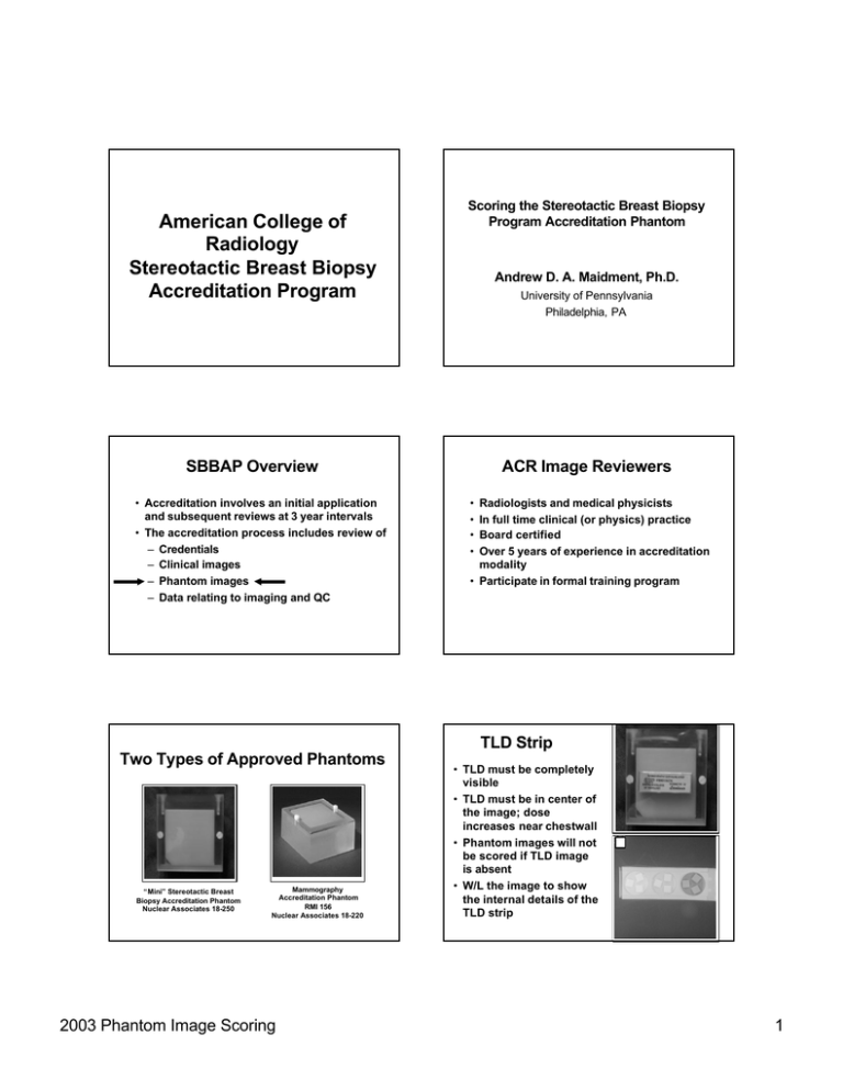
American College of Radiology Stereotactic Breast Biopsy Accreditation Program Scoring the Stereotactic Breast Biopsy Program Accreditation Phantom Andrew D. A. Maidment, Ph.D. University of Pennsylvania Philadelphia, PA SBBAP Overview • Accreditation involves an initial application and subsequent reviews at 3 year intervals • The accreditation process includes review of – Credentials – Clinical images – Phantom images – Data relating to imaging and QC ACR Image Reviewers • • • • Radiologists and medical physicists In full time clinical (or physics) practice Board certified Over 5 years of experience in accreditation modality • Participate in formal training program TLD Strip Two Types of Approved Phantoms “Mini” Stereotactic Breast Biopsy Accreditation Phantom Nuclear Associates 18-250 Mammography Accreditation Phantom RMI 156 Nuclear Associates 18-220 2003 Phantom Image Scoring • TLD must be completely visible • TLD must be in center of the image; dose increases near chestwall • Phantom images will not be scored if TLD image is absent • W/L the image to show the internal details of the TLD strip 1 Viewing Conditions The ACR SBBAP Phantom Image Review Protocol • General lighting low and difuse • Viewboxes should avoid direct or reflected bright light • Viewboxes should be capable of producing a luminance of at least 3000 candela per square meter • Mask the image • Use a magnifying glass of 2X or higher for specks, other test objects, and artifacts as necessary General Scoring SBBAP Testing Criteria Phantom Image Quality • Each object group is scored separately • Count # of visible objects from largest downward until a score of 0 or 0.5 is reached, then deduct for artifacts Fibers MAP Phantom F/S Digital 4.0 5.0 Mini Phantom F/S Digital 2.0 3.0 Speck Groups 3.0 4.0 2.0 3.0 Masses 3.0 3.5 2.0 2.5 • Carefully review images for other artifacts; where appropriate, investigate causes and pursue corrective action Fibers • Each fiber is one point if the full length is visible and location and orientation are correct. • A fiber is 0.5 point if >half fiber is visible, and location and orientation are correct. • Stop after reaching a score of 0 or 0.5 and can no longer determine the full fiber orientation or borders. • Deduct any fiber-like artifact appearing anywhere in the wax insert area from the last “real” half or whole fiber scored if the artifactual fiber is equally or more apparent. Deduct only from the last real fiber, not from additional fibers. 2003 Phantom Image Scoring Fibers 2 Speck Groups • Starting with the largest speck group, each speck group is 1 point if >4 of the six specks in the group are visible in the proper locations. • A speck group as 0.5 if 2 or 3 are visible in the proper locations. • Stop counting when reaching a score of 0 or 0.5. • If noise or speck -like artifacts are visible in the wrong locations within wax insert area deduct them one for one from the individual specks counted in the last whole or half speck group scored. Masses • Each mass is one point if minus density object is visible in correct location and is generally circular. • A mass is 0.5 point if minus density object is visible in correct location but mass is not generally circular. • Stop after reaching a score of 0 or 0.5. • Deduct any mass-like artifact appearing anywhere in the wax insert area from the last “real” half or whole mass scored if the artifactual mass is equally or more apparent. Deduct only from the last real mass. Image Quality Problems • All Modalities – Excessive random noise – Excessive structured noise – Non-uniform background – Excessive blur (unsharpness) – Poor contrast – Extra object(s) on film – Wrong orientation of insert • Film-Screen – OD too light – OD too dark 2003 Phantom Image Scoring Speck Groups Masses Artifacts • All Modalities – Minus/plus density specks – Minus/plus density streaks – Minus/plus density blotches – Patterned background – Excessive Cut-off • Film-Screen – Roller Marks – Wet Pressure Marks – Residual Grid Lines – Grid inhomogeneities 3 Possible Causes of Artifacts • All Modalities – Tube target or filter – Compression paddle – Breast Support Tray Possible Causes of Artifacts • Film-screen or hard-copy film – Dust or Dirt – Processor Developer – Processor rollers – Grid – Screen or cassettes – Film handling – Film fog or light leak Assignment of Final Grade in the Accreditation Review Possible Causes of Artifacts • Digital – Digital Image Receptor – Detector Calibration – Inadequate SNR • Scores from the two reviewers are averaged • Phantom must pass all 3 objects; A fail in one of the areas results in a phantom failure • No unacceptable artifacts – That is, artifacts which obscure or impair clinical information Additional Review • Two reviewers must fail the same object for the phantom to fail immediately • Disagreements will be sent out for a third review (and a fourth if necessary) • The phantom image score is determined by the score from the additional reviewer averaged with that if the initial reviewer making the same pass/fail decision. ACR Stereo Accreditation Program Reasons for Failures – 1st Attempt 2002 10% 0% 10% Phantom Only Dose 18% 62% Clinical & Phantom 2003 Phantom Image Scoring Clinical Only Processor 4 Summary • 2 phantom types; 6 versions • Review images under appropriate conditions • Review each object separately; score until 0.5 or 0, then subtract for artifacts • Passing score dependent upon phantom type and modality (digital vs. S-F) • Technique should be appropriate to modality • Artifact evaluation is critical 2003 Phantom Image Scoring 5

