Lecture #13 – 10/3 – Dr. Wormington
advertisement
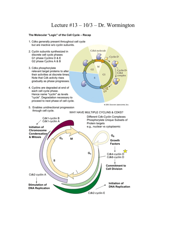
Lecture #13 – 10/3 – Dr. Wormington The Molecular "Logic" of the Cell Cycle – Recap 1. Cdks generally present throughout cell cycle but are inactive w/o cyclin subunits. 2. Cyclin subunits synthesized in discrete cell cycle phases G1 phase Cyclins D & E G2 phase Cyclins A & B 3. Cdks phosphorylate relevant target proteins to alter their activities at discrete times. Note that Cdk activity rises gradually as phase progresses. 4. Cyclins are degraded at end of each cell cycle phase. Hence name "cyclin" as levels "cycle". Degradation necessary to proceed to next phase of cell cycle. 5. Enables unidirectional progression through cell cycle. WHY HAVE MULTIPLE CYCLINS & CDKS? Initiation of Chromosome Condensation & Mitosis Different Cdk-Cyclin Complexes Phosphorylate Unique Subsets of Protein targets e.g., nuclear vs cytoplasmic Growth Factors Commitment to Cell Division Stimulation of DNA Replication Initiation of DNA Replication Making it fit – Human DNA must be compacted >100,000X Packaging achieved by progressive assembly of "naked" DNA Into increasingly compact DNA-protein complexes = Chromatin Nucleosome = Fundamental Unit Core = 2X ea. H2A, H2B, H3, H4 + Charged Histones Neutralize - Charged DNA for "1st order" Compaction HI Linker Outside Core Physiological [K+] Provides Additional Charge Neutralization to Further Compact Nucleosomes Compaction in Interphase Cell Metaphase Chromosome Electron Micrographs of Chromatin 10 nm "Beads on String" 30 nm Condensed Structure "Head-On" View" H2A H2B H3 H4 "Side" View Structure of The Nucleosome Derived by X-ray crystallography Mitosis: An Exquisitely Choreographed Process Comprised of Distinct Events The BIG Picture • Condense the Chromosomes • Generate Mitotic Spindle • Breakdown Nuclear Envelope • Attach Chromosomes to Spindle • Segregate Chromosomes to Daughter Cells • Cytokinesis • Decondense Chromosomes • Depolymerize Spindle Microtubules • Reform Nuclear Envelope THE ENTIRE PROCESS IS "DRIVEN" BY THE ACTIVATION & INACTIVATION OF THE G2-M CDKS Centrosome & DNA Replication Cell is now Tetraploid 4n 4n 2n 2n The Centrosome aka the Microtubule Organizing Center Source of all microtubules within the cell Fluorescent Micrograph of Interphase Mammalian Cells Electron Micrograph of A Centrosome containg its pair of Centrioles (Microtubules (MT) radiating Away from Centrosome are in yellow) Centrosome Migration Nuclear Envelope Breakdown Sister Chromatids Condense Kinetochore Microtubules & Joined at Centromeres "Capture" Chromosomes 1 each/chromatid Sister Chromatids + Centromere aka Spindle Pole Homologous Chromosomes DO NOT Pair in Mitosis Chromosomes Decondense Nuclear Envelope Reforms Cytokinesis Checkpoint Sister Chromatids Aligned towards Opposite Poles Sister Chromatids Separate Fluorescence Micrographs of Mitotic Human Cells DNA Microtubules The Advantages of Sex – Meiosis Promotes Genetic Diversity Haplontic Life Cycle Adults & Gametes (n) Zygote (2n) e.g., fungi Alternation of generations Both (n) and (2n) adult Stages e.g., most plants Diplontic Life Cycle Adult (2n) Gametes (n) e.g., all animals & some plants The Holy Grail of Meiosis Reduce Chromosome # from 2n to n Ensure Each Haploid Product Has Complete Set of Chromosomes Promote Genetic Diversity by Recombination Between Homologous Chromosomes Centrosome & DNA Replication Same as in Mitosis Cell is Tetraploid 4n Homologous Chromosomes Align Along Lengths Synapsis Recombination Between Homologous Chromosomes Occurs 4n 2n 2n Recombination Between Homologous Chromosomes Increases Genetic Diversity Sister Chromatids Sister Chromatids Tetrad or Bivalent e.g. Red = maternal Chromosome 16 Blue = paternal Chromosome 16. Note: There can be multiple Chiasmatas between a pair of homologous Chromosomes! Parental Nonrecombinant Chromatids chiasmata Fluorescent Micrographs of Synapsed Homologous Chromosomes In Meiotic Prophase I in Xenopus Oocytes Note: Sister Chromatids Are So Closely Aligned They Cannot Be Distinguished From Each Other
