LETTER
doi:10.1038/nature14593
Eye-like ocelloids are built from different
endosymbiotically acquired components
Gregory S. Gavelis1, Shiho Hayakawa1,2,3, Richard A. White III4{, Takashi Gojobori3,5, Curtis A. Suttle2,4,6,7, Patrick J. Keeling2,7
& Brian S. Leander1,2,7
Multicellularity is often considered a prerequisite for morphological
complexity, as seen in the camera-type eyes found in several groups of
animals. A notable exception exists in single-celled eukaryotes called
dinoflagellates, some of which have an eye-like ‘ocelloid’ consisting of
subcellular analogues to a cornea, lens, iris, and retina1. These planktonic cells are uncultivated and rarely encountered in environmental
samples, obscuring the function and evolutionary origin of the ocelloid. Here we show, using a combination of electron microscopy,
tomography, isolated-organelle genomics, and single-cell genomics,
that ocelloids are built from pre-existing organelles, including a cornea-like layer made of mitochondria and a retinal body made of
anastomosing plastids. We find that the retinal body forms the central
core of a network of peridinin-type plastids, which in dinoflagellates
and their relatives originated through an ancient endosymbiosis with
a red alga2. As such, the ocelloid is a chimaeric structure, incorporating organelles with different endosymbiotic histories. The anatomical
complexity of single-celled organisms may be limited by the components available for differentiation, but the ocelloid shows that preexisting organelles can be assembled into a structure so complex that
it was initially mistaken for a multicellular eye3. Although mitochondria and plastids are acknowledged chiefly for their metabolic roles,
they can also be building blocks for greater structural complexity.
Many organisms can orient to light. In some single-celled eukaryotes, such as Chlamydomonas and many dinoflagellates, an ‘eyespot’
directs photons onto photoreceptors on the flagellum, allowing the cell
to respond to the intensity and direction of light4,5. A vastly more
complex structure is found in warnowiid dinoflagellates: the eye-like
ocelloid. Ocelloids consist of subcellular components resembling a
lens, a cornea, iris-like rings, and a pigmented cup called the retinal
body6–9, which together so resemble the camera-type eyes of some
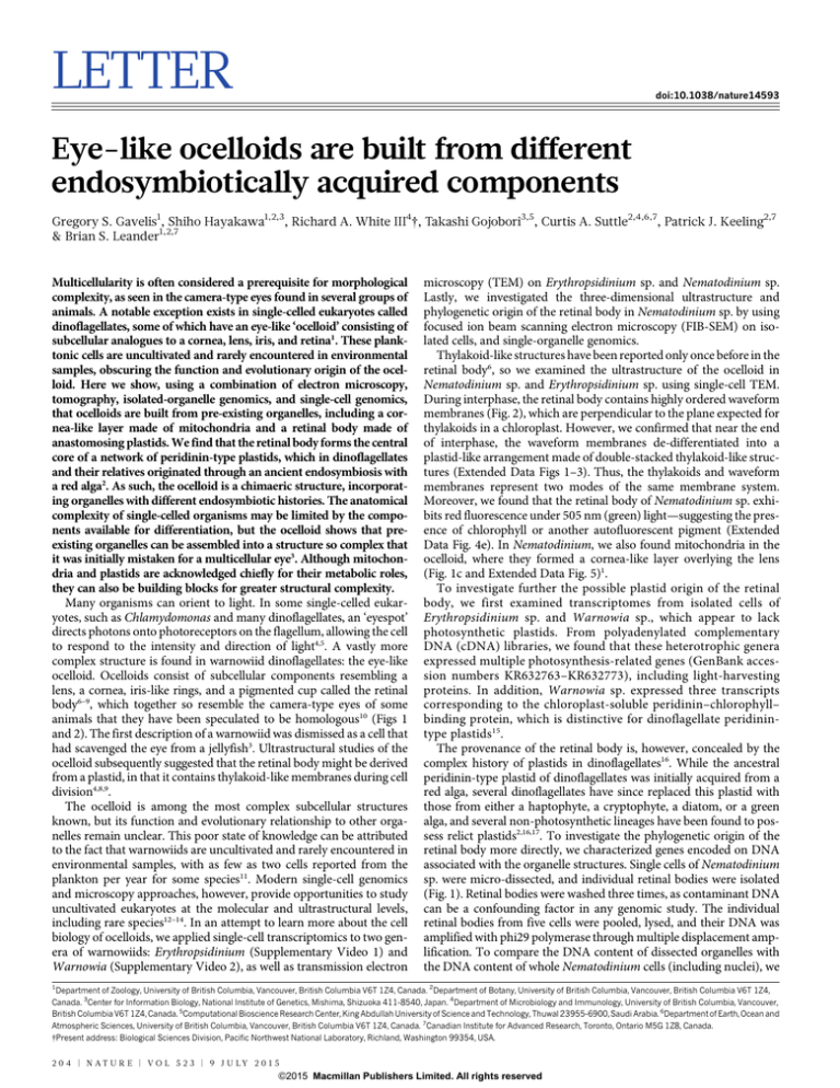
animals that they have been speculated to be homologous10 (Figs 1
and 2). The first description of a warnowiid was dismissed as a cell that
had scavenged the eye from a jellyfish3. Ultrastructural studies of the
ocelloid subsequently suggested that the retinal body might be derived
from a plastid, in that it contains thylakoid-like membranes during cell
division4,8,9.
The ocelloid is among the most complex subcellular structures
known, but its function and evolutionary relationship to other organelles remain unclear. This poor state of knowledge can be attributed
to the fact that warnowiids are uncultivated and rarely encountered in
environmental samples, with as few as two cells reported from the
plankton per year for some species11. Modern single-cell genomics
and microscopy approaches, however, provide opportunities to study
uncultivated eukaryotes at the molecular and ultrastructural levels,
including rare species12–14. In an attempt to learn more about the cell
biology of ocelloids, we applied single-cell transcriptomics to two genera of warnowiids: Erythropsidinium (Supplementary Video 1) and
Warnowia (Supplementary Video 2), as well as transmission electron
microscopy (TEM) on Erythropsidinium sp. and Nematodinium sp.
Lastly, we investigated the three-dimensional ultrastructure and
phylogenetic origin of the retinal body in Nematodinium sp. by using
focused ion beam scanning electron microscopy (FIB-SEM) on isolated cells, and single-organelle genomics.
Thylakoid-like structures have been reported only once before in the
retinal body6, so we examined the ultrastructure of the ocelloid in
Nematodinium sp. and Erythropsidinium sp. using single-cell TEM.
During interphase, the retinal body contains highly ordered waveform
membranes (Fig. 2), which are perpendicular to the plane expected for
thylakoids in a chloroplast. However, we confirmed that near the end
of interphase, the waveform membranes de-differentiated into a
plastid-like arrangement made of double-stacked thylakoid-like structures (Extended Data Figs 1–3). Thus, the thylakoids and waveform
membranes represent two modes of the same membrane system.
Moreover, we found that the retinal body of Nematodinium sp. exhibits red fluorescence under 505 nm (green) light—suggesting the presence of chlorophyll or another autofluorescent pigment (Extended
Data Fig. 4e). In Nematodinium, we also found mitochondria in the
ocelloid, where they formed a cornea-like layer overlying the lens
(Fig. 1c and Extended Data Fig. 5)1.
To investigate further the possible plastid origin of the retinal
body, we first examined transcriptomes from isolated cells of
Erythropsidinium sp. and Warnowia sp., which appear to lack
photosynthetic plastids. From polyadenylated complementary
DNA (cDNA) libraries, we found that these heterotrophic genera
expressed multiple photosynthesis-related genes (GenBank accession numbers KR632763–KR632773), including light-harvesting
proteins. In addition, Warnowia sp. expressed three transcripts
corresponding to the chloroplast-soluble peridinin–chlorophyll–
binding protein, which is distinctive for dinoflagellate peridinintype plastids15.
The provenance of the retinal body is, however, concealed by the
complex history of plastids in dinoflagellates16. While the ancestral
peridinin-type plastid of dinoflagellates was initially acquired from a
red alga, several dinoflagellates have since replaced this plastid with
those from either a haptophyte, a cryptophyte, a diatom, or a green
alga, and several non-photosynthetic lineages have been found to possess relict plastids2,16,17. To investigate the phylogenetic origin of the
retinal body more directly, we characterized genes encoded on DNA
associated with the organelle structures. Single cells of Nematodinium
sp. were micro-dissected, and individual retinal bodies were isolated
(Fig. 1). Retinal bodies were washed three times, as contaminant DNA
can be a confounding factor in any genomic study. The individual
retinal bodies from five cells were pooled, lysed, and their DNA was
amplified with phi29 polymerase through multiple displacement amplification. To compare the DNA content of dissected organelles with
the DNA content of whole Nematodinium cells (including nuclei), we
1
Department of Zoology, University of British Columbia, Vancouver, British Columbia V6T 1Z4, Canada. 2Department of Botany, University of British Columbia, Vancouver, British Columbia V6T 1Z4,
Canada. 3Center for Information Biology, National Institute of Genetics, Mishima, Shizuoka 411-8540, Japan. 4Department of Microbiology and Immunology, University of British Columbia, Vancouver,
British Columbia V6T 1Z4, Canada. 5Computational Bioscience Research Center, King Abdullah University of Science and Technology, Thuwal 23955-6900, Saudi Arabia. 6Department of Earth, Ocean and
Atmospheric Sciences, University of British Columbia, Vancouver, British Columbia V6T 1Z4, Canada. 7Canadian Institute for Advanced Research, Toronto, Ontario M5G 1Z8, Canada.
{Present address: Biological Sciences Division, Pacific Northwest National Laboratory, Richland, Washington 99354, USA.
2 0 4 | N AT U R E | V O L 5 2 3 | 9 J U LY 2 0 1 5
G2015
Macmillan Publishers Limited. All rights reserved
LETTER RESEARCH
a
b
c
m
m
m
(Mitochondria)
‘Cornea’
r
L
L
‘Lens’
(Plastids)
Retinal body
1 μm
1 μm
d
Number of photosystem reads
501,338
Ps
bA
Ps
bB
Ps
bD
Pe
tB
Ps
aB
12
e
D
30
Five whole cells
Total number of reads
Pe
t
Amplification template
Figure 1 | Genomics and structure of organelles
in the ocelloid. a, Illustration of Nematodinium
showing the basic components of the ocelloid with
their putative organellar origins. b, TEM of the
ocelloid of Erythropsidinium, including the lens (L)
and retinal body (r). c, TEM of the ocelloid of
Nematodinium, depicting the edge of the lens (L)
where it is overlain by a cornea-like layer of
mitochondria (m). d, Genomic reads amplified
from five whole cells of Nematodinium; arrow,
retinal body. e, Genomic reads amplified from five
retinal bodies (arrow) after they were microdissected from individual cells of Nematodinium.
364 340
9,798
206
Five isolated retinal bodies
110 80
92
tB
tD
Pe
bD
Pe
bB
Ps
bA
Ps
Ps
Ps
aB
10 μm
also pooled five intact Nematodinium sp. cells and subjected them to
the same procedures for DNA amplification and sequencing. From
sequence databases derived from both samples, we identified genes
that are encoded in the plastid of other dinoflagellates. Overall, six
plastid genes were identified from isolated retinal bodies, PsaB,
PsbA, PsbB, PsbD, PetB, and PetD, spanning photosystems I and II.
These genes grouped strongly with the peridinin-containing plastids of
dinoflagellates in individual and concatenated phylogenetic analysis
(Fig. 3 and Extended Data Figs 6 and 7), and, collectively, plastidencoded genes represented 13% of all reads. By contrast, the proportion of plastid/nuclear DNA in the whole-cell amplification was less
than 0.0001%. The representation of plastid DNA in the retinal body
was, therefore, over 1,600-fold higher than in whole cells (Fig. 1).
While in situ hybridization is required to conclude firmly that plastid
genomic DNA is localized within the retinal body, our findings strongly
suggest that the retinal body is associated with a plastid genome.
Although the genomic data suggest that the retinal body is a derived
plastid, there is another potential source of plastid DNA within the cell.
Our isolates of Nematodinium contained small brown-pigmented
bodies with double-stacked thylakoids typical of peridinin-type plastids. The presence of these plastids in addition to the retinal body raises
the possibility that Nematodinium has two different morphotypes of
peridinin plastid within the same cell. However, the physical relationship between these plastid types was unclear from TEM alone, and the
retinal body retains a distinct pigmentation as well as producing
daughter retinal bodies through binary fission6,8.
To investigate the physical connections between the different components of the ocelloid and surrounding structures, such as peridinintype plastids, we performed FIB-SEM tomography on a single isolated
cell of Nematodinium sp. The three-dimensional reconstructions of
our FIB-SEM data demonstrated that the outer membrane of the
retinal body is fused to a network of adjacent plastids, forming a
membranous web throughout the cell (Fig. 4, Extended Data Fig. 8
and Supplementary Video 3). Therefore, the retinal body appears to be
a differentiated region of a larger, netlike plastid. The fact that this
plastid network was not evident in previous TEM-based studies of
Nematodinium18 suggests that hidden organelle networks could be
widely overlooked in nature. Functional differentiation of discrete
regions of plastids is known in other contexts, such as the pyrenoid—a centralized carbon-fixing region in many plastids—or the
eyespots of some other eukaryotes, which consist of an intra-plastidial
pigment cluster facing the flagellum4,5.
Figure 2 | Ultrastructure of the retinal body in
Nematodinium sp. A composite of 12 electron
micrographs showing a glancing section through
the retinal body, which contains stacked waveform
membranes (white square and inset) enveloped by
pigmented lipid droplets (asterisk). Scale bar, 1 mm.
1 μm
100 nm
9 J U LY 2 0 1 5 | V O L 5 2 3 | N AT U R E | 2 0 5
G2015
Macmillan Publishers Limited. All rights reserved
RESEARCH LETTER
Cyanidioschyzon merolae
Red algae
Cyanidium caldarium
Guillardia theta
Rhodomonas lens
Red algal derived plastids
Karenia brevis
100/1
Karlodinium micrum
85/1
Emiliana huxleyi
Phaeocystis globosa
Pavlova lutheri
Chromera velia
Myzozoa
100/1
Amphidinium carterae
Amphidinium operculatum
75/1
Dinoflagellates
100/1 Neoceratium horridum
Neoceratium fusus
35/0.9
Heterocapsa triquetra
100/1
95/1
Heterocapsa rotundata
80/.99
Heterocapsa niei
Symbiodinium sp.
Polarella glacialis
85/1
Warnowiid
Akashiwo sanguinea
Nematodinium sp.
100/0.9
Vitrella brassicaformis
Ectocarpus siliculosus
Fucus vesiculosus
Nannochloropsis gaditana
Phaeodactylum tricornutum
Thalassiosira pseudonana
65/1 Durinskia baltica
100/1 Kryptoperidinium foliaceum
Cyanophora paradoxa
Glaucophytes
Glaucocystis nostochinearum
Lepidodinium viride
Bigelowiella natans
Green algae
Euglena gracilis
and
Chlamydomonas rheinhardtii
derived
plastids
Zea mays
Anabaena variabilis
Nostoc commune
Lyngbya wollei
Cyanobacteria
Microcystis aeruginosum
Synechococcus elongatus
Prochlorococcus marinus
Tomographic reconstructions also confirmed a close association
between mitochondria and the lens of the ocelloid. The mitochondria
surrounding the lens were interconnected and formed a sheet-like
‘cornea’ layer consistent with TEM data. The corneal layer surrounded
all regions of the lens except for a few minor perforations and the side
facing the retinal body (Fig. 4). The corneal mitochondria appear to
form a continuous network with mitochondria in the nearby cytoplasm. The ocelloid, therefore, represents an intriguing mixture of
components with endogenous and endosymbiotic origins.
a
b
Figure 3 | Phylogeny of retinal-body-encoded
proteins. Six partial plastid genes from the retinal
body of the ocelloid in Nematodinium sp. were
amplified. Photosystem I P700 apoprotein A2,
photosystem II protein D1, photosystem II CP47
protein, photosystem II protein D1, cytochrome b6,
and cytochrome b6/f complex subunit 4 were
translated and concatenated for a 1,618-aminoacid alignment. The tree was inferred by analysing
the 42-taxon alignment using maximum
likelihood. Statistical support for the branches was
evaluated using 500 maximum likelihood
bootstrap replicates and Bayesian posterior
probabilities. Support values are shown for all
branches within the Myzozoa (dinoflagellates and
chromerids).
Before this study, there was little evidence for homology between
the ocelloid and other structures found in dinoflagellates4. On the basis
of its resemblance to camera-type eyes, a relationship was even suggested between the ocelloid and the eyes of some animals10. To the
contrary, our findings indicate that the ocelloid is a conglomerate of
several membrane-bound organelles, including endomembrane vesicles, mitochondria, and plastids. The ocelloid is probably homologous
to the much simpler eyespots found in several other lineages of dinoflagellates (Extended Data Fig. 9), most of which share features in
d
Ocelloid
‘Cornea’a
Lens
Nucleus
‘Lens’
Ocelloid with plastid network
c
Retinal body
(top)
Retinal body
(bottom)
Figure 4 | Three-dimensional reconstruction of the ocelloid of
Nematodinium sp. using FIB-SEM tomography. a, Stack of a halved cell,
showing the nucleus and the ocelloid (box). b, FIB-SEM slice of the ocelloid,
depicting the lens, mitochondria (blue), and retinal body (red). c, Translucent
FIB-SEM stack of the region surrounding the ocelloid, including the lens
(yellow) and full plastid network (red). d, Reconstructions of the ocelloid and its
component parts, including the mitochondrial cornea-like layer, vesicular lens,
and retinal body.
2 0 6 | N AT U R E | V O L 5 2 3 | 9 J U LY 2 0 1 5
G2015
Macmillan Publishers Limited. All rights reserved
LETTER RESEARCH
common with the peridinin plastid4,19,20. Peridinin plastids stem from
an ancient red alga that was incorporated by the common ancestor of
all myzozoans (dinoflagellates, chromerids, and apicomplexans),
many of which (including all apicomplexans) subsequently lost photosynthesis and reduced their plastids to cryptic, morphologically simple structures2,16. While morphological reduction is a common trend
among endosymbiotic organelles, the ocelloid in warnowiids demonstrates that increased complexity can also arise.
To understand the function of the ocelloid, a basic knowledge of the
life history of warnowiid dinoflagellates is required. Understanding
warnowiid behaviour is a difficult problem, however, because their
cells are rarely encountered, have never been cultivated, and degrade
rapidly when removed from the plankton11. Nevertheless, we observed
one important detail of warnowiid life history using TEM of individual
cells isolated directly from the ocean. We found that the food vacuoles
in Nematodinium contained trichocysts (Extended Data Fig. 10),
which are defensive extrusive organelles found in dinoflagellates21.
These data suggest that Nematodinium feeds on other dinoflagellates,
so one hypothesis is that the ocelloid is involved in the detection of
other dinoflagellates as prey. Some dinoflagellates are capable of bioluminescence22, which may be what ocelloids detect, but all dinoflagellates contain a distinctively large nucleus of permanently condensed
chromosomes, and these chromosomes polarize light23. An intriguing
possibility is that the ocelloid can detect polarized light, and, by extension, preferred prey. Testing such a specific phototactic behaviour will
be challenging until warnowiids are brought into culture. Nevertheless,
the genomic and detailed ultrastructural data presented here have
resolved the basic components of the ocelloid and their origins, and
demonstrate how evolutionary plasticity of mitochondria and plastids
can generate an extreme level of subcellular complexity.
Online Content Methods, along with any additional Extended Data display items
and Source Data, are available in the online version of the paper; references unique
to these sections appear only in the online paper.
Received 12 February 2015; accepted 22 May 2015.
Published online 1 July 2015.
1.
2.
3.
4.
5.
6.
7.
Greuet, C. Organisation ultrastructurale de l’ocelle de deux Peridiniens
Warnowiidae, Erythropsis pavillardi Kofoid et Swezy et Warnowia pulchra Schiller.
Protistologica 4, 209–230 (1968).
Janouskovec, J. et al. A common red algal origin of the apicomplexan,
dinoflagellate, and heterokont plastids. Proc. Natl Acad. Sci. USA 107,
10949–10954 (2010).
Kofoid, C. A. & Swezy, O. The free-living, unarmoured dinoflagellates. Mem. Univ.
Calif. 5, 1–562 (1921).
Dodge, J. D. The functional and phylogenetic significance of dinoflagellate
eyespots. Biosystems 16, 259–267 (1984).
Kreimer, G. Reflective properties of different eyespot types in dinoflagellates.
Protist 150, 311–323 (1999).
Greuet, C. Structural and ultrastructural evolution of ocelloid of Erythropsidiniumpavillardi, Kofoid-and-Swezy (dinoflagellate Warnowiidae, Lindemann) during
division and palintomic divisions. Protistologica 13, 127–143 (1977).
Greuet, C. Structure fine de locelle d’Erythropsis pavillardi Hertwig, peridinien
Warnowiidae Lindemann. C.R. Acad. Sci. 261, 1904–1907 (1965).
8.
9.
10.
11.
12.
13.
14.
15.
16.
17.
18.
19.
20.
21.
22.
23.
Hoppenrath, M. et al. Molecular phylogeny of ocelloid-bearing dinoflagellates
(Warnowiaceae) as inferred from SSU and LSU rDNA sequences. BMC Evol. Biol. 9,
116 (2009).
Leander, B. S. Different modes of convergent evolution reflect phylogenetic
distances. Trends Ecol. Evol. 23, 481–482 (2008).
Gehring, W. J. New perspectives on eye development and the evolution of eyes and
photoreceptors. J. Hered. 96, 171–184 (2005).
Gomez, F., Lopez-Garcia, P. & Moreira, D. Molecular phylogeny of the ocelloidbearing dinoflagellates Erythropsidinium and Warnowia (Warnowiaceae,
Dinophyceae). J. Eukaryot. Microbiol. 56, 440–445 (2009).
Yoon, H. S. et al. Single-cell genomics reveals organismal interactions in
uncultivated marine protists. Science 332, 714–717 (2011).
Lasken, R. S. Genomic sequencing of uncultured microorganisms from single
cells. Nature Rev. Microbiol. 10, 631–640 (2012).
Kolisko, M. et al. Single-cell transcriptomics for microbial eukaryotes. Curr. Biol. 24,
R1081–R1082 (2014).
Hofmann, E. et al. Structural basis of light harvesting by carotenoids: peridininchlorophyll-protein from Amphidinium carterae. Science 272, 1788–1791 (1996).
Keeling, P. J. The number, speed, and impact of plastid endosymbioses in
eukaryotic evolution. Annu. Rev. Plant Biol. 64, 583–607 (2013).
Saldarriaga, J. F. et al. Dinoflagellate nuclear SSU rRNA phylogeny suggests
multiple plastid losses and replacements. J. Mol. Evol. 53, 204–213 (2001).
Mornin, L. & Francis, D. Fine structure of Nematodinium armatum, a naked
dinoflagellate. J. Microsc. 6, 759–772 (1967).
Lindberg, K., Moestrup, O. & Daugbjerg, N. Studies on woloszynskioid
dinoflagellates I: Woloszynskia coronata re-examined using light and electron
microscopy and partial LSU rDNA sequences, with description of Tovellia gen. nov.
and Jadwigia gen. nov. (Tovelliaceae fam. nov.). Phycologia 44, 416–440 (2005).
Moestrup, O., Hansen, G. & Daugbjerg, N. Studies on woloszynskioid
dinoflageflates III: on the ultrastructure and phylogeny of Borghiella dodgei gen. et
sp nov., a cold-water species from Lake Tovel, N. Italy, and on B. tenuissima comb.
nov. (syn. Woloszynskia tenuissima). Phycologia 47, 54–78 (2008).
Hausmann, K. Extrusive organelles in protists. Int. Rev. Cytol. 52, 197–276 (1978).
Abrahams, M. V. & Townsend, L. D. Bioluminescence in dinoflagellates: a test of the
burglar alarm hypothesis. Ecology 74, 258–260 (1993).
Liu, J. & Kattawar, G. W. Detection of dinoflagellates by the light scattering
properties of the chiral structure of their chromosomes. J. Quant. Spectrosc. Radiat.
Transf. 131, 24–33 (2013).
Supplementary Information is available in the online version of the paper.
Acknowledgements This work was supported by grants from the Natural Sciences and
Engineering Research Council of Canada (2014-05258 to B.S.L., and 227301 to P.J.K.)
and the Tula Foundation (Centre for Microbial Diversity and Evolution). We thank
G. Owen for his operation of the FIB-SEM and G. Martens for preparing our samples for
tomography. G.S.G. thanks S. Maslakova, C. Young, A. Lehman, and D. Blackburn for
training in developmental biology, marine systems, electron microscopy, and
ultrastructure, respectively. C.A.S., P.J.K. and B.S.L. are Senior Fellows of the Canadian
Institute for Advanced Research.
Author Contributions G.S.G., S.H., P.J.K. and B.S.L. designed the experiments. G.S.G.
performed light microscopy, TEM, FIB-SEM, dissected-organelle and single-cell
genomics, and phylogenetic analyses on specimens he collected in Canada, with
resources and funding from B.S.L. and P.J.K. S.H. performed light microscopy, TEM,
and transcriptomics on specimens she collected in Japan with resources and funding
from T.G., and was supported in Canada by P.J.K. and B.S.L. R.A.W. prepared genomic
libraries for sequencing and participated in single-cell genomics with funding from
C.A.S. G.S.G. and B.S.L. wrote the manuscript and all authors participated in the drafting
process.
Author Information Transcriptomic data from Warnowia sp. and Erythropsidinium sp.
have been deposited in GenBank under accession numbers KR632763–KR632773.
Plastid genomic data from Nematodinium sp. have been deposited in GenBank under
accession numbers KP765301–KP765306. Reprints and permissions information is
available at www.nature.com/reprints. The authors declare no competing financial
interests. Readers are welcome to comment on the online version of the paper.
Correspondence and requests for materials should be addressed to G.G. at
(zoark0@gmail.com).
9 J U LY 2 0 1 5 | V O L 5 2 3 | N AT U R E | 2 0 7
G2015
Macmillan Publishers Limited. All rights reserved
RESEARCH LETTER
METHODS
Collection. From 2005 to 2009, Erythropsidinium sp. and Warnowia sp. were
collected from the marine water column in Suruga Bay (Numaza, Shizuoka),
Japan. On an inverted light microscope, cells of Erythropsidinium sp. were identified on the basis of the presence of an ocelloid and a piston organelle (Extended
Data Fig. 2b and Supplementary Video 1). Cells of Warnowia sp. were recognized
as ocelloid-bearing cells encircled three or more times by a helical groove
(Extended Data Fig. 4a and Supplementary Video 2). cDNA libraries from four
cells of Warnowia sp. and two cells of Erythropsidinium sp. were prepared as
described24. In the summer of 2012 and 2013, Nematodinium sp. was collected
from surface water in Bamfield Inlet, Bamfield, British Columbia, Canada, with a
20 mm plankton net. Cells of Nematodinium sp. were identified on the basis of the
presence of an ocelloid and nematocysts (Extended Data Fig. 4c). Uncultivated
Nematodinium sp. cells containing putative prey organisms (visible as pigmented
vacuoles) were chosen for TEM, so that their feeding habits could be inferred from
intracellular remnants (Extended Data Fig. 10). In total, 12 cells of Nematodinium
sp. were fixed and mounted individually for TEM, and 58 cells of Erythropsidinium
sp. were obtained and mounted for TEM in groups.
Fluorescence and differential interference contrast microscopy. Red epifluorescence of the Nematodinium sp. retinal body was excited with a 505 nm argon
laser on a Zeiss Axioplan inverted microscope (Extended Data Fig. 4a). Differential
interference contrast observations of Nematodinium sp., Warnowia sp., and
Erythropsidinium sp. were performed using the same microscope (Extended
Data Fig. 4).
Single-cell TEM of uncultivated Nematodinium sp. Each isolated cell of
Nematodinium sp. was micropipetted onto a slide coated with poly-L-lysine.
Cells were fixed with 2% glutaraldehyde in filtered seawater for 30 min on ice.
After two washes in filtered seawater, cells were post-fixed in 1% OsO4 for 30 min.
Cells were dehydrated through a graded series of ethanol (50%, 70%, 85%, 90%,
95%, 100%, 100%) at 10 min each, and infiltrated with a 1:1 acetone-resin mixture
for 10 min. Cells were steeped in Epon 812 resin for 12 h, after which the resin was
polymerized at 60 uC for 24 h to produce a resin-embedded cell affixed to the glass
slide. Using a power drill, resin was shaved to a 1 mm3 block, which was removed
from the glass slide with a fine razor. The block, containing a single cell, was
superglued to a resin stub in the desired orientation for sectioning. Thin
(45 nm) sections were produced with a diamond knife, post-stained with uranyl
acetate and lead citrate, and viewed under a Hitachi H7600 TEM.
Isolation of the retinal bodies of Nematodinium sp. In preparation for singleorganelle genomics, five cells of Nematodinium sp. with no visible prey contents
were selected to minimize the chances of genetic contamination. Each cell of
Nematodinium was micropipetted onto a slide in a droplet of TE buffer and affixed
to a patch of poly-L-lysine. Cells were lysed with nuclease-free water. The nucleus
and other cell contents were gently dislodged with rinses of TE buffer, leaving the
retinal body behind for manual isolation (Fig. 1d). Unlike the retinal body, which is
darkly pigmented, the cornea and mitochondria of the ocelloid are much smaller,
transparent, and could not be isolated after cell lysis or tracked through rinse steps.
Five different retinal bodies were isolated and pooled onto a new, sterile slide, and
washed three times with TE buffer to remove as many other cellular remnants as
possible.
Single-organelle genomics of Nematodinium sp. To test for the presence of a
plastid genome in the retinal body, we performed a genomic amplification using
phiX 29 polymerase (Repli-G mini kit, Qiagen) on five individually isolated retinal
bodies that were then pooled together. We performed a control reaction by amplifying a pool of five whole cells of Nematodinium sp. using the same procedures as
for the retinal bodies. The whole-cell amplification provided a measure of overall
plastid DNA concentration, against which the retinal body plastid DNA concentration could be compared. To minimize amplification bias, each reaction was
divided into four aliquots, run in parallel, and pooled after the 15 h amplification
period. Paired end sequencing on an Illumina MiSeq yielded 9,798 reads from the
retinal bodies, versus 501,338 reads from whole cells. From these reads, plastid
genes were assembled using the de novo assembly program Ray25, which fragmented the reads into a variety of hash sizes (‘kmers’), then assembled them. We found
the assembly from 53 base pair (bp) kmers to be optimal, recovering six partial
plastid genes (Fig. 1d, e). To estimate the concentration of plastid reads in the
G2015
whole cell versus isolated retinal body amplifications, we counted plastid reads in
Bowtie26, a read mapping program, then divided them by the total number of reads
sequenced from that reaction (Fig. 1d, e).
Molecular phylogenetic analyses. The six plastid genes, photosystem I P700
apoprotein A2 (PsaB), photosystem II protein D1 (PsbA), photosystem II CP47
protein (PsbB), photosystem II protein D1 (PsbD), cytochrome b6 (PetB), and
cytochrome b6/f complex subunit 4 (PetD) were translated, and their amino acids
aligned with a representative set of eukaryotes in Muscle27, with fast-evolving and
ambiguously aligned regions removed in Gblocks 0.91b28. GenBank accession
numbers are listed in Extended Data Figs 6 and 7. The amino-acid substitution
model (Protein GTR gamma) was estimated from the concatenated alignment of
1,618 amino acids using the Models package in Mega 6.0.5 (ref. 29). A maximum
likelihood phylogeny was run with 500 bootstraps in RAxML30. A second,
Bayesian analysis was run for 10,000 generations in MrBayes 3.2 (ref. 31), using
the high-heating setting of (nchains 5 4), to account for rapid evolution of dinoflagellate plastids. These maximum likelihood analyses were run both for the
multiprotein data set and for each protein individually (Extended Data Figs 6
and 7). A dinoflagellate phylogeny was estimated using 18S and 28S ribosomal
DNA sequences, concatenated as 2,331 nucleotide alignment, across 36 dinoflagellate taxa including published sequences from Nematodinium sp., Warnowia
sp., and Erythropsidinium sp. (Extended Data Fig. 6).
FIB-SEM. Cells of Nematodinium sp. were individually transferred into a droplet
of 20% bovine serum albumin in phosphate buffered saline solution (an osmotically inert solution). Cells were frozen immediately to minimize fixation artefacts,
using a Leica EM HPM 100 high-pressure freezer. Freeze substitution was subsequently used to remove the aqueous content of the cells and replace it with an
acetone solution containing 5% water, 1% osmium tetroxide, and 0.1% uranyl
acetate, at 280 uC for 48 h, 220 uC for 6 h, then graded back to 4 uC over 13 h.
The prepared samples were washed twice in 100% acetone. Two cells were recovered by micropipette. Each cell was placed on a separate Thermonox coverslip,
where it adhered to a patch of poly-L-lysine. In preparation for FIB-SEM, cells were
infiltrated with a 1:1 mix of acetone and Embed 812 resin for 2 h, then 100% resin
overnight. A second Thermonox coverslip was applied, sandwiching each cell in a
thin layer of resin between the coverslips. Resin was polymerized at 65 uC for 24 h.
The top coverslip was then removed with a razor blade to expose the resin face
overlying the cell.
A single cell was imaged by an FEI Helios NanoLab 650 dual-beam FIB-SEM.
The ion beam milled through the cell in 20 nm increments, yielding 190 image
slices. Slices were aligned as a z-stack in Amira 5.5. Features of interest, including
mitochondria and chloroplasts, were semi-automatically segmented: that is,
manually traced in approximately one of every three slices, before automatic
interpolation filled in the volumes between the slices. Images that did not pass
quality screening because of fluctuations in microscope beam power and autofocus
were not directly segmented, but were interpolated from segmentation on neighbouring images, according to the manufacturer’s instructions. Surfaces of the
mitochondria, chloroplasts, and vesicles were generated, smoothed, and colourized to produce a three-dimensional model of the components that form the
ocelloid (Supplementary Video 3).
24. Hayakawa, S. et al. Function and evolutionary origin of unicellular camera-type eye
structure. PLoS ONE 10, http://dx.doi.org/10.1371/journal.pone.0118415
(2015).
25. Boisvert, S. et al. Ray: simultaneous assembly of reads from a mix of highthroughput sequencing technologies. J. Comput. Biol. 17, 1519–1533 (2010).
26. Langmead, B. & Salzberg, S. Fast gapped-read alignment with Bowtie 2. Nature
Methods 9, 357–359 (2012).
27. Edgar, R. C. MUSCLE: Multiple sequence alignment with high accuracy and high
throughput. Nucleic Acids Res. 32, 1792–1797 (2004).
28. Castresana, J. Selection of conserved blocks from multiple alignments for their use
in phylogenetic analysis. Mol. Biol. Evol. 17, 540–552 (2000).
29. Tamura, K. et al. MEGA6: molecular evolutionary genetics analysis version 6.0. Mol.
Biol. Evol. 30, 2725–2729 (2013).
30. Stamatakis, A. RAxML-VI-HPC: maximum likelihood-based phylogenetic analyses
with thousands of taxa and mixed models. Bioinformatics 22, 2688–2690 (2006).
31. Ronquist, F. et al. MrBayes 3.2: Efficient Bayesian phylogenetic inference and
model choice across a large model space. Syst. Biol. 61, 539–542 (2012).
Macmillan Publishers Limited. All rights reserved
LETTER RESEARCH
Extended Data Figure 1 | TEM of thylakoid membranes in Nematodinium
sp. a, A small, peripheral plastid in Nematodinium sp. with typical thylakoids
resembling peridinin plastids in other dinoflagellates. b, Thylakoids in the iris
region of the ocelloid. c, Thylakoids in the iris positioned beside waveform
G2015
membranes (w) of the retinal body, during interphase. d, A retinal body
towards the end of interphase, in which the waveform membranes
de-differentiate and are continuous with the typical thylakoids. Typical
thylakoids are marked by arrows.
Macmillan Publishers Limited. All rights reserved
RESEARCH LETTER
Extended Data Figure 2 | Development in warnowiids a, b, Light
micrographs of several cells of Nematodinium sp., and Erythropsidinium sp.,
progressing from interphase (left) to division (right). Scale bars, 10 mm. c, TEM
of membranes in the retinal body, during differentiated (left, Nematodinium
G2015
sp.), transitional (middle, Erythropsidinium sp.), and de-differentiated
modes (right, Nematodinium sp.). Scale bars, 200 nm. The double arrowhead
marks a typical plastid; arrowheads mark the retinal bodies; arrows mark
lenses that are de-differentiating.
Macmillan Publishers Limited. All rights reserved
LETTER RESEARCH
Extended Data Figure 3 | Transient thylakoids in the retinal body viewed with TEM. a, b, Ocelloid in a cell of Nematodinium sp. near division. c–e, Ocelloid in
cells of Erythropsidinium sp. during division. L, lens; t, thylakoids; asterisks, lipid droplets; arrows, waveform membranes.
G2015
Macmillan Publishers Limited. All rights reserved
RESEARCH LETTER
Extended Data Figure 4 | Light micrographs of warnowiids used in this
study. a, Still frame from a video of Warnowia sp. b, Erythropsidinium sp.
c, Nematodinium sp. with a nematocyst (arrowhead). d, The ventral side of
Nematodinium sp. showing red pigmentation of the retinal body.
G2015
e, Epifluorescence image of the same cell and angle, showing red
fluorescence of the retinal body excited by 505 nm light. f, Nematodinium
sp. showing a bright spot of reflectivity (that is, ‘eyeshine’) (arrowhead) in
the ocelloid. Scale bars, 10 mm.
Macmillan Publishers Limited. All rights reserved
LETTER RESEARCH
Extended Data Figure 5 | TEM of the cornea-like layer of mitochondria in
the ocelloid of Nematodinium sp. a, Low-magnification TEM of the ocelloid,
with rectangles delimiting the areas of higher magnification shown in
G2015
b–d. b–d, High magnifications of structures bordering the lens (L).
Mitochondria, m; pigmented ring, p; retinal body, r.
Macmillan Publishers Limited. All rights reserved
RESEARCH LETTER
Extended Data Figure 6 | Individual ribosomal gene and photosystem
protein gene trees. For c and d, the photosystem genes for Nematodinium sp.
were amplified from the retinal body of the ocelloid. Support values for all
phylogenies were calculated from 100 bootstraps using maximum likelihood
analysis. a, 18S ribosomal DNA gene phylogeny derived from a 1,717-bp
alignment across 33 dinoflagellate taxa. b, 28S ribosomal DNA gene phylogeny
derived from a 970-bp alignment across 43 dinoflagellate taxa. For both a and
G2015
b, warnowiids are highlighted in yellow and Nematodinium sp. is highlighted in
black. c, Photosystem I P700 apoprotein A2 (PsaB) protein phylogeny derived
from a 508 amino acid (AA) alignment across 42 photosynthetic taxa.
d, Photosystem II protein D1 (PsbA) protein phylogeny derived from a 360 AA
alignment across 39 photosynthetic taxa. For c and d, dinoflagellates are shaded
in grey, and Nematodinium sp. is highlighted in black.
Macmillan Publishers Limited. All rights reserved
LETTER RESEARCH
Extended Data Figure 7 | Individual photosystem protein trees. All the
photosystem genes from Nematodinium sp. were amplified from the retinal
body of the ocelloid. Support values for all phylogenies were calculated from
100 bootstraps using maximum likelihood analysis. a, Photosystem II CP47
(PsbB) protein phylogeny derived from a 504 AA alignment across 38
photosynthetic taxa. b, Photosystem II protein D1 (PsbD) phylogeny derived
G2015
from a 342 AA alignment across 42 photosynthetic taxa. c, Cytochrome b6
(PetB) protein phylogeny derived from a 216 AA alignment across 32
photosynthetic taxa. d, Cytochrome b6/f complex subunit 4 (PetD) protein
phylogeny derived from an 161 AA alignment across 31 photosynthetic taxa.
Dinoflagellates are shaded in grey, and Nematodinium sp. is highlighted in
black.
Macmillan Publishers Limited. All rights reserved
RESEARCH LETTER
Extended Data Figure 8 | Continuity between the retinal body and the
plastid network in Nematodinium sp. a, FIB-SEM slice of plastids attached to
retinal body. b, TEM overview of ocelloid in a high-pressure frozen cell.
c, FIB-SEM overview of ocelloid in a high-pressure frozen cell. d, Threedimensional reconstruction of the ocelloid shown halved. e, Three-dimensional
reconstruction of the ocelloid in full. f, Fusion site between plastids joined to
the retinal body as seen in TEM. g, Site where the waveform-membrane
G2015
region of the ocelloid joins to a region with thylakoids as seen in TEM. Inset
shows thylakoids, and corresponds to the box in the main image. h, Fusion site
as seen through FIB-SEM. i, Tracing of membrane continuity in Amira.
j, Partial reconstruction of the ocelloid in Amira. Arrowheads point to fusion
zones between sites bounded by the plastid membrane (reconstructed in red),
blue denotes mitochondria, yellow denotes the surface of the lens. L, lens; w,
waveform membranes; t, thylakoids.
Macmillan Publishers Limited. All rights reserved
LETTER RESEARCH
Extended Data Figure 9 | Dinoflagellate eyespot types within a phylogenetic
context. Diagrams of whole cells and eyespots are shown for all dinoflagellates
for which both ultrastructural descriptions and 18S and 28S ribosomal DNA
sequences have been published. Eyespot diagrams highlight plastid-like
structures (crimson), as well as mitochondria (dark blue), lens-like vesicles
(light blue), lipid droplets (red dots), and crystalline layers (grey dashes). The
phylogenetic tree was inferred from a 2,331-nucleotide alignment of
G2015
concatenated 18S and 28S ribosomal DNA sequences across 36 genera;
statistical support was evaluated with 500 bootstraps using maximum
likelihood and 10,000 generations of Bayesian analysis. Bootstrap values above
60% are shown. For some taxa, 18S and 28S ribosomal sequences were
concatenated from different species within the genus. Only the genus is shown
for these taxa.
Macmillan Publishers Limited. All rights reserved
RESEARCH LETTER
Extended Data Figure 10 | Light micrographs and TEM showing food
vacuoles in Nematodinium sp. a, Differential interference contrast light
micrographs showing a cell with prey (P) visible as green tinted food vacuole.
b, Differential interference contrast light micrographs showing a cell in which
the condensed dinoflagellate-type nuclei (n) are visible as birefringent
chromosomes both in the predator and in the prey. c, Differential interference
contrast light micrographs of a Nematodinium sp. cell containing digested prey
G2015
(arrowhead) and co-occurring with potential prey, a smaller dinoflagellate.
d, TEM showing a food vacuole inclusion consisting of a bolus of discharged
trichocysts. e, TEM of undischarged dinoflagellate-type trichocysts showing
their characteristic square shape in transverse section. f, TEM of discharged
dinoflagellate-type trichocysts showing their characteristic striation pattern in
longitudinal section.
Macmillan Publishers Limited. All rights reserved


