by THE SYSTEM ATIC POSITION OF THE ... AND ITS RELATION TO THE ...
advertisement
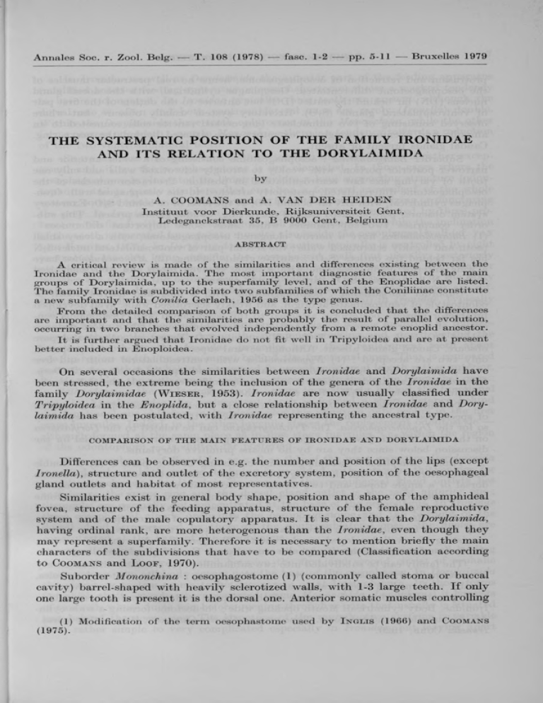
THE SYSTEMATIC POSITION OF THE FAMILY IRONIDAE AND ITS RELATION TO THE DORYLAIMIDA by A. COOMANS and A . V A N D E R H E ID E N Instituut voor Dierkunde, Rijksuniversiteit Gent, Ledeganckstraat 35, B 9000 Gent, Belgium ABSTRACT A critical review is made of the similarities and differences existing between the Ironidae and the Dorylaimida. The most important diagnostic features of the main groups of Dorylaimida, up to the superfamily level, and of the Enoplidae are listed. The family Ironidae is subdivided into two subfamilies of which the Coniliinae constitute a new subfamily with Conilia Gerlach, 1956 as the type genus. From the detailed comparison of both groups it is concluded that the differences are important and that the similarities are probably the result of parallel evolution, occurring in two branches that evolved independently from a remote enoplid ancestor. It is further argued that Ironidae do not fit well in Tripyloidea and are at present better included in Enoploidea, On several occasions the similarities between Ironidae and Dorylaimida have been stressed, the extreme being the inclusion of the genera of the Ironidae in the family Dorylaimidae (W ie s e r , 1953). Ironidae are now usually classified under Tripyloidea in the Enoplida, but a close relationship between Ironidae and Dory­ laimida has been postulated, with Ironidae representing the ancestral type. COMPARISON OF THE M AIN FEATU RES OF IR O N ID A E A N D D O R Y LA IM ID A Differences can be observed in e.g. the number and position of the lips (except Ironella), structure and outlet of the excretory system, position of the oesophageal gland outlets and habitat of most representatives. Similarities exist in general body shape, position and shape of the amphideal fovea, structure of the feeding apparatus, structure of the female reproductive system and of the male copulatory apparatus. It is clear that the Dorylaimida, having ordinal rank, are more heterogenous than the Ironidae, even though they may represent a superfamily. Therefore it is necessary to mention briefly the main characters of the subdivisions that have to be compared (Classification according to Coom ans and L oof , 1970). Suborder Mononchina : oesophagostome (1) (commonly called stoma or buccal cavity) barrel-shaped with heavily sclerotized walls, with 1-3 large teeth. If only one large tooth is present it is the dorsal one. Anterior somatic muscles controlling (1) Modification of the term oesophastome used by (1975). I n g l is (1966) and Coomans protrusion and retraction of oesophagostome, hence no real protractor muscles of the oesophagostome differentiated. Oesophagus cylindrical, with the dorsal gland nucleus (DN) far behind the outlet (DO) but anterior to the outlets of the first pair of ventrosublateral glands (S]0). Excretory system usually obscure, but where observed consisting of two uninucleate long-necked renette cells connected to an ampulla and opening through an excretory pore situated behind the nerve ring. Caudal glands present or absent. Suborder Bathyodontina : oesophagostome consisting of a wider anterior and narrower posterior portion, with weakly to strongly sclerotized w'alls and only one tooth of varying size but ventrosublateral in position. Protractor muscles of the oesophagostome differentiated, posteriorly attached to the oesophageal wall. Oeso­ phagus cylindrical, with DN far behind DO, at the level of or behind SiO. Excretory system obscure, pore situated behind nerve ring. Caudal glands present. This sub­ order comprises two superfamilies which show some important differences : (1) Bathyodontoidea have a narrow elongated oesophagostome, with a very small tooth and weakly sclerotized walls ; the second pair of ventrosublateral nuclei S2N lies far behind the outlets (S2O) ; cardiac glands lacking. (2) Mononchuloidea have a wide anterior oesophagostome with a large, grooved tooth and well sclerotized wralls, S2N lie opposite S2O ; cardiac glands present. Suborder Dorylaimina : oesophagostome with a long and narrow tooth or odontostyle of ventrosublateral origin, and weakly sclerotized walls. Well developed protractor muscles posteriorly attached to the oesophageal wall. Oesophagus con­ sisting of a narrow anterior part and a wider posterior one. DN a short distance behind DO, well anterior to SiO ; SiN opposite SiO. Excretory system and pore obscure. Caudal glands absent. Although several superfamilies have been proposed, only two are accepted : (1) Nygolaimoidea with ventrosublateral tooth and free cardiac glands ; both SiN at about the same level and equally developed. (2) Dorylaimoidea with an axial odontostyle and usually no free cardiac glands ; SiN usually at different levels and S1 N1 often smaller than S1 N2 . Two more suborders (Diphtherophorina and Trichosyringina) show a number of specialised and aberrant characters that obscure their origin. This is especially so for the Diphtherophorina. The Trichosyringina can be related to the Dorylaimina on the basis of their juvenile stages. Both groups however are not essential for the discussion below since they are by no means primitive dorylaims. Family Enoplidae : oesophagostome consisting of two parts : (1) a double walled anterior part with three single, two single ventrosublateral and one double dorsal or a single dorsal and two double ventrosublateral teeth ; (2) an elongated odontophore region. Protractor muscles controlling protrusion of oesophagostome intra-oesophageal ; 4 retractor muscles outside oesophagus. Oesophagus cylindrical with 5 glands, the nuclei of which occur at the basis of the oesophagus ; outlets only known for the anterior three glands : SiO anterior to DO. Excretory system consisting of a well developed, single renette cell, opening medio-ventrally between the first and second circlet of cephalic sense organs. Caudal glands usually present. The family can be subdivided into two subfamilies : (1) Ironinae with anteriorly attenuating body ; relatively narrow mouth opening ; usually flattened spicules, usually with median sclerotization and ventral flange ; gubernaculum with sclero­ tized proximal and lateral margins of the corpus ; and (2) Coniliinae n. subf. : Ironidae. Body cylindrical. Mouth opening wide ; cheilostome forming a wide cylin­ der. Spicules long and tubiform. Type genus : Conilia G e r l a c h , 1956 ; other genus : Ironella C o b b , 1920. In Ironidae as well as Dorylaimida the anterior part of the feeding apparatus (modified anterior feeding apparatus or oesophagostome) shows a marked tendency to become elongated. In Ironidae typically three teeth are present, although the dorsal one is often and both ventrosublateral ones are rarely (Ironella) double. Within the Dorylaimida three teeth only occur in the Mononchina, while the other forms possess one tooth ; even in Mononchina there is a tendency towards a reduction of the two ventrosublateral teeth. So we see that the occurrence of three teeth in the Dorylaimida is rather excep­ tional and, if so, the oesophagostome is not elongated. In those cases where the oesophagostome is elongated its lining provides a long supporting structure (odontophore) and is partly double walled enabling a forward movement of the whole system, so that teeth or tooth can protrude from the mouth for seizing or puncturing the prey. In Ironidae, protraction of the oesophagostome is mediated by three pro­ tractors confined within the oesophageal wall (one per sector), while the inclination of the teeth is operated by separate muscles also inside the oesophageal wall (cf. v a n der H e id e n , 1975). In Dorylaimida tins protraction typically is controlled by eight protractor muscles lying outside the oesophageal wall, but usually posteriorly attached to it. The retraction system is similar in both groups in that the retractor muscles are outside the oesophageal wall, attach to it anteriorly and to the body wall posteriorly. However, the number and position of retractor muscles are dif­ ferent : typically four (two subventral and two laterodorsal) in Ironidae, typically eight submedian ones in Dorylaimida. In both groups the teeth (or tooth) are (is) replaced during moulting by replace­ ment teeth (tooth) formed during the previous moulting and stored behind the functional teeth (tooth). Ironidae - juveniles have their replacement teeth about one lip-region width (or even more) behind the functional ones. That is compared to teeth-size rather far behind, compared to oesophagostomal length rather anterior. In Mononchina the replacement teeth are stored partly inside the functional ones ; in Bathyodontina the replacement tooth occurs immediately behind the functional one ; in Nygolaimoidea the replacement tooth is formed a short distance behind the functional one, whereas in Dorylaimoidea this situation only occurs in the first stage juveniles. Indeed, in the other juvenile stages the replacement odontostyle although formed at the same place as in first stage juveniles, i.e. within the region of the odontophore — is shifted far posteriad. The oesophagus of Enoplidae as well as this of the most primitive Dorylaimida is cylindrical ; in both groups its lining is provided with cuticular thickenings for muscle attachment. In Dorylaimida none of the oesophageal gland outlets lies anterior to the nerve ring and the dorsal gland outlet is the most anterior one ; the nuclei are normally not concentrated at the base of the oesophagus. In Ironidae three oesophageal glands open into the oesophagostome and the opening of the dorsal gland is preceeded by those of the ventrosublateral ones ; the nuclei are concentrated at the base of the oesophagus. The excretory system of Ironidae consists of a longnecked single cell leading to a medioventral pore situated between the first and second circlet of cephalic sense organs ; the cell body occurs near the base of the oesophagus. In Dorylaimida the excretory system seems to be degenerate or at least obscure. In those forms for which the system has been reported (some Mononchida, Longidorus) it consists of two cells whose ducts join before opening through a pore that usually is situated just behind the nerve ring. The structure of the reproductive systems is variable from rather simple to very complicated especially in Ironidae, but leaving apart the secondary complications, the male as well as the female reproductive system of both groups resemble each other in gross morphology. The greatest variation is found in the uterus and although this may be useful to differentiate between the lower taxa, it is not reliable to trace evolutionary lines between higher ones. Until more is known about the cellular anatomy of the systems in both groups com­ parisons are difficult. d is c u s s io n A critical appraisal of the similarities between Ironidae and Dorylaimida leads to the conclusion that they more likely are the result of parallel evolution rather than of close relationship. The mechanism by which teeth, tooth-like structures or spears are protruded by the action of protractor muscles upon a rigid, sclerotized tube has originated independently in several groups of nematodes. The elongation of the anterior feeding apparatus is apparently advantageous for the functioning of such a system. An elongation has been achieved in all Ironidae and concerns the odontophore region, but has only been fully achieved in the more specialised Dorylaimida where it also concerns the tooth and the region around it. The elongation apparently was not present in the ancestral form of the Dorylaimida and originated within the group, probably in two steps ; it was accompanied by a reduction of the teeth to one. The protractor system in Ironidae is clearly of oesophageal origin, that of Dorylaimida may be of somatic origin or derived from the sheath that surround the oesophagus. Tooth formation and especially storage of a replacement tooth at some distance behind a functional one is correlated with tooth-size, thickness of the oesophagostomal wall and with the functioning of the anterior feeding apparatus. The phe­ nomenon occurs also in other groups (cf. Chromadorida), though less pronounced. In any case it is evident that the condition in which the replacement tooth is stored at some distance behind the functional one has been achieved independently in Ironidae and Dorylaimida. indeed, the most primitive Dorylaimida have the replace­ ment tooth inside or immediately behind the functional one. Cuticidar thickenings of the oesophageal lining for muscle attachment are rather rare outside Ironidae and Dorylaimida, they are nevertheless occasionally found in other forms (e.g. Eurystomina and Thoracostoma, cf. Chitw ' ood & C h it ­ w ood, 1950). An important difference seems the position of the nuclei and outlets of the oeso­ phageal glands. Since all Dorylaimida are comparable in having the outlets and nuclei behind the nerve ring this character was probably present in the ancestral form. On the other hand it should be stressed that this difference may not be over­ emphasized. Indeed, no other group has developed this situation and hence it can be considered as something typical for Dorylaimida (a synapomorphy). Little in­ formation is available about the excretory system of Dorylaimida except that it usually is considered to be reduced. If the systems so far described represent the typical situation, it is basically different from that of Ironidae. So, while a number of differences can be attributed to special adaptions within each group, some of them seem to be fundamental. In the past the Ironidae too often have been compared with the more specialised Dorylaimina, while the more primitive Mononchina and Bathyodontina were overlooked. Therefore it seems that at present sufficient knowledge is lacking to say that the Dorylaimida originated from forms near the Ironidae. P o s s ib le P evo l u t i o n = p i e s i omo r p h of Iro n id a e and D o ry la im id a from E n o p lid a an cesto r, A = apom orph A IL o f oesophagostom e; r e d u c t i o n o f t e e t h t o one v e n tro -su b lateral IRONIDAE L engthening o f oesophagostom e ; p rotractor system o f oesop h agos tom e i n s i d e th e s t o m o d e a l w a l l ; r e p l a c e m e n t t e e t h a t some d i s ­ ta n ce beh in d f u n c t i o n a l ones P o s t e r i o r s h i f t o f o e so p h ag e a l glan d o u t l e t s ; DN and SjN f a r b e h i n d DO r e s p . S^O ; rep la cem en t t e e t h im m e d iate ly behind f u n c t i o n a l o n e s ; p r o t r a c t o r s y s t e m o f o e s o p h a g o s to m e o u t s id e th e stom odeal w a l l P a ' ENOPLID ANCESTOR w i t h w i d e o e s o p h a g o s t o m e and t h r e e e q u a l l y d e v e l o p e d t e e t h ; DN and S^N v e r y f a r b e h i n d DO r e s p . S^O IR O N ID A E \ L e n gA?t h e n i n g OF O e s o p h a g o s t o m e w i t h w i d e a n t e r i o r and narrow p o s t e r i o r p a rt ; to o th l a r g e , g r o o v e d ; S2N o p p o s i t e S2O ; 3 c a r d i a c glands T u b u la r oesophagostom e S2N f a r b e h i n d S2O ; tooth sm all POSITION L e n g t h e n i n g and n a r r o w i n g o f an terio r o e s o p h a g o s t o m e and t o o t h ; r e p l a c e m e n t t o o t h some d i s t a n c e b e h i n d fu n ctio n al o n e ; o e s o p h a g u s f l a s k s h a p e d ; DN l e s s f a r b e h i n d DO ; S^N o p p o s i t e S] 0 SYSTEMATIC R eduction of c a r d ia c g la n d s ; o d o n t o s t y l e ; backward s h i f t o f rep lacem en t od on tostyle Figure 1 represents possible evolutionary pathways of Ironidae and Dorylaimida. If this scheme is more substantiated by further findings it will imply some taxo­ nomic changes, but at the moment it is judged to early to do so. Concerning the position of the Ironidae within the Enoplida, there seem to be at least as many arguments for an inclusion in the Enoploidea as in 7 ripyloidea. According to G e r l a c h & R i e m a n n (1974) the Tripyloidea comprise four families : Tripylidae, Prismatolaimidae, Ironidae and Cryptonchidae. The position of the latter family is doubtful (see C o o m a n s & L o o f , 1970). Tripylidae and Pris­ matolaimidae are relatively small forms, mainly from freshwater and soil. Their cuticle is often annulated ; the amphideal fovea occurs at some distance behind the lips instead of immediately behind them ; the oesophagostome is very different from that in Ironidae : a simple collapsed tube with dorsal tooth or funnel-shaped in Tripylidae, barrel-shaped with dorsal tooth in Prismatolaimidae. The oesophageal lining has no cuticular thickenings for muscle attachment. The oesophago-intestinal junction is prominent. According to C h i t w o o d & C h i t w o o d (1950), C l a r k (1961) and D e C o n i n c k (1965) Enoploidea can be differentiated from Tripyloidea mainly bv the duplicate head cuticle, resulting from a fluid filled space (cephalic ventricle of I n g l i s , 1964), although according to I n g l i s (1964, p. 271-290) this structure may be lacking. Some Ironidae as Dolicholaimus and Trissonchulus have a cephalic ventricle. All together it seems that a cephalic ventricle is not a constant character of the E no­ ploidea and that its partial absence in Ironidae cannot be an objection for the inclusion of the latter in the former group. I n g l i s (1964) noted the presence of supplementary sense organs (cephalic slits) in Enoplidae as well as Ironidae. Medioventral preanal supplements are lacking or few in number in males of the Ironidae (in fact only 1 in well documented cases) and this is in agreement with the diagnosis of Enoploidea. Therefore we are inclined to support I n g l i s (1964) in considering Ironidae as closely related to Enoploidea and to remove them from Tripyloidea. The latter superfamily then comprises only 2 or 3 families that need careful re­ examination and Ironidae are included in Enoploidea. REFEREN CES B. G . & C h i t w o o d , M. B. (1950) — An introduction to nematology. Monumental Printing Co., Baltimore, Md. 213 pp. C la r k , W . C. (1961) — A revised classification of the order Enoplida (Nematoda). A .Z. Jl Sei 4. 123-150. C o o m a n s , A . (1975) — Morphology of Longidoridae. In : Nematode vectors of plant viruses (Ed. : Lamberti, F .; Taylor, C. E. & Seinhorst, J. W .). Plenum Press, London, pp. 15-37. C o o m a n s , A . & L o o f , P. A . A . (1970) — Morphology and taxonomy of Bathyodontina (Dorylaimida). Nematologica 16, 180-196. D e C o n i n c k , L. (1965) — Classe des nematodes. Systématique des Nematodes et sousolasse des Adenophorea. In : Traité de Zoologie (Éd. Grassé, P. P.) 4, 586-681. G e r l a c h , S. A . & R i e m a n n , F. (1974) — The Bremerhaven checklist of aquatic nema­ todes. A catalogue of Nematoda Adenophorea excluding the Dorylaimida. Part 2. Verbiff. Inst. Meeresforsch. Bremerh. Suppl. 4, 405-736. H e i d e n , A ., v a n d e r (1975) — The structure of the anterior feeding apparatus in members of the Ironidae (Nematoda : Enoplida). Nematologica 20 (1974), 419-436. I n g l i s , W . G. (1964) — The marine Enoplida (Nematoda) : a comparative study of the head. Bidl. Br. M us. nat. Hist. (Zool.) 11, 263-376. I n g l i s , W . G. (1966) — The origin and function of the clieilostomal complex in the nematode Falcaustra stewarti. Proc. Linn. Soc. Lond. 177, 55-62. W i e s e r , W . (1953) — Free-living marine nematodes. I. Enoploidea. Acta Univ. Lund., N .F. 2, 49, 1-155. Ch it w o o d , ADDENDUM After this paper was written we discovered that Andrâssy (1976) had already subdivided the family Ironidae into two subfamilies and had proposed a new sub­ family Thalassironinae for those forms with 10 (6 + 4) well developed cephalic setae. He listed four genera in alphabetic order under this subfamily, viz. Conilia Gerlach, 1954 ; Ironella Cobb, 1920 ; Parironus Micoletzky, 1930 and Thalassironus de Man, 1889. No type genus was indicated. This taxon is based on synplesiomorphy and considered to be polyphyletic, hence not accepted here. REFE R E N C E I. (1976) — Evolution as a basis for the systematization of nematodes. Pitman Publishing, London. A n drâssy,
