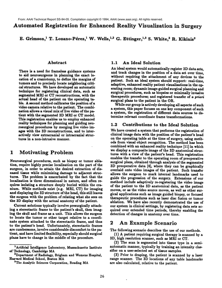
From: AAAI Technical Report SS-94-05. Compilation copyright © 1994, AAAI (www.aaai.org). All rights reserved.
Automated Registration
for Enhanced Reality
E. Grimson, 1 T. Lozano-P~rez, 1 W. Wells, 1,2 G. Ettinger,
Abstract
Motivating
in Surgery
1,s S. White, s ~
R. Kikinis
1.1
An Ideal Solution
Anideal system wouldautomatically register 3Ddata sets,
and track changesin the position of a data set over time,
without requiring the attachment of any devices to the
patient. Suchan ideal system shonid support: real-time,
adaptive, enhancedreality patient visualisations in the operating room; dynamicimage-guidedsurgical planning and
surgical procedures, such as biopsies or minimallyinvasive
therapeutic procedures; and registered transfer of a Fr/Or/
surgical plans to the patient in the OR.
Whileour group is actively developingall aspects of such
a system, this paper focuses on one key componentof such
a system, the registration of different data sources to determine relevant coordinate frame transformations.
There is a need for frameless guidance systems
to aid neurosurgeonsin planning the exact location of a craniotomy,to define the marginsof
tumorsand to precisely locate neighboringcritical structures. Wehave developed an automatic
techniquefor registering clinical data, such as
segmentedMRIor CTreconstructions, with the
actual head of the patient on the operating tsble. Asecondmethodcalibrates the position of a
video camerarelative to the patient. Thecombination allowsa visual mixof live video of the patient with the segmented 3D MR/orCT model.
This registration enables us to employenhanced
reality techniquesfor planning and guiding nellrosurgical procedures by merginglive video images with the 3D reconstructions, and to interactively viewextracranial or intracraniai structures in a non-intrusive manner.
1
Visualization
Problem
Neurosurgical procedures, such as biopsy or tumor ablation, require highly precise localization on the part of the
surgeon, in order to attain the desired extraction of diseased tissue while minimizingdamageto adjacent structures. The problem is exacerbated by the fact that the
Iocalisation is three dimensionalin nature, and often requires isolating a structure deeply buried within the cranium. While methods exist (e.g. MR.I, CT) for imaging
anddisplayingthe 3Dstructure of the head, this still leaves
the surgeonwith the problemof relating what she sees on
the 3D display with the actual anatomyof the patient.
Current solutions typically involve presurgically attaching a stereotactic frame to the patient’s skull, then imaging the skull and frame as a unit. This allows the surgeon
to locate the tumoror other target relative to a coordinate system attached to the stereotactic frame, and thus
to the patient’s head. Unfortunately, stereotactic frames
are cumbersome,
involve considerable discomfort to the patient, andhavelimited flexibility, especiallyshouldsurgical
plan. have to change in the middle of the procedure.
IArtificia] IntelligenceLaboratory,Massachusetts
Institute
of Technology,CambridgeMA
=Departmentof Radiology, Brighamand Womens
Hospital,
Harvard Medical School, Boston MA
3TheAnalytic Sciences Corporation, ReadingMA
26
1.2
Contributions
to the Ideal Solution
Wehave created a system that performsthe registration of
clinical imagedata with the position of the patient’s head
on the operating table at the time of surgery, using methods from visual object recognition. The methodhas been
combinedwith an enhancedreality technique [11] in which
we display a compositeimage of the 3D anatomical structures with a view of the patient’s head. This registration
enables the transfer to the operating roomof preoperative
surgical plans, obtained through analysis of the segmented
3D preoperative data [4], where they can be graphically
overlaid onto video images of the patient. Such transfer
allows the surgeon to mark internal landmarks used to
guide the progression of the surgery. Extensions of our
methodinclude adaptively re-registering the video image
of the patient to the 3D anatomical data, as the patient
moves,or aa the video source moves,aa well as other surgical applications such as imageguided biopsy, or focused
therapeutic procedures such as laser disc fusion or tumor
ablation. Wehave also recently demonstrated the use of
our systemin clinical settings, by registering data sets acquired over extended time periods, thereby enabling the
detection of changes in anatomyover time.
2
An Example Scenario
The following scenario describes the use of our methods.
(1) A patient requiring surgical therapy is scannedby
3D, high resolution scanner, such aa MR/orCT.
(2) The scan is segmentedinto tissue type in a semiautomatic manner,typically by training an intensity classifter on a user-selected set of tissue samples.
(3) Prior to draping, the patient is scannedby a laser
range scanner. The 3D locations of any table landmarks
are also calculated, relative to the patient.
[) The MR/or CT scan is automatically registered to
patient skin surface depth data from the laser ranger.
, provides a transformation from MRI/CTto patient.
,) The position and orientation of a video camerarele,to the l~tient is determined, by matching video images
~e laser points on an object to the actual 3Dlaser data¯
provides a transformation from patient to camera.
) The registered internal anatomy is displayed in en’ed reality visualization [I 1] to "see" inside the patient,
the two computed transformations are used to transthe 3D model into the same view as the video image
Le patient, so that video mixing allows the surgeon to
~oth images simultaneously.
) The patient is draped and surgery is performed. The
~ced reality visualization does not interfere with the
:on, but provides her with additional visualisation ination to greatly expand her limited field of view.
) The location of table landmarks can be continually
:ed to identify changes in the position of the patient’s
ude, relative to the visualization camera. Viewer
ion can also be continually tracked to identify any
ges. Visualisation updates are performed by updathe patient to viewer transformation¯
) In general, the surgery is executed with an accurately
~ered enhancedvisualisation of the entire relevant paanatomy, and thus with reduced side effects.
Details
of Our Approach
I of this scenario is standard practice. Methodsexist
’art 2 [4]. Parts 8-9 are part of our planned future
¯ Here, we focus on parts 3-7, where the key step is
egistration of data obtained from the patient in the
Lting room with previously obtained data.
." use a multi-stage matching and verification of a 3D
set acquired at the time of the surgery with 3D clini¯ ta sets acquired previously. The central ideas are to
laser striping device to obtain 3D data from the p~s skin, and to use a sequence of recognition techniques
~tch this data to segmented skin data from the MR/
t ~ reconstruction. These techniques allow us to accur register the clinical data with the current position of
atient, so that we can display a superimposed image
: 3D structures overlaid on a view of the patient¯
e basic steps of our method are outlined below.
Model input
btain a segmented 3D reconstruction of the patient’s
,my, for example using CTor MRI. The segmentation
~ically done by training an intensity classifier on a
elected set of tissue samples, where the operator uses
ledge of anatomy to identify the tissue type. Once
I training is completed, the rest of the scans can be
~atically classified on the basis of intensities in the
ed images, and thus segmented into tissue types [4].
naticaily removinggain artifacts from the sensor data
¯ used to improve the segmentation [12].
is 3D anatomical reconstruction is referred to as the
1, and is represented relative to a model coordinate
¯ For simplicity, the origin of the coordinate system
e taken as the centroid of the points.
27
Figure I: Exampleof registered laser data (shown as large
dots) overlaid on CTmodel.
Data input
3.2
Weobtain a set of 3D data points from the surface of the
patient’s skin by using a laser striping device. For our purposes, the laser simply provides a set of accurate 3d point
measurements, obtained along a small (5-10) set of planar
slices of the object (roughly 240 points per slice). This
information is referred to as the data, and is represented
in a coordinate frame attached to the laser, which reflects
the position of the patient in a coordinate frame that exists in the operating room. Our problem is to determine a
transformation that will map the model into the data in a
consistent manner.
3.3
Matching data sets
Wematch the two data sets using the following steps.
(1) First, we sample a set of views of the model. For
each view, we use a s-buffer method to extract a sampled
set of visible points of the model. For each such model, we
execute the matching process described below.
(2) Next, we separate laser data of the patient’s head
from background data. Currently we do this with a simple
user interface. Note that this process need not be perfect,
we simply want to remove gross outliers from the data.
From this data, we find three widely separated points that
come from the head.
(3) Weuse constrained search [6] to match triples of visible sampledmodel points to the three selected laser points.
The method finds all such matches consistent with the relative geometry of each triple, and for each we compute
the coordinate frame transformation that maps the three
laser points into their corresponding model points. These
transformations form a set of hypotheses. Note that due to
sampling, these hypothesized transformations are at best
approximations to the actual transformation.
In examples such as Figure 1, there are typically ~ 1000
laser sample points, and the model has typically 40,000
sample points. Given a view, and a coarsely sampled zbuffer, there are typically I000 model points in the sampled
view. In principle, there are ~ 10Is possible hypotheses,
but ,i|ng simple distance constraints, there are usually
I00, 000 possible hypotheses that remain.
(4) Weuse the Alignment Method[7] to filter out those
hypotheses, by transforming all the laser points by the bypothesised transformation, and verifying that the fraction
of the transformed laser points without a corresponding
model point within some predefined distance is less than
some predefined bound. Wediscard those hypotheses that
do not satisfy this verification.
(5) For each verified hypothesis, we refine as follows:
(5.1) Evaluate the current pose. Thus, if ~ is a vector
representing a laser point, mj is a vector representing a
model point, and T is a coordinate frame transformation,
then the evaluation function for a particular pose is
ICT) = 2_-,2..,e
,., .
(I)
j
This objective function is similar to the posterior marginal
pose estimation (PMPE) method used in [I0]. This Gaussian weighted distribution is a methodfor roughly interpolating between the sampled model points to estimate the
nearest point on the underlying surface to the transformed
laser point. Because of its formulation, the objective function is generally quite smooth, and thus facilitates "pulling
in ~ solutions from moderately removed locations in parameter space. As well, it bears some similarity to the radial
basis approximation schemes used for learning and recognition in other parts of computervision (e.g. [2]).
(5.2) Iteratively maximizethis evaluation function using
Powelrs method. This yields an estimate for the pose of
the laser points in model coordinates.
(5.3) Execute this refinement and evaluation process using a multiresolution set of Gaussians.
(5.4) Using the resu]tlng pose of this refinement, repeat
the pose evaluation process, now using a rectified least
squares distance measure. In particular,
perform a second sampling of the model from the current viewpoint, using a finer sampled s-buffer. Relative to this finer model,
evaluate each pose by measuring the distance from each
transformed laser point to the nearest model point, (with
a cutoff at some predefined maximumdistance). Evaluate
the pose by summingthe squared distances of each point.
Minimize using Powel]’s method to find the least-squares
pose solution. Here the evaluation function is
E2(T)-~min{d2m,x,m~.nlT/.i-mjl
2 } (2)
$
where ~x is some preset maximumdistance. This objective function is essentially the same as the MAPmatching scheme of [10], and acts much llke a robust chamfer
matching scheme (e.g. [8]). This second objective function
is more accurate locally, since it is composedof saturated
quadratic forms, but it is also prone to sticking in local
minima. Hence we add one more stage.
(5.5) To avoid locai minima traps, randomly perturb
the solution and repeat the ]east squares refinement. We
continue, keeping the new pose if its associated RMSerror
is better than our current best. Vv’e terminate this process
28
..........
~....
~_.i
.......
"........
~........
.........
~........
~........
~........
,,,, ....... ~
s.o e.i
~ .... ~ ....
~o s.s
..s
m.~
Figure 2: Histogramof residual errors for pose of Figure 1.
when the number of such trials that have passed since the
RMSvalue was last improved becomes larger than some
threshold.
(5.6) The final result (Figure I) is a pose, and a measure
of the residual deviation of the fit to the model surface.
Wecollect such solutions for each verified hypothesis,
over nil legal view samples, and rank order them by smallest P_MSmeasure. The result is a highly accurate tra~sformation of the MRIdata into the laser coordinate frame.
3.4
Camera Calibration
Oncewe have such a registration, it can be used for surgical
planning. A video camera can be positioned in roughly the
viewpoint of the surgeon, i.e. looking over her shoulder.
If one can calibrate the position and orientation of this
camera relative to the laser coordinate system, one can
then render the aligned MR/or CT data relative to ~he
view of the camera. This rendering can be mixed with the
live video signal, giving the surgeon an enhanced reality
view of the patient’s anatomy [11]. This can be used for
tasks such as planning a craniotomy or a biopsy, or defining
the margins of an exposed tumor for minimal excision.
Wehave investigated two methods for calibrating the
camera position and orientation, one using a calibration
object of knownsize and shape, and one using an arbitrary
object (such as the patient’s head). In each case, matching
3D laser features against video images of those features
allows us to solve for the position of the camera.
4
Testing
the Method
Wehave run a series of controlled experiments, in which
we have registered a CTreconstruction of a plastic skull
with laser data extracted for a variety of viewpoints. In all
cases, the system finds a correct registration, with typical
residual RM$errors of 1.6 millimeters.
Wehave also run a series of trials with actual neurosurgery patients. An example registration of the laser data
against an MRImodel of the patient is shown in Figure 3.
Note that in this case, while most of the scalp had been
shaved for surgery, a patch of hair was left hanging down
over the patient’s temple. As a result, laser data coming
from the hair cannot be matched against the segmented
skin surface in the MRImodel, and this showsup as a set of
points slightly elevated above the patient’s skin surface in
the final registration. Wecan automatically remove these
re 3: Exampleof registered laser dsts (shown as large
overlaid on an ~ model. This is n case of registration
actual neurosurgical case, with the patient fully prepped
srgery before the laser data is acquired.
Figure 4: Usins the results of Figure 3, and given a calibration
of a video camerarelative to the laser, we can overlay parts of
the MRImodelon top of s video view of the patient, providing
an enhancedreality visualization of the tumor. In this figure,
the tumor is shownin green, and the ventricles are displayed
as a landmark
inblue.
~s, and reregister the remaining data. Also displayed
he internal positions of the tumor and the ventricles.
RMSerror in this case was 1.9ram. Finally, given the
tration between the patient and the model (by matchhe laser data in this manner) we can transform the
:l into the coordinate system of a second video camand overlay this model on top of the camera’s video
¯ This is shownin Figure 4.
mentioned earlier, the method has applications for
ca] planning and guidance, including tumor excision
)iopsy. The method has broader application, however,
ding the registration of multiple clinical data sets such
RI versus CT. A companion paper [5] discusses the
cation of our methodto change detection studies for
ing lesion growth in patients with multiple sclerosis.
Related
~
Imagesof the HeadUsing Probability and ConnectiVity.
;~AT’ ~4(e):10~7-~045.
[5] Ettinger, G.J. , W.E.L. Grimson, and T. Losano-P~res,
1994 "Automatic 3D Image Registration for Medical
Change Detection Applications ~, AAA/1994Spring SymposinmSeries, .4pp|ica~on~ of ComputerV, ion in ~d~edica] ImageProcessing.
[6] Grimson, W.E.L., 1990, Object RecoFnlt~onb~l Computer:
The ro|e of geometr/c constraints, MITPress, Cambridge.
[7] Hutten]ocher, D. and S. Ullman, 1990, "itecognizing Solid
Objects by Alignmentwith an Image,~ Int..I. Comp.Vi:.
[8] 3lung, H., It.A. Itobb and K.S. Holton, 1992, UANewApproach to 3-D Registration of Multimodallty Medical Images by Surface Mat~M~g",
in Vbualiza~ion in Biomedical
Computing- SPIE: 196-213.
[9] Peli~sarl, C.A.; Chen, G.T.Y.; Spelbring, D.R.; Weichselbanm, R.R.; Chin-Tu
Chen, "Accurate three-dimensional
registration of CT, PET,and/or MRimages of the brain",
3CAT13(1):20-26, 1989.
W.M., 1993, ~ta~istical Object Recognitlon~Ph.D.
[10] Wells,
TheWs, MIT. (MIT AI Lab Tit 1398)
[11] W. Wells, R. I(;H~, D. Altobelli, W. Loreneen, G. Ettinger, H. Cline, P.L. Gleasonand F. Jolesz, 1993, "Video
Registration using Fiducials for Surgical EnhancedReality" JSth Gon£IEEEEng. Med. Biol. Soc..
[12] Wells, W.M.,R. Kit~,;~, F.A. Joless, and W.E.L. Grimson, 1993, "Statistical Gain Correction and Segmentation
of MagneticResonanceImaging Data", in preparation.
Work
al other groups have reported methods similar to
Of particular interest are three such approaches.
other groups use alternative least squares minimiza~ethods [9; 3] with some operator input to initialize
o guide the search. A third group [I] perform regis,n by matching ridge lines on surfaces.
’erences
kyache,
N.,.I.D.
Boissonnst,
L.Cohen,
B.Geiger,
J.Levy/ehel, O. Mongs,P. Sander, "Steps Towardthe Automatic
nterpretation of ~-DImages", In ~DImagingin J/’edicine,
:dited by H. Fuchs, K. Hohne,S. Piser NATO
ASI Series,
|prinser-Verlaa, 1990, pp 107-120.
]runelli,R and T. Pogglo, 1991, "HyberBFNetworksfor
teal Object Recognition", IJCAI, Sydney, Australia.
~hampleboux,
G.; Lsvallee, S.; Sseliski, It.; Brunie, L.,
:From accurate range imaging sensor calibration to acurate model-based ~D object local~stion’, IE~ Conf.
7omp.Via. Putt. Recog.:83-89, 1992
;line, H.E. and W.E. Lorenscu and R. I~i~-~, and F.
oless, 1990, "Three-Dimensional Segmentation of MR
29


