From: ISMB-93 Proceedings. Copyright © 1993, AAAI (www.aaai.org). All rights reserved.
Transmembrane
Sholom
M. Weiss
t
Segment Prediction
Sequence Data*
Dawn M.
Cohen
t
from Protein
Nitin
Indurkhya
Department of Computer Science,
Rutgcrs University
New Brunswick,
New Jersey
08903, USA
:~ Telecom Australia
Research Laboratories
770, Blackburn
Rd, Clayton,
Vic 3168, AUSTRALIA
Abstract
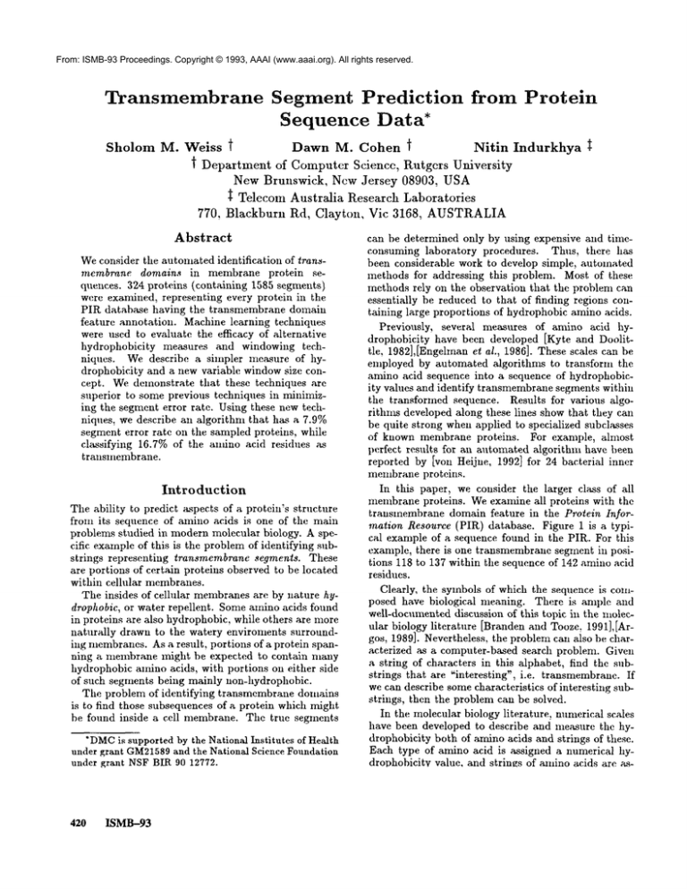
Weconsider tile automated identification of transmembrane domains in membrane protein sequences. 324 proteins (containing 1585 segrrmnts)
werc examined, representing every protein in the
PIR database having the transmembrane domain
feature annotation. Machine learning techniques
were used to evaluate the efficacy of alternative
hydrophobieity measures and windowing techniques. We describe a simpler measure of taydrophobicity and a new variable windowsize concept. Wedemonstrate that these techniques are
superior to some previous techniques in minimizing the segment error rate. Using these new techniques: we describe an algorithm that has a 7.9%
segment error rate on the sampled proteins, while
classifying 16.7% of the anfino acid residues as
transmembrane.
Introduction
The ability to predict aspects of a protein’s structure
from its sequence of amino acids is one of the main
problems studied in modern molecular biology. A specific exampleof this is the problem of identifying substrings representing transmembrane segments. These
are portions of certain proteins observed to be located
within cellular membranes.
The insides of cellular membranesarc by nature hydrophobic, or water repellent. Someamino acids found
in proteins are also hydrophobic, while others are more
naturally drawn to the watery enviroments surrounding membranes.As a result, portions of a protein spanning a membrane might be expected to contain many
hydrophobic aHfino acids, with portions on either side
of such segments being mainly non-hydrophobic.
The problem of identifying transmcmbrane domains
is to find those subsequences of a protein which might
be found inside a cell membrane. The truc segments
*DMC
is supported by the National Institutes of Health
under grant GM21589
and the National Science Foundation
under grant NSFBIR 90 12772.
420
ISMB-93
can be determined only by using expensive and timeeo1~suming laboratory procedures. Thus, there has
been considerable work to develop simple, automated
methods for addressing this problem. Most of these
methods rely on the observation that the problem can
essentially be reduced to that of finding regions containing large proportions of hydrophobic amino acids.
Previously, several measures of amino acid hydrophobieity have been developed [Kyte and Doolittle, 1982],[Engelmanet al., 1986]. These scales can be
employed by automated Mgorithms to transform the
amino acid sequence into a sequence of hydrophobicity values and identify transmembrane segments within
the transformed sequence. Results for various algorithms developed along these lines show that they can
be quite strong when applied to specialized subclasses
of known membrane proteins.
For example, almost
perfect results for an automated algorithm have been
reported by lyon Heijne, 1992] for 24 bacterial inner
membrane proteins.
In this paper, we consider the larger class of all
membraneproteins. Weexanfine all proteins with the
transmembrane domain feature in the Protein Information Resource (PIR) database. Figure 1 is a typical example of a sequence found in the PIR. For this
example, there is one transmembrane segment in positions 118 to 137 within the sequence of 142 amino acid
residues.
Clearly, the symbols of which the sequence is composed have biological meaning. There is ample and
well-docmuented discussion of this topic in the molecular biology literature [Branden and Tooze. 1991],lArgos, 1989]. Nevertheless, the problem can aiso be characterized as a computer-based search problem. Given
a string of characters in this alphabet, find the substrings that are ~interesting’, i.e. transmembranc. If
we can describe somecharacteristics of interesting substrings, then the problem can bc solved.
In the molecular biology literature, numerical scales
have been developed to describe and measure the hydrophobicity both of amino acids and strings of these.
Each type of amino acid is assigned a numerical hydrophobicitv value, and strings of a~nino acids are a~s-
5
1
31
61
91
121
PNIQN
SQTNV
NSAVA
SPESS
FRILL
10
PDPAV
SQSKD
WSNKS
CDVKL
LKVAG
15
YQLRD
SDVYI
DFACA
VEKSF
FNLLM
20
SKSSD
TDKTV
NAFNN
ETDTN
TLRLW
25
KSVCL
LDMRS
SIIPE
LNFQN
SS
30
FTDFD
MDFKS
DTFFP
LSVIG
Figure 1: Sequence for PIR entry Pirl:Rwhuac. Transmembrane segment is shown in bold.
signed a hydrophobicity value that is a function of
the individual hydrophobicities of their components.
Transmembrane segments have a tendency towards
large average hydrophobicities, despite the presence of
non-hydrophobic residues within them. (Table 1 shows
the hydrophobicity scales that have been used in this
study. Discr: a discretization of Kyte-Doolittle, sugested in [Arikawa e$ al., 1992]; K-D: Kyte-Doolittle
yte and Doolittle, 1982]; Eng: Engelman [Engelman
e~ el., 1986]; Heijne: [von Heijne, 1992])
The most common approach used in the molecular biology literature to the problem of identifying
transmembrane regions, has been to select a fixed
windowsize, e.g. 20 characters, and to sequentially
segment the string looking for regions where the average hydrophobicity exceeds some threshold. Researchers have developed varying indexes [Kyte and
Doolittle, 1982],[Engelmanet al., 1986] and classification algorithms [von Heijne, 1992], implemented in
computer programs, to predict transmembrane segments for newly sequenced, but unsegmented data.
Performance has been evaluated by comparing the predicted segments with the true segments of knownprotein sequences.
While the traditionM molecular biology approach
has been to program an algorithm directly based on
experience and reports in the research literature, there
has been at least one attempt to apply computerintensive machine learning techniques to a large volumeof segmented sequence data [Arikawa et al., 1992].
In that study, all transmembrane segments in the PIR
database were collected. However, samples of nontransmembrane segments were obtained by randomly
selecting segments of length 30 from all proteins (membrane and non-membrane) in the PIR database. The
size of the non-transmembrane set was very large about 28 times the size of the transmembrane set. Using these samples, they attempted to learn decision
trees which would separate the transmembrane from
non-transmembrane segments. In that study, decision
tree attributes were not based directly on hydrophobicity indexes, but rather on a restricted class of regular expressions 1 appearing in one class, but not the
other. Further experiments were considered by mapping each of the 20 aminoacids into one of three groups
based on the hydrophobieity index, Discr, of Table 1.
~K
I Patterns that contained don’t care subpatterns were
considered.
However,overall, their results are substantially weaker
than others reported in the molecular biology literature for hydrophobicity-based classification.
Moreover, while their experiments may provide someinsight
about characteristics of transmembrane regions, they
cannot be used directly to locate such regions in new
proteins.
In this paper we consider a different machinelearning approach for sequence analysis. Instead of regular
expressions, we return to the standard hydrophobicity
analysis. Weapply classifier learning techniques. This
requires that we find methods for transforming variable length segment training examples into fixed length
feature vectors. In this way, we can identify rules for
classifying whether a given sequence is or is not transmembrane.Weexamine the efficacy of alternative hydrophobicity measures and windowing techniques for
learning useful rules. Wedevelop a new, simpler measure of the hydrophobicity of a sequence and a new
variable window size concept. We demonstrate that
these techniques are superior to previous techniques,
in mininfizing the segment error rate. Finally, we show
how the results of simulation experiments can be used
to formulate a new segmentation algorithm that performs well on the membraneproteins in the large PIR
dataset. (The final step is necessary, because a good
solution to the problem of classifying segments does
not immediately provide a method for segmenting an
unlabeled new sequence. It is useful, however, for providing a relatively unbiased framework for comparing
alternative hydrophobicity measures, using computerbased train and test evaluations.)
Methods
The central goal of this work is to develop an algorithm
that can segment unlabeled new sequences of amino
acids of membraneproteins. The input is a string of
characters and the output is a list of the positions of
the transmembrane segments. Wewill describe several
experimental procedures that are compatible with this
goal. Machinelearning techniques are used to evaluate
and compare performance of alternative measurements
and techniques for determining if segments are transmembrane.
Computer-based techniques that learn from data
have several key advantages over strictly
humanderived solutions:
¯ The computer can search over a large space of pos-
Weiss
421
Code
I
V
L
F
C
M
A
G
T
S
W
Y
P
H
Q
N
E
D
K
R
Discr Scale
high
high
high
high
high
high
high
neutral
neutral
neutral
neutral
neutral
neutral
low
low
low
low
low
low
low
K-D Scale
4.5
4.2
3.8
2.8
2.5
1.9
1.8
-0.4
-0.7
-0.8
-0.9
-1.3
-1.6
-3.2
-3.5
-3.5
-3.5
-3.5
-3.9
-4.5
Eng. Scale
3.1
2.6
2.8
3.7
2.0
3.4
1.6
1.0
1.2
0.6
1.9
-0.7
-0.2
-3.0
-4.1
-4.8
-8.2
-9.2
-8.8
-12.3
He~jne Scale
0.971
0.721
0.623
0.427
1.806
0.136
0.267
0.160
-0.083
o0.119
-0.875
-0.386
-0.451
-2.189
-1.814
-1.988
-2.442
-2.303
-2.996
-2.749
Table 1: Hydrophobicity Scales Used In This Study
sibilities
timal.
and find a solution that is near or even op-
¯ The computer can hide some data, train on the remainder and test on the hidden data. Thus it can
sinmlate predictions for new sequences.
For our experiments, it is most appropriate to make
use of two related classification techniques: decision
trees or decision rules. A standard classification problern is formulated in terms of relating class labels to
particular features or measurements. In our experiments, the features are hydrophobicity values and positional information. Rules or trees identify ranges
of these variables appropriate for distinguishing the
classes "transmembrane" and "non-transmembranc’.
Numerical variables are searched over their entire
ranges in the data set to find cutoffs of minimumclassification error.
Several alternative methodsare available for obtaining good thresholds. Decision tree learners such as
CAI~T[Breiman et al., 1984] or C4 [Quinlan, 1987]
perform this search over a single variable at a time.
In this study, a system capable of performing more
extensive search was used, namely, the rule induction
program Swap-1 [Weiss and Indurkhya, 1991].
Rule induction methods, such as Swap-l, attempt to
find a compact covering rule set that completely separates the classes. The covering set is found by heuri~
tically searching for a single best rule that covers cases
for only one class. Having found a best conjunctive
rule for a class C, the rule is addedto the rule set, and
the cases satisfying it are removedfrom further consid422
ISMB-93
eration. The process is repeated until no cases remain
to be covered. Unlike decision tree induction programs
and other rule induction methods, Swap-1 h~ an advantage in that it uses optimization techniques to revise and improve its decisions. Once a covering set
is found that separates the classes, the induced set of
rules is further refined by either pruning or statistical
techniques. Using train and test evaluation methods,
the initial coveringrule set is then scaled to back to the
most statistically accurate subset of rules. For a more
detailed description of the Swap-1 learning method,
the reader is referred to [Weiss and Indurkllya, 1991].
Someexperiments involved only a single variable,
and the results for decision tree and rule induction
are identical.
Whenmore than one numerical variable is necessary, Swap-1 should give a better answer
because it is capable of optimizing nmltiple variables
and thresholds. It is also more likely to find a compact
solution which is amenable to algorithmic description.
A comparison of Swap-1 with CARTand other algorithms on several real-world applications can be found
in [Weiss and Indurkhya, 1991].
Just as important as the method of learning from
data is the technique, for evaluating performance. We
measured performance in terms of error rates estimated by 10-fold crossvalidation [Stone, 1974]. For
10-fold crossvalidation, the data are randomly partitioned in 10 groups of 10%of the cases. The computer
trains in turn on 90% of the cases and tests on the
remaining 10%, for each partition. The reported error
rate is the average of the 10 trials. This gives a relatively unbiased estimate of future performance [Stone,
1974].
Hydrophobicity
Indexes
There are two well-known hydrophobicity scales: [Kyte
and Doolittle, 1982] and [Engelman e~ al., 1986]. In
addition, a hydrophobicity scale has recently been described, derived specifically for bacterial inner niembranes [yon Heijne, 1992]. Weuse these scales, which
assign hydrophobicities to individual amino acids, in
order to define hydrophobicity indezes for strings of
amino acids. Wecompare the results for indexes derived from each of the scales.
In the simplest form of hydrophobicity index, i.e.
equation 1, the average hydrophobicity is computed
over a string, where n is the length of the string, and
hpi is the hydrophobicityof the ith residue in the string
(from Table 1). Current algorithms almost exclusively
search over a fixed windowsize n, for example a windowof size 21 as in [von Heijne, 1992]. This approach
can be modified slightly by assigning different weights
to each residue in a window,as a function of position
within the window (see equation 2). This method has
been used, giving the central region full weight, but
lower weights for the outer regions.
hpa~e--Ein_-i
hpi
~t
hpwa~e = En=lW, * hpi
n
(1)
(2)
With several different indexes of andno acid hydrophobicity that vary substantially, one might speculate that interpretation of the indexes over strings is
rather subjective. Thus, we proposed a new, simpler
measure that is described in equation 3. As in [Arikawa
et al., 1992], we mapthe 20 character alphabet into 3
groups: high, neutral, or low. These are listed in Table
1. Given a string coded in this 3-character alphabet,
we define a new measure hPdif , which is the difference
between the fraction of high values and low values in
the string. In equation 3, Nh is the number of high
characters and N1 is the number of low characters.
hpdi] =
N~- N1
(3)
The goal of this calculation is the same as the other
hydrophobicity indexes: to search for regions where
high values are found in greater abundance than low
values, hPdif can also be interpreted as a weighted
average hydrophobicity of the segment by assuming a
1/0/-1 weighting for the Discr Scale of Table 1. Unlike
the weighting of Equation 2 though, hp~f does not
assign weights according to position within the segment but simply depends on the number of high and
low characters in the segment. Beyondsimplicity, the
computation of hPdif has another interesting property.
Whensearching over a space of variable length n, its
maximumvalue occurs when the first and last characters of the substring are high.
hp Index
Kyte
Engelman
Heijne
Dif
.Error rate %
2.5
2.7
2.8
2.5
Table 2: Results of Experiment I
Experiments
and Results
Weperformed several computationally intensive experiments to determine which measures are best suited for
transmembrane domain identification,
using hydrophobicity information. For these experiments, we included
all proteins in the PIR database having at least one
~ransmembrane domain feature annotation. We found
324 such proteins, containing 635 (40%) transmembrahe segments and 950 (60%) non-transmembrane
(some of the proteins had more than one transmembrahe segment).
Experiment
I: Hydrophobicity
of
Segmented
Sequences
In this experiment, we compare samples of transmembrane segments and non-transmembrane segments to
test whether hydrophobicity indexes can discriminate
these two classes. Werepresent each segment by its
hydrophobicity index value, and input them to Swap1. Wecompare the accuracy of the rules learned using
each of the three indexes derived from the literature,
as well as the Dif index defined in the previous section. Predictive performance is measured by 10-fold
crossvalidation.
The results of experiment I are listed in Table 2. All
error rates are measured by crossvalidation. In this
experiment we found that Dif did as well as the index
based on the Kyte-Doolittle scale of hydrophobicity.
While the index based on Heijne’s scale was reported
to give almost perfect results for bacterial inner membrane proteins, it is slightly weakeron the larger class
of all membraneproteins.
By considering the classification of segmented sequences, this experiment does not exactly correspond
to the real-world problem where the segment boundaries are not knownfor a new sequence, but nevertheless the results of this experiment are useful because
they set an upper bound on potential performance.
Experiment
II:
Maximum Hydrophobicity
of Variable
Length Segmented Sequences
The strong results of experiment I indicate that it may
be possible to distinguish knownsegments by their hydrophobicities. However, there may be subsegments of
the knownsegments of each class which have hydrophobicities morecharacteristic of the other class. (The limiting case, of course, is a single hydrophobicaminoacid
in a non-traasmembrane segment, which considered by
itself would appear to be transmembrane.) The real
Weiss
423
Max liP Index Method
fixed window-He~ne
fixed window-Engelman
variable window-Heijne
variable window-Kyte
variable window-Engelman
variable window-Dif
Table 3: Results
Error rate
15.6
13.0
15.2
11.7
10.9
10.8
%
of Experiment II
hp greater than
2.5
2.0
1.5
1.0
.5
.0
Table 4: Results
sition 1
Predictive value %
93.5
89.4
79.0
69.5
62.3
56.7
of Experiment III:
Cases
covered
31
302
587
809
965
1079
Beginning at Po-
problem is to find the correct segmentation. Thus, we
must consider some measure that sunm:arizes a search
over the complete space of possible segmentations. The
ma,xinmm hydrophobicity
found for any substring of a
labeled segment can be used to characterize
that segment. If the hydrophobicity
for substrings of transmembrane segments were always greater than those for
non-transmembrane
segments, we would have the answer: look for segments that exceed a certMn threshold
and label these as transmembrane.
The standard technique for generating and evaluating substrings is to use a fixed window size with tapered edges. In our fixed window-size experiments, we
used the window specifications
of [von Heijne, 1992]
which have also been used by other researchers,
hidependently of the fixed window methods, we introduce
a variable window-size concept. Within a known segment, we search every substring of length between 10
and 70 and record the nlaxinmm hydrophobicity
index
as a feature of the string.
Positional information is also needed. "Signal segments", usually found at the beginning of the string,
minfic the hydrophobicity of transmembrane segments,
but should be classified
a.~ non-transmembrane. As the
rule induction program soon discovers,
the position
of tim maximum hydrophobicity
subsegment is quite
useful in differentiating
the signM and true transmembrane segments.
We use Swap-1 with the known segments,
represented by these two features,
namely the maximum
hydrophobicity
within a segment and the starting
position
of the maxinmm substring.
In this way,
we attempt to distinguish
transmembrane from nontransmembrane
segments, using data that would be
more readily available for new proteins.
In addition
to evaluating tile performance of different hydrophobicity indexes, we compare variable and fixed window
techniques.
The results of experiment II are listed in Table 3.
They clearly demonstrate the superiority
of variable
windows over fixed windows. The results
of these experiments nmve us much closer to the real-world problem.
drophobicity threshold above which all segments were
unambiguously transmembrane. If such a method held
for all membrane proteins,
then one could use such
thresholds to separate out the segments that could be
unequivocally labeled as transmembrane, leaving the
labeling of those with lower hydrophobicity values for a
more complicated analysis.
Experiment III is designed
to test whether such thresholds
can be obtained for
our larger set of proteins. The effectiveness
of possible thresholds as candidates for such filtering
can
be assessed by computing the positive predictive value
which measures the percentage of transmembranc segments in the set of segments that exceed the threshold. A value of 100% predictive value for a threshold
would indicate that M1 seginents above the threshold
are transmembrane. If it also covered a relatively large
number of M1 segments, it would allow us to generalize
the hypothesis in lyon Heijne, 1992] to all membram;
proteins.
We used the maximum hydrophobicity
within a segment to characterize
it and considered the predictive
value of using the hydrophobicity index at different
thresholds. Because of the significance
of signal segments, the experiments were also performed with a
starting position that would tend to exclude such segments.
The results of experiment III are listed in Tables 4
and 5. Here we take the full dataset and consider the
predictive value, i.e. the percentage of correct predictions, and the number of cases that are covered when
the fixed window index exceeds the specified thresholds. The results
would appear to indicate that for
our larger group of segments, thresholding on the hydrophobicity value of a segment alone cannot be used
to identify transmembrane segments. This is in contrast with the results of lyon Heijne, 1992] for the
smaller set of 24 bacterial
proteins which suggested
that segments with hydrophobicity above some threshold could bc definitely labeled as transmembrane, leaving those at lower values to a more complicated analysis.
Experiment
Predictive
For the set of
Heijne, 1992],
Segmentation
of Membrane
Proteins
After reviewing these results, we specified an algorithm
to segment unlabeled sequences.
This algorithm is
presented in Figure 2. The algorithrn uses a variable
length window and our hPdif index.
424
ISMB-93
III:
Hydrophobicity
Value
24 bacterial proteins considered in [volt
it was possible to a find a sequence hy-
hp greater than
2.5
2.0
1.5
1.0
.5
.0
Predictive value 70
93.5
93.9
88.6
81.1
71.5
64.3
Table 5: Results of Experiment III:
sition 25
Cases covered
31
279
491
651
793
899
Beginning at Po-
Starting with position 1, The algorithm searches for
a substring of length 10 to 70 that exceeds a ratio of
.712. It exanrines the longer strings first, and if necessary examines every possible substring. If the ratio is
exceeded, the substring is labeled as translncmbrane,
and the algorithm continues at the position following
the end of the substring.
Strings that exceed the .71 threshold are typically
short, on the order of length 10 to 15 characters. Experiment I showed that the correctly labeled segments
typically have an hPdif greater than .31. This naturally leads to an expansion routine that attempts to
expand the string with a ratio greater than .71 to a
longer string with a lower ratio. In the algorithm, an
expansion threshold of .35 was used 3. The expansion
routine is described in Figure 4. It can be efficiently
coded using dynamic progranmfing concepts.
The expansion process is equivalent to measuring another feature of the variable length segment under consideration. One might wonder how this feature would
perform in some of the earlier experiments. Werepeated Experiment II for a variable windowsize and
hPdif. We added a new feature: the length of the
maximurn hydrophobicity string when expanded to a
threshold of .4. The measured error rate was 9.97%,
surpassing the results for the alternative features.
The segmentation algorithm has two parameters
that can be adjusted: the substring threshold and the
expansion thresholds. Good values for these were obtained by an examination of the rules learned by Swal>1 in the earlier experiments. Wetried several different
values around the Swap-1 suggested values and obtained best results by setting these two thresholds to
.71 and .35 respectively.
Segmentation
Algorithm:
Evaluation
Scoring the performance of an algorithm that segments
an unlabeled sequence is not identical to the evaluation of the highly controlled laboratory experiments
described earlier that deal with classification of presegmented sequences. In our segmentation algorithm,
2This value was determined from previous experiments
with Swap-1.
ZExperimentI indicated that a value of .31 would be
helpful. Weexperimented with several values around this
and obtained best results with .35
the search does not take place solely within the boundaries of a knownlabeled segment. Rather, the objective is to identify and label the segments themselves.
In order to assess the quality of such a procedure several metrics are necessary to reflect the different kinds
of errors that might arise:
¯ False Positive Segments: In the sample protein
sequence in Figure 1, a negative segment (that is,
non-transmembrane segment) is known to extend
from position 1 to 117. If the algorithm predicts a
transmembrane segment from position 15 to 45, then
we record a False Positive Error for the negative segment. It is important to note that in order to record
a false positive error, the positive segment identified mus~ lie completely within the boundaries of a
known negative segment (as in the example above)
or extend beyond the boundaries of the known negative segment (for example, a positive segment 1-121,
would result in a false positive error). Scoring of
partial matches is discussed later.
* Multiple False Positives:
In the same example above, suppose the algorithm predicts another
transmembrane segment from position 65 to 95.
Thus, within the negative segment 1-117, our algorithm has predicted two separate transmembrane
segments. While the false positive errors reflect the
numberof negative segments incorrectly classified as
positive segments, we also record as Multiple False
Positives the number of incorrect positive segments
generated by the algorithm. Thus, for this exanlple,
we would record one false positive segment and two
multiple false positives.
¯ False Negative Segments: In the Figure 1 example, there is a transmembrane segment that extends
from position 118 to 137. If the algorithm does not
identify any portion of this segment as a transmembrane segment, then we record this as a False Negative Error.
¯ Scoring Partial Matches: In reality, we will often
have to contend with partial matches. For example,
for the Figure 1 sequence, suppose the algorithm
identifies a transmembrane segment from position
110 to 130. This partially matches the knowntransmembrane segment 118-137. While several methods
for scoring partial matches can be used, we experimented with two possible schemes for scoring partial
matches:
1. PM Scheme: In the PM Scheme we did not
record any error as long as some portion of the
identified positive segment matched some portion
of a knownpositive segment. By this scheme, all
partial matches would be scored as correct. One
criticism of this is that it might be too lenient as a
scoring scheme. As an extreme example, consider
a protein sequence of length 300 where a positive
segment is knownto be from position 100 to 150,
Weiss
425
Input: PS~ a protein sequence in which each amino acid is
coded by its hydrophobic category (high, neutrM, or low)
Output: TM, a set of transmembra~e segments
TM:---- {}
m :--- index to first position in PS
n := rain(m+69, index to last position in PS)
segl := segment(re,n)
while (length(segl)
>= 10)
if xdif(segl) >= 0.71 then
seg2 := expand(segl)
if (length(seg2) >= 25) then
TM := TM U {seg2}
m := index to first position in PS after set2
n := min(m+69, index to last position in PS)
set1 := segment(ram)
ABe
segl := segment(re,n-i)
endif
else
segl := segment(re,n--I)
endif
endwhile
output TM
Figure
2: Segmentation
Algorithm
Xdif(seg)
xp :--- numberof kigh’s in seg
xn :-- number of low’s in seg
len := length of seg
xdif := (xp - xn)/len
return xdif
Figure
3: XDIF Hydrophobicity
Function
Input: seg, a segment of sequence PS
Output: ezpseg, an expanded segment
let seg have endpoints m and n so that seg= segment(m,n)
ml := n -- 69
nl := m + 69
maxseg := segment(ml,nl)
expseg := seg
for (each subsegment, subset, of maxseg)
let subset = segment(m2,n2)
if length(subset) > 70 then move to next subsegment
if seg is not a subsegment of subseg then move to next subsegment
if m2 < 23 then move to next subsegment
if xdif(subseg) < 0.35 then move to next subsegment
if length(subseg) > length(expseg) then expseg := subset
endfor
return expseg
Figure
426
ISMB-93
4: EXPANDSegment
Function
Matching
Metrics
PM
Proteins
324
Positive
Segments
635
Negative Segments
95O
Total SegmentBrrors
125
False Negatives
66
59
False
Positives
Multiple
FalsePositives115
7.9%
Sesment
Error-Rate
True Positive
Rate
7.5%
Predicted Positive Rate 16:7%
Scheme
MPM
324
635
950
137
78
59
115
8.6%
7.5%
16.7%
Table 6: Evaluation of Segmentation Algorithm
and the algorithm identifies a positive segment
from position 149 to 298. By the PM Scheme,
no errors would be recorded for this protein sequence. But clearly, in this case, the algorithm’s
prediction is not of good quality.
2. MPMScheme-" To correct the bias in the PM
Scheme, in the MPMScheme we require that at
least half the predicted positive segment match
a known positive segment. Scoring by the MPM
Schemecould result in some partial matches being
recorded as false negative errors.
A comparison of the number of false negatives under
the PM Scheme and the MPMScheme would also
give an indication of the quality of partial matches.
If the false negative errors did not increase substantially when the PM Scheme is replaced by the MPM
Scheme, this would indicate that the partial matches
are of good quality.
. Predicted Positive Rate: An important metric
to judge the specificity of the segmentation procedure, is to measure the Predicted Positive Rate the percentage of the entire protein sequence that
is predicted as transmembrane. For example, for
the sequence in Figure 1, suppose a positive segment is predicted from position 110 to 130, then the
predicted positive rate is 14.78%. For the same sequence, the True Positive Rate is 14.08%. The predicted positive rate measures the specificity of the
segmentation procedure. For the same number of
errors, a predicted positive rate that is closer to the
true positive rate is more desirable.
These metrics were used in evaluating the segmentation Mgorithmwhich was applied to the full dataset
of 324 proteins representing every transmembrane protein in the PIlL Dataset. The results are listed in Table 6. Wereport the total segment error-rate which is
the percentage of all segments misclassified (false positive and false negative errors). Wealso report results
with both the PM and MPMschemes of scoring partial matches. Under the MPMScheme, the error-rate
increases by less than 1% over the PMScheme. This
is an indication that the predicted positive segments
are mostly of good quality, even under the more lenient PMScheme. Our algorithm misclassified 7.9%
of the segments, while classifying as transmembrane
16.7% of the amino acids. In actuality, 7.5% of the
amino acids belong to transmembrane segments. A
higher expansion threshold is effective in bringing the
predicted positive rate closer to the true positive rate,
but this also makes the segment error-rate muchhigher
(at higher expansion thresholds, the number of false
negatives rises sharply). In our experiments with different threshold values, we attempted to minimize the
segment error-rate.
Discussion
Wehave performed a number of computer-intensive
experiments to determine which measures and procedures are best for identifying transmembrane segments
within membraneproteins using sequence data. Weexamineda large collection of data to obtain these results
and rigorously tested the predictive capability of these
approaches. Experiment I showed that correctly segmented sequences could be distinguished with about
a 2-3% error rate. This could be viewed as an upper
bound on the performance of classifiers based on hydrophobicity indexes. Alternatively, the error might be
reduced with improved indexes or perhaps the labels
assigned in the PIR by the original experimenters are
incorrect.
Experiment II supports several conclusions, among
them:
¯ The Engehnan hydrophobicity index performs the
best amongthe indexes studied here. In [von Heijne, 1992] the Engelman index was used but a modified index was also developed based on the sequence
data. In our experiments, this modified index was
weaker than the others, most likely because it is derived for a very specific class of data: bacterial inner
membrane proteins.
¯ The variable windowis superior to the fixed window.
While one might naively guess that its computation
is intractable for real-world problems, our algorithm
disproves this hypothesis, taking on average only . 1
seconds on a Sparcstation-2 to segment a protein
sequence.
Experiment III disproves the hypothesis that there
exists a hydrophobicity threshold for guaranteeing that
a string is transmembrane. While it may be true for
specialized groups of proteins, such as in [von Heijne,
1992] where a hydrophobicity greater than 1 was sufficient to label a segment transmembrane, this hypothesis does not seem to hold for the more general case.
Examiningthese results, we combined the best techniques inLo a unified algorithm for segmenting unlabeled membranesequences. The results were surprisingly good. The thresholds used in the algorithm can
readily be varied resulting in a tradeoff of false positive or negative errors. Having considered numerous
Weiss
427
hydrophobicity indexes, these experimental results appear to suggest that any further improvementsare less
likely to come from new hydrophobicity indexes than
from supplementary sequence measures and descriptors.
Several underlying assumptions in this work may
have introduced some bias into the transmembrane segment identification algorithms developed here. First of
all, our training sample contained only proteins that
had transmembrane segments. Clearly, it might be difficult to distinguish between (non-transmembrane) hydrophobic core regions of proteins and transmembrane
regions, which both contain large proportions of hydrophobic residues. As a result, methods implemented
in this study can most appropriately be applied to proteins known to contain transmembranc, where we only
wish to knowwhich subsequences are actually found in
the membrane.It might be interesting in the future to
study whether, with minor changes, our methods could
bc extended to distinguish the transmembrane regions
from the more general clo,ss of core regions. Second, we
considered all proteins from the PIR containing transmembrane segments. Wedid not attempt to account
for homologousor related sequences, so that proteins
from families of closely related sequences may have
been given undue importance. Third, in this study,
wc relied exclusively on hydrophobicity measures for
learning transmembrane segment classifiers.
Clearly
there arc manyother factors which could influence the
exact placement of amino acids in or out of a membrane, and it may be useful to include some of these
as additional features to improve on our results.
References
Argos, P. 1989. Predictions of protein structure from
gene and amino acid sequences. In Creighton, T.,
editor 1989, Protein Structure: A Practical Approach.
IRL Press at Oxford University Press.
Arikawa, S.; Kuhara, S.; Miyano, S.; Shinohara, A.;
and Shinohara, T. 1992. A learning algorithm for elementary formal systems and its experiments on identification of transmembrane domains. In Hawaii International Conference on System Sciences. 675-684.
also Japanese Knowledge Acquisition Workshop.
Branden, C. and Tooze, 3. 1991. Introduction to Protein Structure. Garland Publishing, NewYork.
Breiman, L.; Friedman, J.; Olshcn, R.; and Stone,
C. 1984.
Classification
and Regression Tress.
Wadsworth, Monterrey, Ca.
Engelman, D.; Steitz, T.; and Goldman, A. 1986.
Identifying nonpolar transbilaycr helices in amino
acid sequences of membrane proteins.
Annu. Rev.
Biophys., Biophys. Chem 15:321-353.
Kyte, J. and Doolittlc, R. 1982. A simple method for
displaying the hydropathic character of protein. J.
Mol. Biol. 157:105-132.
428 ISMB--93
Quinlan, J. 1987. Simplifying decision trees. International Journal of Man-MachineStudies 27:221-234.
Stone, M. 1974. Cross-validatory choice and asses~
ment of statistical predictiolls. Journal of the Royal
Statistical Society 36:111-147.
Heijne, G.von 1992. Membraneprotein structure prediction: Hydrophobicity analysis and the positiveinside rule. J. Mol. Biol. 225:487-494.
Weiss, S. and Indurkhya, N. 1991. Reduced complexity rule induction. In Proceedings o/IJCAI-91, Sydney. 678-684.

