Harvard-MIT Division of Health Sciences and Technology HST.071: Human Reproductive Biology
advertisement
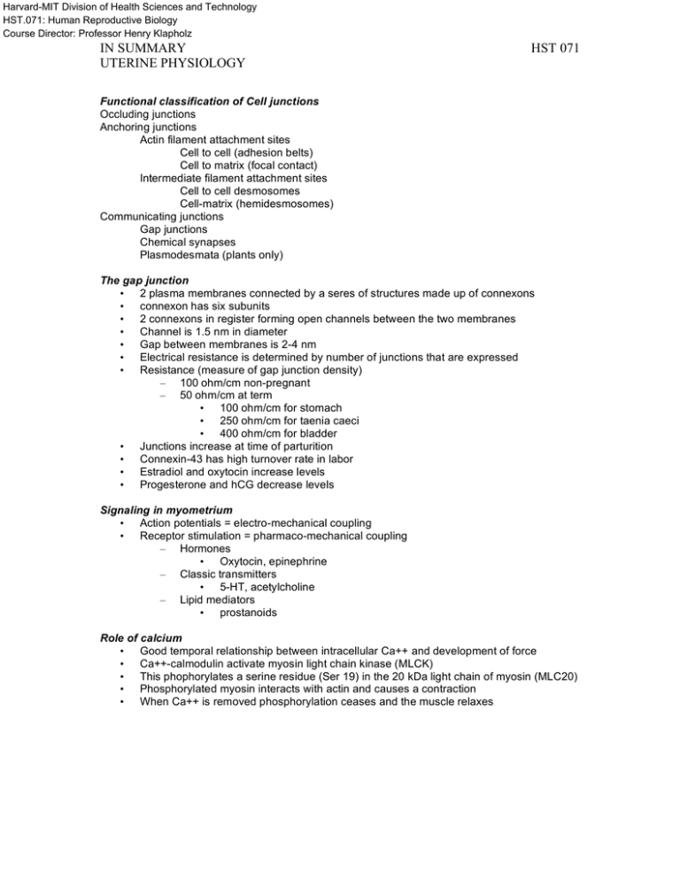
Harvard-MIT Division of Health Sciences and Technology HST.071: Human Reproductive Biology Course Director: Professor Henry Klapholz IN SUMMARY UTERINE PHYSIOLOGY HST 071 Functional classification of Cell junctions Occluding junctions Anchoring junctions Actin filament attachment sites Cell to cell (adhesion belts) Cell to matrix (focal contact) Intermediate filament attachment sites Cell to cell desmosomes Cell-matrix (hemidesmosomes) Communicating junctions Gap junctions Chemical synapses Plasmodesmata (plants only) The gap junction • 2 plasma membranes connected by a seres of structures made up of connexons • connexon has six subunits • 2 connexons in register forming open channels between the two membranes • Channel is 1.5 nm in diameter • Gap between membranes is 2-4 nm • Electrical resistance is determined by number of junctions that are expressed • Resistance (measure of gap junction density) – 100 ohm/cm non-pregnant – 50 ohm/cm at term • 100 ohm/cm for stomach • 250 ohm/cm for taenia caeci • 400 ohm/cm for bladder • Junctions increase at time of parturition • Connexin-43 has high turnover rate in labor • Estradiol and oxytocin increase levels • Progesterone and hCG decrease levels Signaling in myometrium • Action potentials = electro-mechanical coupling • Receptor stimulation = pharmaco-mechanical coupling – Hormones • Oxytocin, epinephrine – Classic transmitters • 5-HT, acetylcholine – Lipid mediators • prostanoids Role of calcium • Good temporal relationship between intracellular Ca++ and development of force • Ca++-calmodulin activate myosin light chain kinase (MLCK) • This phophorylates a serine residue (Ser 19) in the 20 kDa light chain of myosin (MLC20) • Phosphorylated myosin interacts with actin and causes a contraction • When Ca++ is removed phosphorylation ceases and the muscle relaxes IN SUMMARY UTERINE PHYSIOLOGY HST 071 Uterine Smooth Muscle • Bundle of myometrial cells embedded in a matrix of connective tissue • Matrix enables transmission of individual contractile forces • ripening – Increased collagen solubility – Alteration in ground substance • Ripening occurs in the cervix and corpus • Cytoplasm – Myosin (thick filaments) • Hexamer – 2 identical 200kDa heavy chains – 4 light chains (two 20 kDa chains and two 15-17 kDa chains) • Enzyme capable of converting ATP mechanical energy • Head – Actin and myosin interact Figure removed due to copyright restrictions. Please see: – ATPase sites located • Tail Figure 3-5 in Speroff, Leon, Robert H Glass, and Nathan G Kase. – Formation of myosin filaments "Structural similarities of Oxytocin and Vasopressin." In Clinical Gynecologic Endocrinology and Infertility. Oxytocin Baltimore, MD: Williams & Wilkins, 1989. ISBN: 0683078976. • Nonapeptide as shown • First peptide ever synthesized - Nobel Prize • High affinity, low capacity receptors – 80-fold increase in number by term – marked increase in sensitivity • Act by raising intracellular free calcium levels • Calcium stimulates actomyosin formation and muscular contraction • Maternal and fetal source • • • • High affinity, low capacity receptors – 80-fold increase in number by term – marked increase in sensitivity Act by raising intracellular free calcium levels Calcium stimulates actomyosin formation and muscular contraction Maternal and fetal source Uterus Oxytocin receptors distributed in a gradient from fundus (maximum) to cervix (few) Receptor concentration begins to rise early in pregnancy and exponentially increases to term IN SUMMARY UTERINE PHYSIOLOGY HST 071 Prostaglandins • Prostaglandins (PG) and thromboxane • Products of arachidonic acid metabolism. • Produce numerous physiologic and pathophysiologic effects • Regulating cellular processes in nearly every tissue • Local hormones – act in the vicinity of their site of production – Function in an autocrine and/or paracrine manner • Widely distributed • Can be formed by nearly every tissue and cell type • Ability to provoke different responses in various tissues • Five physiologically important prostanoids – PGE2 – PGF2a – PGI2 – TXA2 – PGE2 • Wide spectrum of physiological and pharmacological actions – Immune – Endocrine – Cardiovascular – Renal – Reproductive – Contraction and relaxation of smooth muscle Exert their effects through GTP-binding protein (G protein)-coupled, rhodopsin-type receptors • Receptors for PGE2 are termed EP, which include EP1, EP2, EP3 and EP4 subtypes • High specific PGE2 binding has been observed in the brain, kidney, uterus, liver, thymus and the adrenal medulla • PGE2 has versatile and opposing actions due to multiple EP receptor subtypes and the coupling of EP receptor isoforms to a variety of signal transduction pathways G-proteins • adrenaline • glucagon • luteinizing hormone (LH) • parathyroid hormone (PTH) • adrenocorticotropic hormone (ACTH • prostaglandins • Evidence suggests – PGE2 affects gene transcription – Regulates growth and cell proliferation. • Endogenous PGE2 regulates the growth of epithelial cells • PGE2 exhibits mitogenic activities in bone cells and stimulates DNA synthesis • PGE2 is involved in the growth and metastasis of tumors • Inhibition of prostaglandin synthesis has been shown to result in growth retardation of tumors in experimental animals • Decreased risk of colon cancer with ASA • IN SUMMARY UTERINE PHYSIOLOGY HST 071 Cervical Collagen • Major protein of the extracellular matrix – 25% of mammalian protein – Stiff triple stranded helical structure – α chains (1000 amnio acids long) – Proline and glycine bonds create the left handed helical configuration – More than 20 types of collagen – I, II, III are fibrillar collagens – Assemble into fibrils and then fibers • • • • • • • • • • • Collagen polypeptides formed on membrane bound ribosomes and injected into the ER as pro-α chains (have extra amnio-acids called pro-peptides at ends) In lumen of ER hydroxyproline allows bonding of three pro-α chains to form procollagen Propeptides of type I, II, and III removed by extracellular enzymes converting them to tropocollagen (1.5 nm diameter) Tropocollagen assembles to form fibrils (10-200 nm) Type I and type III Spaces between bundles dilate @ 8-14 weeks Total collagen increases but concentration goes down 30-50% – Water and non-collagen proteins increase – Fibrils decrease in size Stains for collagen polymer show reduced amount (lower numbers of intact fibers) Spaces between bundles dilate @ 8-14 weeks Total collagen increases but concentration goes down 30-50% – Water and non-collagen proteins increase – Fibrils decrease in size Stains for collagen polymer show reduced amount (lower numbers of intact fibers) Ground substance • Glucosamine - naturally occurring amino sugar found in glycosoaminoglycans (mucopolysaccharides) • Integral components of the proteoglycans • Proteoglycans - large carbohydrate rich structures – Resiliency – Load distribution – Shock-absorbing – Compressive – Lubricating • Dietary glucosamine – An immediate precursor for glycosaminoglycan synthesis – Stimulates incorporation of other precursors into the connective tissue matrix • • • • GAG (glycosaminoglycan) – Total increases throughout pregnancy – Concentration remains constant – Helps loosen the collagen network HA (hyaluronic acid) – Increases 12 fold at 2-3 cm dilation – HA may bind water and help hydration of tissue and hence deformability CS (chondroitin sulfate) – Decrease – Results in decreased rigidity of cervical tissue Elastase acts on the telopeptide non-helical domains of collagen IN SUMMARY UTERINE PHYSIOLOGY • • • • • • • • HST 071 – Can degrade collagen, elastin, and proteoglycans – Synergistically with collagenase – Polys from blood – Cervical fibroblasts Amount of soluble collagen (degraded) increases in parallel with enzyme activity More immature crosslinks Collagen with many x-links replaced with collagen with fewer x-links Up to several thousand sugar residues Repeating sequence of non-sulfated disaccharide units Variable amounts in all tissues (esp embryos) Earliest evolutionary form of glycosaminoglycans (GAG) Has some function in cell migration Cervical ripening • Decrease in total collagen content • Increase in collagen solubility • Increase in collagenolytic activity – Collagenase – Leukocyte elastase • Rapid turnover of extracellular matrix • Strong correlation free hydroxyproline • Similar to inflammatory response – IL-8, other cytokines, eosinophils, mast cells, macrophages, neutrophils • • • Sex steroid hormones – Estrogen • Collagen degradation • IV estradiol produces cervical ripening Progesterone – Blocks estrogen induced collagenolysis – Antagonists produce ripening – IL-8 in rabbits is down regulated by progesterone Changes are gradual and antecede labor by several weeks FUNDAMENTAL QUESTIONS 1. 2. 3. 4. Describe the types of junctions seen between cells. What kinds of junctions are seen in myometrial cells? What is the structure of a gap junction? What happens to the expression of oxytocin receptors as gestation advances? 5. What type of connexon is seen in myometrium? 6. Describe the signaling mechanisms in the myometrium. 7. What is oxytocin, its structure and function? 8. What is the role of prostaglandin in myometrial contractility? 9. How do G-proteins work? 10. Describe the cervical ground substance. How is it suited for labor? 11. What factors influence how soft the cervix becomes before the onset of labor? 12. Describe the process of “ripening” in detail. IN SUMMARY UTERINE PHYSIOLOGY HST 071
