Values of the elastic or Young’s modulus for various materials
advertisement
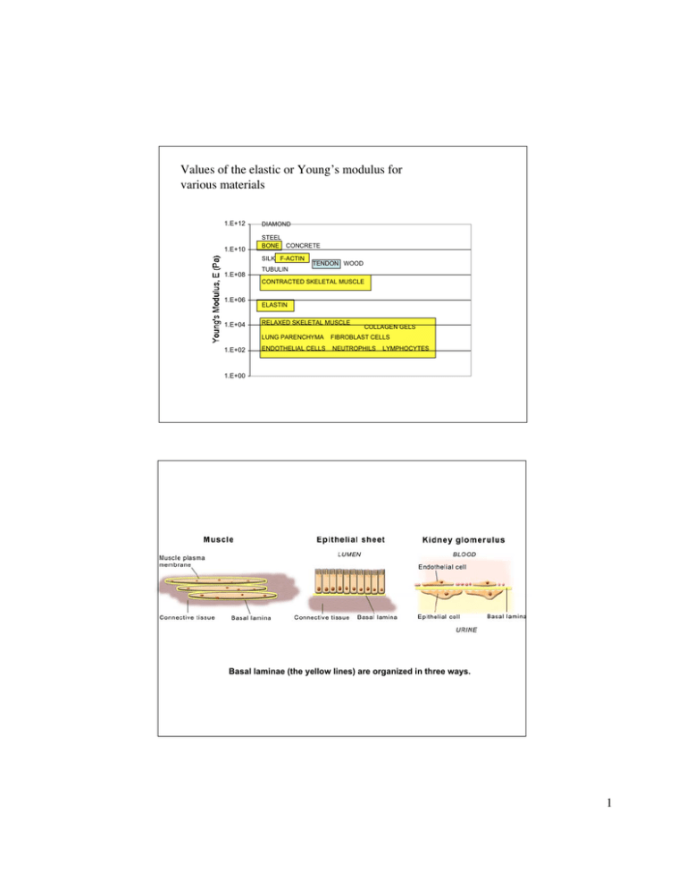
Values of the elastic or Young’s modulus for various materials 1.E+12 1.E+10 DIAMOND STEEL BONE CONCRETE SILK F-ACTIN 1.E+08 TUBULIN TENDON WOOD CONTRACTED SKELETAL MUSCLE 1.E+06 1.E+04 1.E+02 ELASTIN RELAXED SKELETAL MUSCLE COLLAGEN GELS LUNG PARENCHYMA FIBROBLAST CELLS ENDOTHELIAL CELLS NEUTROPHILS LYMPHOCYTES 1.E+00 Basal laminae (the yellow lines) are organized in three ways. 1 Right tibia (somewhat nonisotropic and nonlinear) Longitudinal direction Radial direction Striated muscle A - resting B - max contraction Microscopic sarcomere level C - active component Macroscopic view 2 3 Image removed due to copyright considerations. See Figure 19-41 in: Alberts, Bruce, et al. Molecular Biology of the Cell. 4th ed. New York: Garland Publishing, 2002. Image may be viewed online at the NIH's PubMed Bookshelf. http://www.ncbi.nlm.nih.gov/entrez/query.fcgi?db=Books Events involved in the formation of a collagen fibril. 4 Image removed due to copyright considerations. See Figure 7.3:4 in: Fung, Y. C. Biomechanics: Mechanical Properties of Living Tissues. New York: Springer-Verlag, 1993. Striated appearance of collagen fibrils 5 Collagen fiber arrangement in skin and cornea with alternating directions Image removed due to copyright considerations. See Figure 19-46 in: Alberts, Bruce, et al. Molecular Biology of the Cell. 4th ed. New York: Garland Publishing, 2002. Image may be viewed online at the NIH's PubMed Bookshelf. http://www.ncbi.nlm.nih.gov/entrez/query.fcgi?db=Books 6 ACL -- different strain rates Range of linearity. E = d(stress)/d(strain) = 109 Pa 7 Collagen derives its stiffness, not from the single molecule characteristics of collagen, but rather from the straightening of “wavy” collagen fibers Image removed due to copyright considerations. Cornea, M. Johnson, J. Ruberti 8
