Available online at www.sciencedirect.com
Scripta Materialia 61 (2009) 403–406
www.elsevier.com/locate/scriptamat
Internal nitride formation during gas-phase thermal
nitridation of titanium
P.K. Ajikumar,a,* M. Kamruddin,a P. Shankar,a Ramakrishna Gouda,b
A.K. Balamurugan,a R. Nithya,a A.K. Tyagi,a V. Jayaram,b S.K. Biswasb and Baldev Raja
a
Metallurgy and Materials Group, Indira Gandhi Centre for Atomic Research, Kalpakkam 603102, India
b
Indian Institute of Science, Bangalore 560012, India
Received 13 February 2009; revised 16 April 2009; accepted 17 April 2009
Available online 22 April 2009
Titanium nitride surface layers were prepared by gas-phase thermal nitridation of pure titanium in an ammonia atmosphere at
1373 K for different times. In addition to the surface nitride layer, nitride/hydride formation was observed in the bulk of the specimen. The cross-section of the specimen was characterized by various techniques such as optical microscopy, scanning electron
microscopy, energy dispersive X-ray spectroscopy, secondary ion mass spectrometry and nanomechanical testing, and the mechanism of formation of these phases is discussed.
Ó 2009 Acta Materialia Inc. Published by Elsevier Ltd. All rights reserved.
Keywords: Titanium nitride; Hardness; SIMS; Nanoindentation; SEM
Titanium and its alloys are extensively used in
the aerospace and automobile industries because of their
high specific strength and good mechanical properties.
However, one of the major constraints in the wider
application of titanium alloys is their poor galling resistance in sliding contact with other metals under load.
Surface engineering is a well-established technique to
overcome this problem and to improve the tribological
properties of titanium and its alloys [1].
Titanium nitrides possess high hardness and elastic
modulus combined with low coefficient of friction and
good corrosion resistance [2]. These properties of TiN
have prompted the use of nitrided Ti surfaces for enhanced performance under erosion or erosion–corrosion
service conditions. In addition, nitrided Ti surfaces have
good biocompatibility and hence have potential as implant materials, particularly for components subjected
to erosion wear conditions [3,4]. The presence of TiN/
Ti2N layers at the surface of the nitrided specimens is reported to result in better adhesion of fibroblast cells
compared to an untreated Ti substrate [5,6].
The strong affinity between nitrogen and titanium
provides the driving force to form TiN and Ti2N phases
* Corresponding author.
pkajikumar@gmail.com
Tel./fax:
+91
4427480081; e-mail:
at the surface on nitridation. From the Ti–N phase diagram it is known that Ti exhibits a large solubility for
nitrogen at high temperatures [7]. Several techniques,
including plasma nitriding, ion implantation, gas nitriding, laser nitriding and electrochemical surface nitriding,
have been employed for nitriding titanium and titanium
alloys [8–13]. Among these, gas nitriding and plasma
nitriding are considered to be promising methods for
engineering and biomedical applications because these
processes are independent of the geometry of the samples. Furthermore, using these processes it is possible
to achieve large nitriding depths, compared to other
methods, giving a higher reserve for wear applications.
In the present study, gas-phase thermal nitridation
using ammonia gas as the nitrogen source in a thermogravimetric analysis–mass spectrometry (TGA–MS)
system was carried out. Detailed analysis by X-ray diffraction (XRD), scanning electron microscopy (SEM), energy
dispersive X-ray spectroscopy (EDX), secondary ion
mass spectrometry (SIMS) and nanoindentation studies,
with more emphasis on the examination of the cross-section of the specimens, was undertaken.
The nitridation experiments were carried out in a
home assembled TGA–MS system [14]. The detailed
experimental procedure was reported earlier [10]. For
the sake of completeness, a brief description is given
here. Pure Ti specimens were polished and cleaned
1359-6462/$ - see front matter Ó 2009 Acta Materialia Inc. Published by Elsevier Ltd. All rights reserved.
doi:10.1016/j.scriptamat.2009.04.029
404
P. K. Ajikumar et al. / Scripta Materialia 61 (2009) 403–406
before being exposed to a nitriding environment. These
samples were subjected to nitridation at 1373 K for periods of 5 and 10 h in an ammonia atmosphere with a flow
rate of 15 sccm. The weight gain of the samples during
nitridation was monitored continuously. The nitrided
specimens were characterized by XRD (STOE, Germany) for phase identification. The XRD data were collected in powder mode (PXRD) and glancing incidence
mode (GIXRD) for bulk and surface analysis, respectively. Surface and cross-sectional microstructures were
studied by SEM (Philips GX 30 ESEM). The Ti:N ratio
at different nitrided zones was estimated by cross-sectional EDX, attached to the scanning electron microscope. Elastic modulus and hardness along the crosssection of the specimens were measured by nanoindentation (Hysitron, USA). Loading and unloading curves
are plotted to assess the elastic recovery at different
nitridation zones along the cross-section of the specimen. Titanium, nitrogen and hydrogen imaging was carried out over the cross-section by SIMS (Cameca,
France).
The weight gain of the specimens during nitridation
was measured online by TGA. The specimen (initial
weight 1141.9 mg) treated at 1373 K for 10 h had a
weight gain of 1.86% and the one treated for 5 h gained
1.46%. This gain in weight during the process indicates a
substantial intake of gaseous species into the sample (i.e.
nitrogen and hydrogen, the cracking products of ammonia). PXRD patterns [10] clearly identified that the
nitrided surface layer predominantly consists of TiN
phase with a minor contribution from Ti2N. GIXRD
[10] studies, however, indicated that the submicron surface layer is almost pure TiN.
In order to study the cross-sectional microstructure
and the mechanical properties of the phases present,
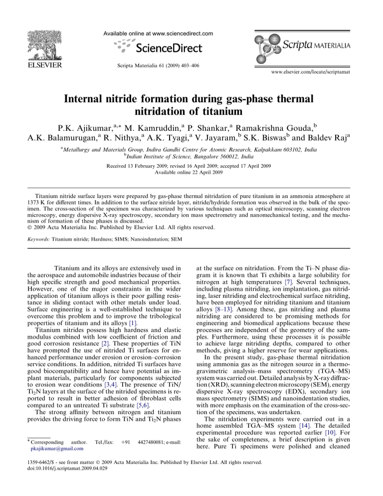
optical microscopy, SEM, EDX, SIMS and nanoindentation techniques were employed. Figure 1 shows the
optical micrograph of the cross-section from a sample
nitrided for 10 h. It reveals a nitrided surface layer,
which is bright and around 75 lm thick. Below the outer
nitride layer a darker phase running almost parallel to
the surface and channeling towards the interior (as indicated by small arrow marks) is seen. A two-phase microstructural contrast is clearly observed in the bulk
beyond the interface. The area fraction, and hence the
volume fraction, of the different phases was estimated
using image analysis software, and it was found that
the darker phase comprises around 5% of the surface
area. Similar observations were also made for the 5 h
Figure 1. Cross-sectional optical micrograph of specimen nitrided at
1373 K for 10 h.
treated specimen except for the thickness of the outer
layer, which was measured to be about 55 lm. Further
investigations were carried out on the 10 h treated
specimen.
Nanoindentation studies were also carried out along
the cross-section of the specimen. Figure 2a illustrates
the variation in hardness as well as elastic modulus
along the interior region. The optical micrograph of
the corresponding regions is shown below (indicated
by the dotted line within the box). At the lighter regions
the hardness and elastic modulus were around 10 and
135 GPa, respectively. Although these values are lower
than previously reported for TiN, it can be assumed
that these are partially nitrided regions [15]. In the darker region, these values fall to 3.5 and 100 GPa, respectively. This hardness value matches well with the
reported hardness value of titanium hydride [16]. Figure 2b illustrates similar nanoindentation measurements covering a wider region, i.e. the nitrided outer
layer, the partially nitrided region and the darker
phase. The outer nitrided layer shows hardness and
elastic modulus values of around 15 and 180 GPa,
respectively. This is comparable to the reported data
[17,18], though these values vary over a wide range
depending upon the methods of preparation and the
process parameters. The observations on the other
two regions are identical to that of Figure 2a. The
load–displacement curves obtained during nanoindentation at different regions are shown in Figure 2c.
The difference in hardness of the regions is apparent
from the large difference in the depth of indentation
for the same applied load and from the varying degrees
of elastic recovery during unloading. At the hydrided
zone, the indentation depth was around 240 nm with
the maximum applied load of 5000 lN followed by a
minimum elastic recovery (20%) during unloading.
In the partially nitrided region, the depth was around
160 nm for the same applied load and there was a substantial elastic recovery (35%) during unloading. For
the completely nitrided uppermost region, the penetration depth was only about 140 nm and almost 50%
recovery upon unloading was observed.
The cross-section of the specimen was subjected to
elemental image mapping by SIMS. A small area covering the partially nitrided zones and narrow dark channels was imaged for nitrogen and hydrogen (Fig. 3a
and b, respectively). The images of nitrogen and hydrogen are complementary to each other. The corresponding line scan as indicated in the image was also plotted
against distance (Fig. 3c). This observation clearly indicated the formation of nitrogen-rich and hydrogen-rich
regions along the cross-section.
In order to substantiate the findings from the nanoindentation and SIMS measurements, these regions were
further investigated by EDX line scan analysis. The
SEM image and the corresponding concentration profile
of titanium and nitrogen up to a depth of 300 lm are
shown in Figure 4a and b. The line profiles indicate that
at the outer surface of the specimen, the concentrations
of Ti and N are almost same, suggesting the presence of
stoichiometric TiN (this was also evident from GIXRD
[10]). The concentration of nitrogen decreases rapidly
towards the interior, showing the formation of sub-stoi-
P. K. Ajikumar et al. / Scripta Materialia 61 (2009) 403–406
405
Figure 2. Elastic modulus and hardness measured using nanoindentation (a) at bulk, (b) towards the surface of the specimen and (c) force–
displacement curves at different regions of the nitrided specimen.
Figure 3. Elemental imaging by SIMS for (a) nitrogen and (b)
hydrogen over a 250 lm area and (c) the corresponding intensity
profile along the dotted lines marked in the images.
chiometric TiN or Ti2N or the precipitation of these
compounds in a Ti matrix.
From the above observations, it is confirmed that a
three-phase region—a completely nitrided outer layer,
partially nitrided interior regions and narrow channels
of titanium hydride—are formed during the nitridation
process. For the specimen nitrided for 10 h, the total
weight gain is 21.28 mg. Bearing in mind the volume
fraction of hydride (5%) from the image analysis and
the ratio of atomic weight of hydrogen to nitrogen, it
can be safely assumed that this weight gain is due to uptake of nitrogen. Converting this weight gain into the
number of nitrogen atoms gives a value of 9.2 1020
atoms (sample size 12.5 10 2 mm3). This would result in a very high concentration of nitrogen at the surface, exceeding the solubility limit and providing a very
strong driving force for surface nitridation. In addition,
a substantial amount of nitrogen and hydrogen would
diffuse into the bulk at the nitriding temperature.
The titanium–nitrogen system has been extensively
studied by Strafford et al. [19], who reported a parabolic
Figure 4. (a) Cross-section SEM micrograph and (b) the corresponding EDX line graph from the surface of the sample.
weight gain rate for the entire exposure period. From
the Ti–N binary phase diagram [7], it is clear that the N
solubility in a-Ti is quite high at high temperatures (about
23 at.% of N above 1323 K) and drops at low temperatures (about 5 at.% 773 K). On heating pure Ti specimen
to the nitriding temperature of 1373 K, a -Ti transforms
to b-Ti. The required nitrogen concentration to maintain
the a phase is 5 at.% at 1373 K [7]. The total weight gain
over the entire nitriding period of 10 h (21.28 mg) corresponds to 6 at.% for the entire Ti specimen. Considering
the formation of the nitride layer at the surface and the
resulting slow inward diffusion of nitrogen, the bulk of
the specimen would remain mostly in b phase at the nitriding temperature since the presence of nitrogen will be
much lower than 5 at.%. Upon cooling it reverts back to
a-Ti and it is likely that the dissolved nitrogen in the bulk
could result in the partial nitridation of the bulk material
since the solubility of nitrogen in titanium is drastically reduced at lower temperatures. Thus the measured weight
406
P. K. Ajikumar et al. / Scripta Materialia 61 (2009) 403–406
gain should be the sum of two different contributions,
namely the external scaling and partial internal nitridation in the bulk. Strafford et al. [19] concluded that the
mechanism of nitridation involves an inward diffusion
of nitrogen and this process is limited by the rate of diffusion of nitrogen which is slower once a nitride layer forms
at the surface. Buscaglia et al. [20] studied the high-temperature behaviour of Nb–Ti alloys of various concentrations in a nitrogen atmosphere. Their Ti(90)–Nb(10)
sample showed, over and above the complete nitridation
of the outer layer, precipitation of titanium nitride needles
throughout the interior region. McDonald et al. [21] also
studied the reaction of nitrogen with titanium at high temperatures. They concluded that the inward diffusion of
nitrogen is the dominant mass transfer process during
nitridation, and that a nitrogen concentration gradient
and a corresponding change in hardness exist across all
phases after nitridation. A systematic investigation of
the titanium–nitrogen system and the phase formation
in the sub-nitride regions was carried out by Lengauer
[22]. The outermost layer was made of TiN1 x at all temperatures. He has also observed Ti4N3 x, Ti3N2 x and
Ti2N phases at different depths. There was no trace of
Ti2N in the specimens treated at and above 1353 K. This
confirms that the Ti2N observed in our sample must have
formed during cooling. In addition, Lengauer estimated
the nitrogen content in the nitrides, which varied from
30% for Ti2N to 37% for TiN1 x.
Considering these observations [7,19–22] and a careful
examination of our nanoindentation, SIMS and EDX results, we were able to deduce the mechanism of formation
of three regions upon nitridation of Ti in NH3 atmosphere. The continuous diffusion of N in Ti, at high temperature, leads to the formation of a nitride layer at the
surface as well as its dissolution in the bulk. SEM and nanoindentation results show that the nitrided surface layer is
around 75 lm thick. A gradient in hardness as well as a
change in concentration of Ti and N (Figs. 2b and 4b) exist even along this layer, indicating that this layer is also
not single phase. We assume that this phase consists of a
thin layer of TiN at the outer surface, followed by a mixture of sub-stoichiometric TiN and Ti2N. If we assume
that the nitrogen requirement on average is 33 at.% (from
Ref. [22]) for the outer 75 lm layer, the nitrogen requirement is of the order of 7 1020 atoms for a specimen of
dimensions 12.5 10 2 mm3. The total nitrogen intake
is calculated to be around 9.2 1020 atoms (neglecting the
weight of hydrogen) from the TGA weight gain measurement. This means that of the order of 2.2 1020 excess
nitrogen atoms are dissolved in the bulk, which are available for internal nitriding. They are unlikely to form the
nitride phase at 1373 K since the solubility of nitrogen
in Ti is around 23 at.% at that temperature [7]. The nitride
formed inside the bulk is probably due to precipitation
upon cooling since the solubility of N in Ti reduces drastically at lower temperatures [7]. The dissolved nitrogen in
the sample will be used up for the nucleation and growth
of nitride phases upon cooling. Simultaneously the dissolved hydrogen also became segregated in narrow channels to form titanium hydride.
In conclusion, nitridation of titanium was carried out
in an ammonia atmosphere at 1373 K for 5 and 10 h in a
TGA–MS system. In addition to the TiN/Ti2N surface
layer, titanium nitride/hydride phase formation was observed in the bulk of the specimens. Cross-sectional
examination of the specimen by optical microscopy,
SEM, EDX, SIMS and nanoindentation revealed a
three-phase microstructure with a completely nitrided
outer layer at the surface followed by a narrow hydride
phase and partially nitrided and hydrided precipitates
throughout the bulk. The measured hardness and elastic
modulus also complement the above inferences of the
formation of a completely nitrided outer layer and partially nitrided internal regions separated by narrow hydride channels.
[1] T. Bell, H. Dong, in: Proceedings of the International
Conference on Advances in Surface Treatment: Research
& Applications (ASTRA), 2003, Hyderabad, India, p. 12.
[2] Shanyong Zhang, Weiguang Zhu, J. Mater. Process.
Technol. 39 (1993) 165.
[3] Y. Tamura, A. Yokoyama, F. Watari, T. Kawasaki,
Dent. Mater. J. 21 (4) (2002) 355.
[4] S. Piscanec, L. Colombi Ciacchi, E. Vesselli, G.
Comelli, O. Sbaizero, S. Meriani, Acta Mater. 52
(5) (2004) 1237.
[5] E. Czarnowska, T. Wierzchoń, A. Maranda-Niedbała, J.
Mater. Process. Technol. 92–93 (1999) 190.
[6] B. Groessner-Schreiber, A. Neubert, W.-D. Muller, M.
Hopp, M. Griepentrog, K.-P. Lange, J. Biomed. Mater.
Res. A 64A (2003) 591.
[7] H.A. Wriedt, J.L. Murray, in: T.B. Massalski (Ed.),
Binary Alloy Phase Diagrams, vol. 3, ASM, Metals Park,
OH, 1986, p. 2705.
[8] S.C. Mishra, B.B. Nayak, B.C. Mohanty, B. Mills, J.
Mater. Process. Technol. 132 (1–3) (2003) 143.
[9] Y. Kasukabe, J.J. Wang, T. Yamamura, S. Yamamoto,
Y. Fujino, Thin Solid Films 464–465 (2004) 180.
[10] P.K. Ajikumar, M. Kamruddin, R. Nithya, P. Shankar,
S. Dash, A.K. Tyagi, Baldev Raj, Scripta Mater. 51
(2004) 361.
[11] Y. Fu, A.W. Batchelor, Wear 214 (1) (1998) 83.
[12] T. Goto, M. Tada, Y. Ito, Electrochim. Acta 39 (8/9)
(1994) 1107.
[13] A. Zhecheva, W. Sha, S. Malinov, A. Long, Surf. Coat.
Technol. 200 (7) (2005) 2192.
[14] M. Kamruddin, P.K. Ajikumar, S. Dash, A.K. Tyagi,
Baldev Raj, Bull. Mater. Sci. 26 (4) (2003) 449.
[15] Te-Hua Fang, Sheng-Rui Jian, Der-San Chuu, Appl.
Surf. Sci. 228 (2004) 365.
[16] J.J. Xu, H.Y. Cheung, S.Q. Shi, J. Alloys Compds. 436
(2007) 82.
[17] G.B. de Souza, C.E. Foerster, S.L.R. da Silva, F.C.
Serbena, C.M. Lepienski, C.A. dos Santos, Surf. Coat.
Technol. 191 (2005) 76.
[18] C.-H. Ma, J.-H. Huang, H. Chen, Surf. Coat. Technol.
200 (2006) 3868.
[19] K.N. Strafford, J.M. Towell, Oxid. Metals 10 (1) (1976)
41.
[20] V. Buscaglia, A. Martinelli, R. Musenich, W. Mayr, W.
Lengauer, J. Alloys Compds. 283 (1999) 241.
[21] N.R. Mcdonald, G.R. Wallwork, Oxid. Metals 2 (3)
(1970) 263.
[22] W. Lengauer, Acta Metall. Mater. 39 (12) (1991) 2985.


