Handheld Sample Preparation for Complex Samples
advertisement

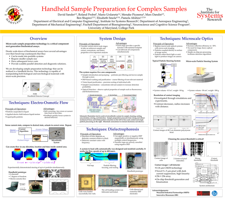
Handheld Sample Preparation for Complex Samples David Sander1,2, Roland Probst3, Mario Urdaneta1,4, Menake Piyasena4, Marc Dandin2,5, Ben Shapiro2,3,5, Elisabeth Smela4,5,6, Pamela Abshire1,2,5,6 Department of Electrical and Computer Engineering1, Institute for Systems Research2, Department of Aerospace Engineering3, Department of Mechanical Engineering4, Fischell Department of Bioengineering5, Neuroscience and Cognitive Science Program6, University of Maryland, College Park Overview System Design Micro-scale sample preparation technology is a critical component next generation biochemical assays. Drastic scale-down of biochemical assays have several advantages over their bench top counterparts that they: • Can be made into portable systems • Require smaller sample size • Have subsequent lower costs • Provide scalable parallel detection and diagnostic solutions. We are developing sample preparation technology that can be realized in a handheld device. This technology is capable of manipulating both biological and non-biological materials with micro-scale precision. Techniques: Microscale Optics Advantages Principle of Operation • Cascade various micro-scale stages to take an unknown sample and extract, concentrate, and detect properties of its important analytes • Each stage provides a specific function with minimal footprint • Can fabricate all components simultaneously Principle of Operation Advantages • Replace macro-scale optical systems with micro-scale imaging • Particles coupled directly to surface of image sensor • Particles either block light or emit light to produce images on camera • Increase collection efficiency to ~50% • No need for large, heavy optical components • Simple on-chip pre-processing facilitates detection algorithms Typical Particle Steering System Micro-scale Particle Steering System The system requires five core components • Sample introduction and pumping – performs pre-filtering and moves sample through system • DEP-based washing and purification – coarse filtering of non-relevant material • Vision based purification – precision sorting of individual cells • Sample Concentration – concentrate relevant analyte to increase detection probability • Optical Detection – observe optical properties of sample such as fluorescence, and bioluminescence • System volume ~0.5 m3, weight ~50 kg • System volume ~50 cm3, weight ~300 g • Investigated through simulations and experiments. • Contrast decreases, radius increases with distance. Techniques: Electro-Osmotic Flow Advantages •Particles suspended in liquid •Applied electric field induces liquid motion •Liquid pulls particles •Self correcting. Any errors at current time fixed at next time. •Feedback quickly forces system to desired behavior. Sense Pixilated Images Microfluidic System 10 0 Schematic illustration (not to scale) of microfluidic system for sample cleaning, sorting, concentration, mixing, and detection. Sample preparation proceeds from strong and robust mechanical forces and initial particle extraction on the left to fine-scale control and single particle processing on the right. (Electrode connections to external electronics not shown.) Advantages • Direction of force depends on dielectric constant relative to fluid. • Dielectric constant varies with frequency. • Can apply positive or negative DEP sequentially or simultaneously using multiple control frequencies. • Parasitic traps are actively cancelled using negative DEP. -4 -3 -4 Contact images of 48.5 μm diameter polystyrene bead. Principle of Operation -5 10 10 10 Distance btw object & sensor (m) 10 284.5 m (exp) 48 m (exp) 5 m (sim) -5 10 Techniques: Dielectrophoresis Actuator Voltages 284.5 m (exp) 48 m (exp) 5 m (sim) 10 -6 10 Sense current state, compare to desired state, actuate to correct error. Repeat. Control Algorithms 2 10 1 Radius (m) Principle of Operation: Contrast Resolution of contact imaging Handheld Detector Concept Desired Behavior 3 10 -6 10 -4 10 Distance btw object & sensor (m) Choosing the correct threshold is critical! Can create flow in any direction, location and time inside control area. A system to load cells automatically was designed and modeled carefully. It works for flow speeds of up to 400 mm/s. Contact image of cells If want: = (a lot of) + (a little of) Vial trap Experimentally demonstrated steering of up to 4 particles simultaneously. Handheld prototype • 8 channels • 7 degrees of freedom • Electro-osmotic flow Wrong threshold Correct threshold Contact imager / cell detector Particle filtering, focusing, and routing Handheld prototype 2 clusters of pixel intensities electr ode channel Images • 0.18 μm CMOS technology • Novel 5 x 5 μm pixel with dark current suppression, high linearity • 256 × 256 array • On-chip threshold generation and binarization System architecture Voltages Cells were loaded successfully into vials. The cell loading system was fabricated and tested. Cells filtered and routed by DEP electrodes Acknowledgements: Maryland Industrial Partnerships (MIPS) Innovative Biosensors (IBI) -2 10


