Document 13309414
advertisement
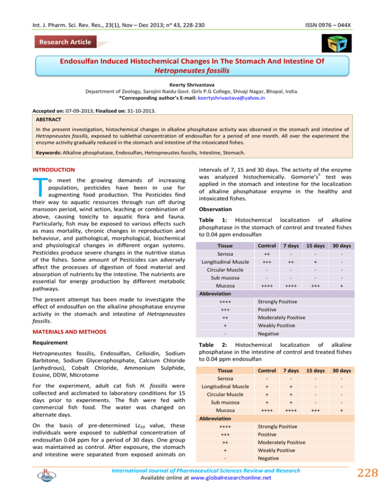
Int. J. Pharm. Sci. Rev. Res., 23(1), Nov – Dec 2013; nᵒ 43, 228-230 ISSN 0976 – 044X Research Article Endosulfan Induced Histochemical Changes In The Stomach And Intestine Of Hetropneustes fossilis Keerty Shrivastava Department of Zoology, Sarojini Naidu Govt. Girls P.G College, Shivaji Nagar, Bhopal, India. *Corresponding author’s E-mail: keertyshrivastava@yahoo.in Accepted on: 07-09-2013; Finalized on: 31-10-2013. ABSTRACT In the present investigation, histochemical changes in alkaline phosphatase activity was observed in the stomach and intestine of Hetropneustes fossilis, exposed to sublethal concentration of endosulfan for a period of one month. All over the experiment the enzyme activity gradually reduced in the stomach and intestine of the intoxicated fishes. Keywords: Alkaline phosphatase, Endosulfan, Hetropneustes fossilis, Intestine, Stomach. INTRODUCTION T o meet the growing demands of increasing population, pesticides have been in use for augmenting food production. The Pesticides find their way to aquatic resources through run off during mansoon period, wind action, leaching or combination of above, causing toxicity to aquatic flora and fauna. Particularly, fish may be exposed to various effects such as mass mortality, chronic changes in reproduction and behaviour, and pathological, morphological, biochemical and physiological changes in different organ systems. Pesticides produce severe changes in the nutritive status of the fishes. Some amount of Pesticides can adversely affect the processes of digestion of food material and absorption of nutrients by the intestine. The nutrients are essential for energy production by different metabolic pathways. The present attempt has been made to investigate the effect of endosulfan on the alkaline phosphatase enzyme activity in the stomach and intestine of Hetropneustes fossilis. MATERIALS AND METHODS intervals of 7, 15 and 30 days. The activity of the enzyme was analyzed histochemically. Gomorie’s5 test was applied in the stomach and intestine for the localization of alkaline phosphatase enzyme in the healthy and intoxicated fishes. Observation Table 1: Histochemical localization of alkaline phosphatase in the stomach of control and treated fishes to 0.04 ppm endosulfan Tissue Control 7 days 15 days 30 days Serosa Longitudinal Muscle Circular Muscle Sub mucosa Mucosa ++ +++ ++++ ++ ++++ + +++ + Abbreviation ++++ +++ ++ + - Requirement Hetropneustes fossilis, Endosulfan, Celloidin, Sodium Barbitone, Sodium Glycerophosphate, Calcium Chloride (anhydrous), Cobalt Chloride, Ammonium Sulphide, Eosine, DDW, Microtome For the experiment, adult cat fish H. fossilis were collected and acclimated to laboratory conditions for 15 days prior to experiments. The fish were fed with commercial fish food. The water was changed on alternate days. On the basis of pre-determined Lc50 value, these individuals were exposed to sublethal concentration of endosulfan 0.04 ppm for a period of 30 days. One group was maintained as control. After exposure, the stomach and intestine were separated from exposed animals on Strongly Positive Positive Moderately Positive Weakly Positive Negative Table 2: Histochemical localization of alkaline phosphatase in the intestine of control and treated fishes to 0.04 ppm endosulfan Tissue Control 7 days 15 days 30 days Serosa Longitudinal Muscle Circular Muscle Sub mucosa Mucosa + + + ++++ + + + ++++ +++ + Abbreviation ++++ +++ ++ + - Strongly Positive Positive Moderately Positive Weakly Positive Negative International Journal of Pharmaceutical Sciences Review and Research Available online at www.globalresearchonline.net 228 Int. J. Pharm. Sci. Rev. Res., 23(1), Nov – Dec 2013; nᵒ 43, 228-230 ISSN 0976 – 044X 5 RESULTS AND DISCUSSION In the present study, alkaline phosphatase activity was observed in stomach and intestine of healthy and intoxicated fishes. In the stomach of normal fishes mucosa showed strongly positive reaction while longitudinal and serosa exhibited positive and moderately positive reaction respectively. The enzyme activity was not seen in circular muscle and sub-mucosal layer. After 7 days intoxication, there was no change in circular muscle, sub-mucosa and mucosa. But enzyme activity was reduced in serosal and longitudinal muscle layers. The enzyme activity was gradually inhibited in longitudinal muscle and mucosa after longer exposure. At the end of the experiment enzyme activity was seen only in mucosal layer (Table 1, Figure 1-3). gomori’s test. It was also observed that reduction in enzyme activity was increased with the long term exposure of endosulfan. (Table 2, Figure 4-6) Figure 3: Photomicrograph of stomach of heteropneustes fossilis after 30 days exposure to 0.04 ppm endosulfan showing varying response to Gomori’s Test. (Transverse Section 150X) Figure 1: Photomicrograph of Control Stomach of Heteropneustes fossilis showing alkaline phosphatase activity with Gomori’s Test. (Transverse Section 60X) Figure 4: Photomicrograph of control intestine of heteropneustes fossilis showing alkaline phosphatase activity with Gomori’s Test. (Transverse Section 200X). Figure 2: Photomicrograph of stomach of heteropneustes fossilis after 15 days exposure to 0.04 ppm endosulfan showing varying response to Gomori’s Test. (Transverse Section 60X) In controlled intestine, intense enzyme activity was seen only in mucosal layer, whereas, remaining layers showed weakly positive reaction except serosa which exhibited no reaction. After one week, there was no change in enzyme activity. No further change was noticed in all the layers except mucosa which showed minimum reaction with Figure 5: Photomicrograph of intestine of heteropneustes fossilis after 15 days exposure to 0.04 ppm endosulfan showing varying response to Gomori’s Test. (Transverse Section 200X). International Journal of Pharmaceutical Sciences Review and Research Available online at www.globalresearchonline.net 229 Int. J. Pharm. Sci. Rev. Res., 23(1), Nov – Dec 2013; nᵒ 43, 228-230 ISSN 0976 – 044X Our finding are in accordance in the Sastry & Sharma11(1979), Sastry & Malik9-10 (1979,1981), Goel et al.4 (1982), Lata & Shrivastava8 (1983), Arora & 2 1 14 Kulshrestha (1985), Arora (1986), Virk et al. (1987), 6 3 13 Gupta & Sharma (1987), Gill et al. (1990), Thaker et al. (1997). CONCLUSION Figure 6: Photomicrograph of intestine of heteropneustes fossilis after 30 days exposure to 0.04 ppm endosulfan showing varying response to Gomori’s Test. (Transverse Section 150X) The various zoologists have studied the effect of pollutants on alkaline phosphatase activity in digestive 11 tract of various animals Sastry & Sharma , Sastry & Malik9-10, Goel et al.4, Thaker et al.13, Singh & Bhatnagar12, Arora & Kushrestha2, Gill et al.3, Keerty et al.7 etc. Sastry & Sharma (1979) 11 observed inhibited alkaline phosphatase activity in Channa-punctatus due to endrin intoxication. Sastry and Malik (1979, 1981) 9-10 studied the effect of dimecron and diazinon respectively on digestive system of Channa-punctatus and reported depletion in alkaline phosphatase activity in both investigations. Goel et al.4 (1982) observed the effect of azodyes on alkaline phosphatase activity in intestine of Heteropneustes fossilis and Clarias batrachus and found significant depletion. Lata & Shrivastava8 (1983) also reported depletion in enzyme activity in Puntius sophore after the exposure of carpet dying chemicals. Arora & Kulshrestha2 (1985) studied the effect of carbaryl and endosulfan on alkaline phosphatase activity in intestine of Channa-striatus and reported inhibition of enzyme activity. Arora1 (1986) also showed decreased activity of alkaline phosphatase in intestine of metal intoxicated finger lings of Cyprinus carpio. Virk et al.14 (1987) noticed diminished activity of alkaline phosphatase in stomach and intestine of Mystus tengara after endrin and carbaryl intoxication. Gupta & Sharma6 (1987) also recorded declined enzyme activity in instetine of Colisa fasciata due to the exposure of chrysophenine-G and direct deep black. Gill et al.3 (1990) observed decreasing trend in alkaline phosphatase in gut of Puntius conchonius after the intoxication with phosphamidon and 13 aldicarb. Thaker et al. (1997) also investigated the hexavalent chromium toxicity on activity of alkaline phosphatase in intestine of Periophthalmus dipes and found inhibition. From the present result, it may be conclude that endosulfan caused destruction in tissues and hydrolytic enzymes of stomach and intestine, thus disturbing the metabolic progress in these organs. The inhibited activity of alkaline phosphatase in intestine may be due to a decreased rate of transphosphorylation and transport across membranes, as tissues of the intestine were progressively damaged in the endosulfan exposure. Hence reduction in the activity of this important enzyme in the intestine tissue obviously indicates an impaired nutrients assimilation and absorption. REFERENCES 1. Arora N, Alkaline and Acid phosphatase activities in the intestine of fingerlings od Cyprinus Carpio on exposure dto sublethal doses of Iron and Zinc. Abst., All India Semi. On the role of Zoology in the development of M.P, 1986, P 28. 2. Arora N, Kulshrestha SK, Effects of Chronic exposure to sublethal doses of two pesticides on alkaline and acid phosphatase activities on the intestine of a fresh water teleost, Channa striatus (Channidae) Acta, hydrochim. Hydrobiol, 13(5), 1985, 619-624. 3. Gill TS, Pande J, Tewari H, Enzyme modulation by sublethal concentrations of aldicarb, phosphamidon and endosulfan in fish tissues. Pesticide-Biochemistry and Physiology, 38(3), 1990, 231-244. 4. Goel KA, SK Tyagi, AK Awasthi, Ind. Jour. Zool, 23, 1982, 33. 5. Gomori GJ, Cell. Comp. Physiol, 17, 1941, 71. 6. Gupta RC, Sharma D, Him J. Env. Zool, 1, 1987, 28. 7. Keerty S, Sarita K, Asha M, Sudha S, Indian J. Applied & Pure Bio, 22(2), 2007, 265-269. 8. Lata S, VMS Srivastava, Comp. Physiol. Ecol, 8, 1983, 124. 9. Sastry KV, PV Malik, Arch. Environ. Contam, Torxcol, 8 1979, 397-407. 10. Sastry KV, PV Malik, J. Environ. Biol, 2(3), 1981, 18-28. 11. Sastry KV, SK Sharma, Bull. Environ. Contam. Toxicol, 22, 1979, 4. 12. Singh R, N. Bhatnagar, Indian J. Applied & Pure Bio, 19(3) 2004, 287-290. 13. Thaker J, J Chhaya, S Nuzhat, R Mittal, A P Mansuri, R. Kundu, Indian J. Exp. Biol, 33, 1997, 397-400. 14. Virk S, K. Kaur, S. Kaur, Indian J. Ecol., 14(1), 1987, 14-20. Source of Support: Nil, Conflict of Interest: None. International Journal of Pharmaceutical Sciences Review and Research Available online at www.globalresearchonline.net 230
