Document 13308407
advertisement
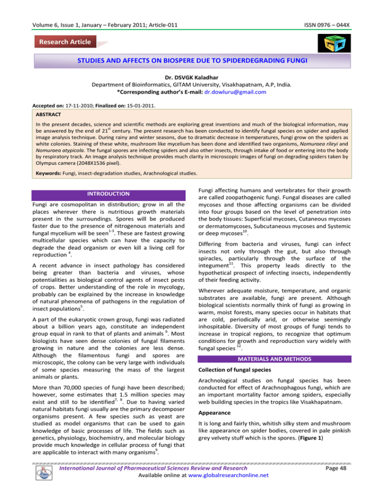
Volume 6, Issue 1, January – February 2011; Article-011 ISSN 0976 – 044X Research Article STUDIES AND AFFECTS ON BIOSPERE DUE TO SPIDERDEGRADING FUNGI Dr. DSVGK Kaladhar Department of Bioinformatics, GITAM University, Visakhapatnam, A.P, India. *Corresponding author’s E-mail: dr.dowluru@gmail.com Accepted on: 17-11-2010; Finalized on: 15-01-2011. ABSTRACT In the present decades, science and scientific methods are exploring great inventions and much of the biological information, may st be answered by the end of 21 century. The present research has been conducted to identify fungal species on spider and applied image analysis technique. During rainy and winter seasons, due to dramatic decrease in temperatures, fungi grow on the spiders as white colonies. Staining of these white, mushroom like mycelium has been done and identified two organisms, Nomuraea rileyi and Nomuraea atypicola. The fungal spores are infecting spiders and also other insects, through intake of food or entering into the body by respiratory track. An image analysis technique provides much clarity in microscopic images of fungi on degrading spiders taken by Olympus camera (2048X1536 pixel). Keywords: Fungi, insect-degradation studies, Arachnological studies. INTRODUCTION Fungi are cosmopolitan in distribution; grow in all the places wherever there is nutritious growth materials present in the surroundings. Spores will be produced faster due to the presence of nitrogenous materials and fungal mycelium will be seen1-3. These are fastest growing multicellular species which can have the capacity to degrade the dead organism or even kill a living cell for reproduction 4. A recent advance in insect pathology has considered being greater than bacteria and viruses, whose potentialities as biological control agents of insect pests of crops. Better understanding of the role in mycology, probably can be explained by the increase in knowledge of natural phenomena of pathogens in the regulation of 5 insect populations . A part of the eukaryotic crown group, fungi was radiated about a billion years ago, constitute an independent 6 group equal in rank to that of plants and animals . Most biologists have seen dense colonies of fungal filaments growing in nature and the colonies are less dense. Although the filamentous fungi and spores are microscopic, the colony can be very large with individuals of some species measuring the mass of the largest animals or plants. More than 70,000 species of fungi have been described; however, some estimates that 1.5 million species may exist and still to be identified7, 8. Due to having varied natural habitats fungi usually are the primary decomposer organisms present. A few species such as yeast are studied as model organisms that can be used to gain knowledge of basic processes of life. The fields such as genetics, physiology, biochemistry, and molecular biology provide much knowledge in cellular process of fungi that 9 are applicable to interact with many organisms . Fungi affecting humans and vertebrates for their growth are called zoopathogenic fungi. Fungal diseases are called mycoses and those affecting organisms can be divided into four groups based on the level of penetration into the body tissues: Superficial mycoses, Cutaneous mycoses or dermatomycoses, Subcutaneous mycoses and Systemic or deep mycoses10. Differing from bacteria and viruses, fungi can infect insects not only through the gut, but also through spiracles, particularly through the surface of the integument11. This property leads directly to the hypothetical prospect of infecting insects, independently of their feeding activity. Wherever adequate moisture, temperature, and organic substrates are available, fungi are present. Although biological scientists normally think of fungi as growing in warm, moist forests, many species occur in habitats that are cold, periodically arid, or otherwise seemingly inhospitable. Diversity of most groups of fungi tends to increase in tropical regions, to recognize that optimum conditions for growth and reproduction vary widely with fungal species 12. MATERIALS AND METHODS Collection of fungal species Arachnological studies on fungal species has been conducted for effect of Arachnophagous fungi, which are an important mortality factor among spiders, especially web building species in the tropics like Visakhapatnam. Appearance It is long and fairly thin, whitish silky stem and mushroom like appearance on spider bodies, covered in pale pinkish grey velvety stuff which is the spores. (Figure 1) International Journal of Pharmaceutical Sciences Review and Research Available online at www.globalresearchonline.net Page 48 Volume 6, Issue 1, January – February 2011; Article-011 ISSN 0976 – 044X Figure 1: Young spider with mushroom like fungal colony after infection Figure 2: Fungal staining (a)Fungal specimen before staining (b) Fungal species on spider (c) Stained specimen Microscopic examination Phylum: Ascomycota Preparation of reagent: Class: Ascomycetes /Hyphomycetes 15% Potassium Hydroxide Solution Order: Hypocreales (a) Potassium hydroxide 15 g Family: Clavicipitaceae (b) Glycerol 20 ml Genus: Nomuraea (c) Distilled water 80 ml Species: rileyi Procedure b. Description (1) Place the material to be examined onto a clean glass microscope slide. Nomuraea rileyi is an important fungus that attacks spiders. An outbreak of this fungus was reported in 1985 on larvae of rice insects, when it prevented an increase in the population of Spodoptera sp. Nomuraea rileyi is composed of pale green to gray-green conidiophores on a white basal felt of mycelium. The conidia are broadly ellipsoid and in dry chains. They are 3.5-4.5 x 2-3 µm long. The conidiophores have branches. Each branch contains 2-5 phialides or conidial chains. The early infective stage of N. rileyi is a white mass of fungus covering the spider. After a few days, the spores are formed and the host becomes pale green. Nomuraea rileyi also attacks the larvae of stems borers, leaffolders, armyworms, and caseworms. (2) Add a drop of 15% KOH to the material and mix. (3) Place a cover glass over the preparation. (4) Allow the KOH preparation to sit at room temperature until the material has been cleared. (5) Observe the preparation at 10x, 45x and 100x microscopy. Image analysis The images from microscopy has been analysed using image analysis softwares such as Image analyzer v1.30 and Pixcavator 2.4.1 softwares. Figure 3: Image analysis: Nomuraea rileyi RESULTS AND DISCUSSION Fungi have been attached to the bodies of the spiders, appeared as white mycelium and the stained parts of the fungi have been visualized as greenish-blue. Both filamentous and individual species with spores has been visualized and is provided in Figure 2. 2. Nomuraea atypicola (Figure 4) 1. Nomuraea rileyi (Figure 3) a. Classfication a. Classfication Kingdom: Fungi Kingdom: Fungi Phylum: Ascomycota International Journal of Pharmaceutical Sciences Review and Research Available online at www.globalresearchonline.net Page 49 Volume 6, Issue 1, January – February 2011; Article-011 Class: Ascomycetes ISSN 0976 – 044X Figure 4: Image analysis: Nomuraea atypicola Order: Hypocreales Family: Clavicipitaceae Genus: Nomuraea Species: atypicola b. Description The conidia of N. atypicola to be cylindrcal and curved, 46 x 1.2-1.5 µ.m; the conidia of the present fungus are ellipsoid and 4.5-5 x 2-2.5 µ.m. Figure 5 provides the clarity of images by using image analysis software’s. Figure 5: Image clarity using Pixcavator Figure 6 has provided the description of the stained specimen. There are 10 dark and 1 light objects visualized. Dark objects have more number of round objects (10 to 45) than the light object (3). Figure 6: Object analysis using pixcavator During winter season, the round, white mycelium forming on the young spiders may appear due to the higher humidity present in the air, making a favorable temperature and condition to kill the host and develop mycelium from the spores. Hence the destruction and growth of lager species due to micro organisms are being studying from decades. Fighting against microorganisms by larger species can be done by immune-pathogen mechanisms, where the activities are less in insects such as spiders. Due to this lack of active immune system, most of the fungal species such as Nomuraea rileyi and N.atypicola are growing actively by utilization of nitrogenous compounds from spiders on biosphere. CONCLUSION In the tropical countries such as India, there is a huge climatic variations occurring during the present century. Temperatures are extremely increased and there is uneven rainfall and winter seasons. Due to this change, new and emerged microbial organisms are forming which are mostly affecting smaller species such as insects (spiders, ants, mosquitoes), small fishes, reptiles and aves. The new species also may exist on these spider webs and the identification of these species is presently focused due to degradation of various types of insects and other species on the spider web. Fungi act as pathogens and kills young spiders during winter season due to having adequate temperature for the growth of fungal spores. Further studies can provide relationship of insect-parasitism or insect-degradation mechanisms. Isolation of metabolites from these species may kill plasmodium species, and also used as insecticides and pesticides. Phylogenetic analysis of Arachnomycetus fungi can be provided. Further studies can identify new fungal species on insects and new emerging fungal species due to biotic and climatic changes. Acknowledgement: The author would like to thank GITAM University for providing computational and wet lab facility and access to e-journals to carry out this research. International Journal of Pharmaceutical Sciences Review and Research Available online at www.globalresearchonline.net Page 50 Volume 6, Issue 1, January – February 2011; Article-011 REFERENCES 1. Wyatt MK, Parish ME, Spore germination of citrus juice-related fungi at low temperatures, Food Microbiology, 12, 1995, 237-243. 2. Norman AG, The biological decomposition of plant materials. III. Physiological studies on some cellulose decomposing fungi, Ann.Applied Biol., 17, 1930, 575-613. 3. Gaskill JO, Gilman JC, Role of nitrogen in fungous thermogenesis, Plant Physiol., 14, 1939, 31-53. 4. Cooke RC, Whipps JM, The evolution of modes of nutrition in fungi parasitic on terrestrial plants, Biological Reviews, 55(3), 1980, 341-362. 5. Ferron P, Biological control of insect pests by entomogenous fungi, Ann.Rev. Entomol., 23, 1978, 409-442 6. Mitchell LS, Jeffrey DS, Evolution of the protists and protistan parasites from the perspective of molecular systematic, International Journal for Parasitology, 28(1}, 1998, 11-20. ISSN 0976 – 044X 7. Hawksworth DL. The fungal dimension of biodiversity: magnitude, significance, and conservation, Mycological Research, 95, 1991, 641655. 8. Hawksworth DL, Kirk PM, Sutton BC, Pegler DN, Ainsworth and Bisby's Dictionary of the Fungi, 8th Ed, CAB International, Wallingford, United Kingdom, 1995. 616. 9. Taylor JW, Bowman B, Berbee ML, White TJ, Fungal model organisms: phylogenetics of Saccharomyces, Aspergillus and Neurospora, Systematic Biology, 42, 1993, 440-457. 10. Gerhard L, John WB, Nicholas JC, Anthony LJC, Murray HGM, Hirsutide, a Cyclic Tetrapeptide from a Spider-Derived Entomopathogenic Fungus, Hirsutella sp., J. Nat. Prod., 68 (8), 2005, 1303–1305. 11. Kaya HK, Gaugler R, Entomopathogenic Nematodes, Annual Review of Entomology, 38, 1993, 181-206. 12. David LH, The magnitude of fungal diversity: the 1.5 million species estimate revisited, Mycological Research, 105(12), 2001, 1422-1432. **************** International Journal of Pharmaceutical Sciences Review and Research Available online at www.globalresearchonline.net Page 51

