Identification of the Histidine Ligands to the Binuclear Metal ... Phosphotriesterase by Site-Directed Mutagenesis?
advertisement
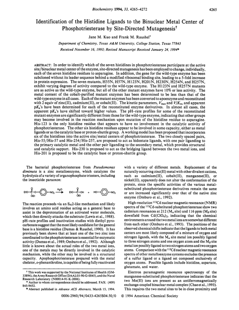
Biochemistry 1994, 33, 4265-4272 4265 Identification of the Histidine Ligands to the Binuclear Metal Center of Phosphotriesterase by Site-Directed Mutagenesis? Jane M. Kuo and Frank M. Raushel' Department of Chemistry, Texas A&M University, College Station, Texas 77843 Received November 16, 1993; Revised Manuscript Received January 24, 1994" ABSTRACT: In order to identify which of the seven histidines in phosphotriesterase participate a t the active site/binuclear metal center of the enzyme, site-directed mutagenesis has been employed to change, individually, each of the seven histidine residues to asparagine. In addition, the gene for the wild-type enzyme has been subcloned without its leader sequence behind a modified ribosomal binding site, leading to a 5-fold increase in protein expression. The seven mutants, H55N, H57N, H123N, H201N, H230N, H254N, and H257N, exhibit varying degrees of activity compared to the wild-type enzyme. The H123N and H257N mutants are as active as the wild-type enzyme, but all of the other mutant enzymes have 10% or less activity, The metal content of the cobalt-purified mutant enzymes has been determined to be less than that of the wild-type enzyme in all cases. Each of the mutant enzymes has been converted to apoenzyme and reconstituted with 2 equiv of zinc(II), cadmium(II), or cobalt(I1). The kinetic parameters, V,,, and V/Km,and apparent pKa's have been determined for each of the reconstituted enzyme derivatives. In almost all cases, the apparent pK,'s have shifted toward higher values. The pH-rate profiles for some of the reconstituted mutant enzymes are significantly different from those for the wild-type enzyme, indicating that other groups may become involved in the reaction mechanism upon mutation of the histidine residue to asparagine. His-123 is the only histidine residue that appears to have no involvement in the catalytic activity of phosphotriesterase. The other six histidine residues appear to be involved in some capacity, either as metal ligands or as the catalytic base or proton-shuttle group. A working model has been proposed that incorporates six of the histidines into the active site/metal centers of phosphotriesterase. The two closely spaced pairs, His-55/His-57 and His-254/His-257, are proposed to act as bidentate ligands, with one pair liganding to the primary catalytic metal and the other pair liganding to the secondary metal, which provides structural and catalytic support. His-230 is proposed to act as the bridging ligand between the two metal ions, and His-201 is proposed to be the catalytic base or proton-shuttle group. The bacterial phosphotriesterase from Pseudomonas diminuta is a zinc metalloenzyme, which catalyzes the hydrolysis of a variety of organophosphate triesters, including the insecticide paraoxon: The reaction proceeds via an S~2-likemechanism and likely involves an amino acid residue acting as a general base to assist in the deprotonation of an activated water molecule, which then directly attacks the substrate (Lewis et al., 1988). pH-rate profiles and inactivation studies with diethyl pyrocarbonate suggest that the most likely candidate for the general base is a histidine residue (Dumas & Raushel, 1990). It has previously been shown that at least one of the two zinc ions coordinated to the phosphotriesterase is essential for enzymatic activity (Dumas et al., 1989; Omburo et al., 1992). Although little is known about the actual roles of the two metal ions, one of the metals may be directly involved in the catalytic mechanism, while the other may be involved in a structural capacity. Apophosphotriesterase prepared with the metal chelator, o-phenanthroline, is capable of being fully reactivated t This work was supported by the National Institutes of Health (GM33894), the Army ResearchOffice (DAAL03-90-G-0045),and the Naval Research Laboratory (N00014-9 1-K-2006). * Author to whom correspondenceshould be addressed. FAX: (409) 845-9452. Abstract published in Advance ACS Abstracts, March 15, 1994. @ 0006-2960/94/0433-4265$04.50/0 , I , with a variety of different metals. Replacement of the naturally occurring zinc(I1) metal with other divalent cations, such as cadmium(II), cobalt(II), manganese(II), or nickel(II), apparently does not alter the conformation of the protein, since the specific activities of the various metalsubstituted phosphotriesterase derivatives remain the same or are increased significantly over that of the native zinc enzyme (Omburo et al., 1992). High-resolution '13Cd nuclear magnetic resonance (NMR) spectra of the 113Cd-substitutedphosphotriesterase show two cadmium resonances at 212 (Ma site) and 116 ppm (MBsite) downfield from Cd(C10&, indicating that the chemical environments around the two metal ions are somewhat different from each other (Omburo et al., 1993). The positions of the observed chemical shifts indicate that the ligands to both metal centers are most likely composed of a mixture of oxygen and nitrogen ligands, with the Ma site metal ion possibly ligated to three nitrogen atoms and one oxygen atom and the MBsite metal ion possibly ligated to two nitrogen atoms and two oxygen atoms. Comparison with the ll3Cd nuclear magnetic resonance spectra of other metalloenzyme systems excludes the presence of a sulfur ligand or a ligand set composed exclusively of oxygen atoms. Possible ligands include histidine, aspartate, glutamate, and water. Electron paramagnetic resonance spectroscopy of the manganese-substituted phosphotriesterase indicates that the two Mn(I1) ions are present as an antiferromagnetically exchange-coupled binuclear metal complex (Chae et al., 1993). This requires the two metal sites to be in close proximity and 0 1994 American Chemical Society Kuo and Raushel 4266 Biochemistry, Vol. 33, No. 14, 1994 suggests that the two metal ions are bridged by a common ligand. The most obviouscandidates among the amino acid residues in phosphotriesterase to act as ligands for the metal centers are histidine, aspartate, and glutamate. Histidine is a common metal center ligand in several zinc metalloenzymes (Vallee & Auld, 1990). The activated water molecule proposed in the chemical mechanism of phosphotriesterase is likely to be an oxygen ligand to one of the two metal centers. Since histidine residues have been implicated in both the catalytic mechanism and metal ligation, it seems likely that some, if not all, of the seven histidines in phosphotriesterase are associated with the active site in some capacity. In order to identify which of the histidine residues may be involved in the chemical mechanism and/or metal center ligation, site-directed mutagenesis has been employed to change, individually, each of the seven histidine residues to asparagine. The substitution of a histidine with an asparagine should make that position less effective, both as a ligand and as a general base, but at the same time, the change should be structurally conservative enough not to cause drastic alterations in the conformation of the active site or in the overall structure of the protein. Kinetic studies with these histidine mutants provide further insight into the possible roles of these histidine residues in the structure and function of the phosphotriesterase. MATERIALS AND METHODS Materials. All chemicals were purchased from Sigma Chemical Co., Aldrich Chemical Co., Fisher Scientific, or United States Biochemical Co. Bacto-tryptone and Bactoyeast extract were purchased from Difco Laboratories. Ultrogel AcA54 was purchased from IBF Biotechnics. GeneAmp DNA amplification kits were purchased from Perkin-Elmer Cetus. Sequenaseversion 2.0 DNA sequencing kits were purchased from United States Biochemical. All enzymes used in the molecular biology experiments were purchased from Promega, United States Biochemical, Stratagene, or New England Biolabs. Geneclean DNA purification kits were purchased from Bio 101. Magic Minipreps DNA purification kits and Magic PCR Preps DNA purification kits were purchased from Promega. All oligonucleotides used in this work were synthesized by the Gene Technology Laboratory of the Biology Department of Texas A&M University. Bacterial Strains and Plasmids. The Escherichia coli strains used for this study were XL1-Blue (recAl, endAI, gyrA96, thi-I, hsdR17, supE44, relAI, lac, [F' proAB, lacIqZMI5, Tn 10 (tet')]) (Bullock et al., 1987) and BL21 (F-ompTrB-mB-) (Studier et al., 1990). ThepBSphagemid (Stratagene) was used as the vector in all cases. The opd gene from Pseudomonas diminuta, which was previously subcloned along with its leader sequence into the pBS+ phagemid at the BamHI site and used under the name of pDDO2, was used as the template for further subcloning experiments. Reconstruction of the opd Gene. The plasmid pDD02 containing the opd gene was used as a template for the polymerasechain reaction (PCR). Primer A (shown in Table 1) was a 44-base oligonucleotide composed of a BamHI site, an optimized E. coli ribosomal binding site (Skoglund et al., 1990), and the codons for the first six amino acid residues of the mature phosphotriesterase. Primer D (shown in Table 1) was a 33-base oligonucleotide composed of a BamHI site, three successive stop codons, and the codons for the last five amino acid residues of the mature phosphotriesterase. The Table 1: Flanking Primers Used in PCR primer A Me1 Ser BamHl /le Gly Thr Gly Asp W ~ C G G A T C C G G A G G ~ A A A AATGTCG A A A T ATCGGCACAGGCGATCY primer D BamHl Ser Ala Arg Leu Thr SCGCGGATC(ITCATCATCATGACGCCCGCAAGGT~ PCR reaction mixture consisted of 200 pM each of the dideoxynucleotide triphosphates, 2.5 units of AmpliTaq DNA polymerase, 1 pM each of primers A and D, 1 ng of pDD02 DNA template, and GeneAmp reaction buffer for a final volume of 100 pL. Two drops of mineral oil were added to prevent evaporation during thermocycling. An initial melt at 95 OC for 2 min was followed by 28 cycles at 94 OC for 1 min, 60 OC for 1 min, and 72 OC for 1 min, and ended with slow cooling to4 OC. After phenol/chloroform extraction to remove the mineral oil, the 105 1-base-pair (bp) fragment was digested with BamHI to yield a 1039-bp fragment, which was concentrated with the Geneclean system and purified by electrophoresis in a 1% agarose gel. The DNA was extracted from the agarose gel with the Geneclean system. The pBS+ vector was also cut with BamHI and purified as above. Vector and insert were combined and concentrated with the Geneclean system before ligation with T4 DNA ligase. The recombinant DNA was transformed into XL1 -Blue cells, and transformants were selected using the blue/white selection feature of XL1-Blue. Plasmids isolated from the selected colonies were screened for the insertion of the opd gene in the correct orientation behind the lac promoter in pBS+ through the following series of restriction digests: BamHI alone (should yield 1039- and 3204-bp fragments), EcoRV/PstI (should yield 264- and 3979-bp fragments), and EcoRV/EcoRI (should yield 8 11- and 3432-bpfragments). Thereconstructed opd gene encoding just the mature phosphotriesterase was designated pJKOl and transformed into BL21 cells. Site- Directed Mutagenesis. The seven histidine residues in phosphotriesterase at positions 55, 57, 123, 201, 230, 254, and 257 were individually changed to asparagine using the method of overlap extension PCR (Ho et al., 1989). The 20-base oligonucleotide primers used to make these mutants are listed in Table 2. Each pair of B and C primers was used to introduce a single base change into the wild-type sequence. Overlap extension PCR consisted of performing the reactions with two separate sets of primers. The first set of primers, primer A and a selected B primer, was used to make an AB fragment. The second set of primers, consisting of the corresponding C primer and primer D, was used to make a CD fragment. The plasmid pJKOl was used as the template in both cases. The AB and CD fragments were purified by agarose gel electrophoresis, followed by either the Geneclean system or the Magic PCR Prep DNA purification system. The two fragments were combined and used in a third PCR, where they acted as both primer and template for each other, since their sequences overlapped in the region around the desired mutation site. Primers A and D were also included to further amplify the resulting AD fragment. The AD fragment was purified from agarose gel, cut with BamHI, concentrated with BamHI-digested pBS+, ligated with T4 DNA ligase, and transformed into XL1-Blue cells. Colonies were screened as above for those vectors containing inserts in the desired orientation. The plasmids for the seven mutants, H55N, H57N, H123N, H201N, H230N, H254N, and H257N, were designated pJKO2-pJKO8, respectively, and transformed into BL21 cells. Histidine Ligands to a Binuclear Metal Center Biochemistry, Vol. 33, No. 14, 1994 4261 enzymes was determined by atomic absorption spectrophotometry. The cobalt-purified enzymes were dialyzed against H55N Primer B YGA T G T G C T C ~ A G T C AGTG? metal-free 50 mM HEPES (pH 8.5) buffer, with several buffer H55N Primer C TACTGACTUGAGCACATC~ changes over a 24-h period at room temperature, to remove the CoC12 that was included in the purification buffer and any SGCCGCA G A T ~ C T C G T G AG’ loosely bound metals from the enzymes. The metalH57N Primer B reconstituted enzymes were passed through Pharmacia PD~ C T ACG C AGMATCTGCGGC~ H57N Primer C 10 Sephadex G-25 desalting columns to remove any loosely bound metals. All samples were analyzed on a Perkin-Elmer H123N Primer B ~GCCACGATAITAACGTCG~ 2380 atomic absorption spectrophotometer. The errors in H123N PrimerC %CCGACGTTA,QATCGTGC~ metal concentration are estimated to be f 15%. Determination of Enzyme Activity. The activity for each H2OlN Primer B %CTGCCGTGITAGTGGTTAC? of the enzyme derivatives was measured by monitoring the H201N Primer C %TAACCACTA&ACGGCAK~ appearance of p-nitrophenol produced by the hydrolysis of paraoxon. The assay mixture consisted of 1 mM paraoxon ~ T ATCGCTDACC C AATAC~3 H230N Primer B in 3 mL of 100 mM CHES (pH 9.0) buffer. In the cases of the cobalt-substituted H55N, H57N, H201N, H230N, and 5TGTATTGGTMAGC.GATGA’ H230N Primer C H254N mutants, it was necessary to add extra CoClz to the assay mixtures in order to obtain maximum activities (50,80, H254N Primer B ~TGCGGGAT~GTCTAGACC’ 200, 10, and 500 pM CoC12, respectively). All spectrophoH254N Primer C ~GGTCTAGACMATCCCGCA~ tometric measurements were made at 25 OC with a Gilford Model 260 spectrophotometer. Rates were determined by H257N Primer B SAATCGCACTQTICGGGATGT~ monitoring the production of p-nitrophenol at 400 nm for 1 H257N Primer C s ACATCCCGMAGTGCG ATT? min, and the values for Vmaxand V/Km were calculated as described previously (Caldwell et al., 1991). The extinction Sequencingof Mutants. The opd gene in each of the eight coefficient for p-nitrophenol at 400 nm was 17 000 M-’ cm-l. recombinants, pJKO1-pJK08, was completely sequenced using pH-Rate Profiles. The apparent PKa values were deterthe dideoxynucleotide chain termination method (Sanger et mined for the Zn(II), Cd(II), and Co(I1) forms of the wildal., 1977) to ensure that only the desired base changes were type and mutant enzymes. Each enzyme derivative was present. Double-stranded plasmid DNA (dsDNA) was assayed over the pH range 5.0-1 1.O at increments of 0.25. isolated using the Magic Miniprep DNA purification system The Vm,, values at each pH were approximated by using a and cut 3’ to the opd gene with EcoRI. The linear dsDNA saturating amount of substrate in each assay. The assay was then incubated with the T7 Gene 6 5‘-3‘ exonuclease to mixtures consisted of 4 mM paraoxon in 3 mL of 100 mM make single-stranded DNA (ssDNA), which was then buffer. The buffers used were sodium acetate, 2-(N-morsequenced using the Sequenase version 2.0 DNA sequencing pho1ino)ethanesulfonic acid (MES), piperazine-N,N’-bis(2kit. ethanesulfonic acid) (PIPES), N- (2-hydroxyethyl)piperazineGrowth Conditions. Overnight cultures of the transformed N’-(2-ethanesulfonic acid) (HEPES), N- [tris(hydroxymethyl)cells grown in Luria-Bertani (LB) broth (Maniatis et al., methyl] -3-aminopropanesulfonic acid (TAPS), 2-(N-cyclo1982) were used to inoculate modified LB medium (Post et hexy1amino)ethanesulfonic acid (CHES), and 3-(cyclohexyal., 1990) containing 1 mM CoC12 and 50 pg/mL ampicillin. lamino)- 1-propanesulfonic acid (CAPS). Other assay conThe cobalt-supplemented cultures were incubated at 30 “ C ditions were the same as those given above. The apparent and induced with isopropyl D-thiogalactopyranoside (IPTG) extinction coefficient for p-nitrophenol at each pH value was in the early log phase. Cells were harvested in the stationary calculated using the equation phase. ‘app = €/(lOpKa-PH 1) Purification of Wild- Type and Mutant Enzymes. Phos(1) photriesterase wild-type and mutant enzymes were purified where the extinction coefficient is 17 000 M-I cm-l and the from BL21 /pJKOl-BL21 /pJK08 according to the procedures pKa value for p-nitrophenol is 7.14. given by Omburo et al. (1992), with the exception that the The pK values for each metal-substituted mutant were purification buffer contained 100 pM CoC12. SDS-PAGE calculated using one of the following equations: and Western blots indicated that all of the mutants were of the same size as the wild-type protein. log V = log(C/(l + 10-pH/K,)) (2) Preparation of Apoenzyme. Apoenzyme was prepared by incubating enzyme with 2 mM o-phenanthroline (OP) at room or temperature for 90 min until there was less than 1% residual activity left. The o-phenanthroline was removed by diafillog V = log(C/(l + K2/10-pH)) (3) tration with metal-free 50 mM HEPES (pH 8.5) buffer over an Amicon PMlO membrane until there was less than 0.5 pM or OP remaining. log V = log(C(1 10-pH/K,)/(l (10-pH/K2)(1 Reactivation of Apoenzyme with Zn(II), Cd(II), and Co(II). Apoenzyme at a concentration of between 0.5 and 2 lo-pH/K,))) (4) mg/mL was reconstituted with the desired metal by adding 2 equiv of either ZnCl2, CdS04, or CoCl2 per equivalent of where Vis the pH-dependent value of Vmaxand C is the pHenzyme and incubating the mixture at room temperature for independent value of Vmax, or at least 12 h. Determination of Metal Content. The metal content of the log V = log((? + Yh(Kl/lO-pH))/(l + Kl/lO-pH)) ( 5 ) cobalt-purified and metal-reconstituted wild-type and mutant or Table 2: Primers Used for Site-Directed Mutagenesis + + + + 4268 Biochemistry, Vol. 33, No. 14, 1994 ~ Kuo and Raushel ~~ Table 3: Activities and Metal Content of Cobalt-Purified Wild-Tvue and Mutant Enzvmes enzyme (mg of enzyme)/ (g of cell paste)" Km (pM)* V, (units/mg) k,,/K, (M-1 s-l) metal contentC Zn(II)/enzyme Co(II)/enzyme Fe(II)/enzyme WT 3.0 110 9600 5.0 x 10⁷ 2.1 0.4 0.1 0.5 110 34 1.8 x 10⁵ 0 0.02 H55N H57N 2.3 430 350 4.9 x 10⁵ 0 0.08 8000 4.9 x 10⁷ 1 .o 0.2 0.03 H123N 2.3 99 1 .o 0.2 0.01 1.4 9 16 1 . 1 x 10⁶ H201N 1.1 0.1 0.04 1.2 320 240 4.5 x 10⁵ H230N 670 9.7 x 10⁶ 0.5 0.1 0.06 H254N 1.7 41 1 .o 0.6 0.06 1.2 430 4700 6.6 X lo6 H257N 4 Cells were grown in CoC12-supplementedmedium. Assays were done at pH 9.0 and 25 O C with paraoxon as the substrate. The cobalt-purified proteins were dialyzed against metal-free buffer before atomic absorption was performed. where Vis the pH-dependent value of V,,,, K is the minimum pH-independent value of Vmax,and Yh is the maximum pHindependent value of V,,,. Circular Dichroism Studies. Wild-type and mutant enzymes were exchanged into 50 mM potassium phosphate (pH 8.0) buffer using Pharmacia PD- 10 Sephadex G-25 desalting columns and scanned over the far-UV range (250-190 nm) in a 0.2-mm cell using a Jasco 5-600 spectropolarimeter. RESULTS Construction, Expression, and Purification of Recombinants. The opd gene from Pseudomonas diminuta was subcloned without its leader sequence into an E. coli expression system. In the reconstructed opd gene, the codon for Ser-30, which is the first amino acid residue in the mature phosphotriesterase, was placed immediately after the Met start codon. Substitution of the P. diminuta ribosomal binding site with an optimized E. coli ribosomal binding site increased the expression of phosphotriesterase by 5-fold. The seven histidine residues of phosphotriesterase were individually mutated to asparagines by site-directed mutagenesis, employing the method of overlap extension PCR. The DNA sequences for the wild-type, H55N, H57N, H201N, and H230N recombinants contained no extraneous base changes. The sequences for the H123N, H254N, and H257N recombinants, however, in addition to the desired mutations, contained a few silent base changes. In the H 123N gene, the codons for Arg-9 1 and Ala-126 were altered (AGA-AGG and GCG-GCA). In the H254N gene, the codons for Ala-281 and Ser-307 were altered (GCT-GCC and TCG-TCA). Finally, the codon for Ile-260 in the H257N gene was altered (ATT-ATA). The mutant proteins were expressed and purified to homogeneity by the same methods used for the wild-type protein; however, none of the mutants was expressed as well as the wild-type protein (Table 3). The expression of the H55N mutant was especially low, with a yield of less than 20% compared to the wild-type enzyme. The metal content, as well as the kinetic parameters, V,,, and V/Km,of the cobaltpurified mutant enzymes ranged in value from less than 1% to 100%of the wild-type values (Table 3). The H55N and H201N mutants had less than 1% activity, and the H57N, H230N, and H254N mutants had less than 10% activity, while the H123N and H257N mutants had at least 50% activity compared to wild-type phosphotriesterase. All of the cobaltpurified mutants bound fewer metals than the wild-type enzyme, with the H55N and H57N mutants showing no bound metals at all after dialysis with metal-free buffer. 0 2 4 6 6 MetallEnzyme 1 0 FIGURE1 : Reactivation of mutants H254N and H230N with varying amounts of metal. The apoenzymes were incubated with 0-10 equiv of ZnCl2 (e),CdS04 (m), or CoCl2 (e),and activities (units/mg) were measured after 12 h of incubation at room temperature. (A) The H254N mutant required only 1 equiv of metal per enzyme to be fully reactivated. (B) The H230N mutant required an excess of metals to be fully reactivated. Reactivation ofthe Apoenzymes. The native Zn(I1) metal in the mutant enzymes could be replaced with other divalent metals, such as Cd(I1) and Co(I1). Apoenzyme was prepared through incubation with the metal chelator, o-phenanthroline. Reactivation experiments were conducted with varying amounts of ZnCl2, CdS04, and CoC12 to determine how many equivalents of metal were required to reactivate each enzyme fully. Apoenzyme was incubated at room temperature with 0-10 equiv of metal and assayed for activity every 2 h up to 12 h and again after 24 h of incubation. All of the enzymes were fully reactivated by 12 h. Typical reactivation curves of the H230N and H254N mutant enzymes with ZnC12, CdS04, and CoC12 at 12 h are shown in Figure 1. The H254N and H257N mutants required only 1 equiv of metal per equivalent of enzyme to achieve maximal activity under the conditions of the reconstitution experiment. The H123N and H201N mutants required between 1 and 2 equiv of metal, while the H55N, H57N, and H230N mutants required an excess of metal to be fully reactivated. Metal Content of Zinc-, Cadmium-, and Cobalt-Reconstituted Mutants. In order to determine whether either of the metal binding sites in phosphotriesterase was affected by any of the histidine replacements, atomic absorption spectrophotometry was conducted with each metal-substituted Biochemistry, Vol. 33, No. 14, 1994 4269 Histidine Ligands to a Binuclear Metal Center Table 4: Metal Content of Reconstituted Enzymes" ~ enzyme Zn(II)/enzyme wild-type H55N H57N H 123N H2O 1N H230N H254N H257N 1.5 0.6 1.4 1.5 1.5 1.6 1.6 1.6 Cd(II)/enzyme Co(II)/enzyme 1.6 1.3 0 0 1.3 0.8 1.1 0 0 1.6 1.o 1.2 1.6 1.7 1.2 1.3 Apoenzymes were reconstituted with 2 equiv of the indicated metal and desalted through Sephadex G-25 before atomic absorption spectrophotometry was performed. mutant. Apoenzyme was incubated at room temperature with 2 equiv of ZnClz, CdS04, or CoClz for 12 h. The metalreconstituted enzymes were then passed through G-25 Sephadex desalting columns to remove any loosely bound metals before atomic absorption spectrophotometry was performed. The zinc-reconstituted wild-type and histidine mutant enzymes, except for H55N, all appeared to have approximately 1.5 equiv of zinc bound per enzyme. The H55N mutant bound less than 1 equiv of zinc per enzyme. In the case of the cadmium-reconstituted enzymes, the wild-type, H123N, H254N, and H257N mutants indicated at least 1.6 equiv of cadmium bound per enzyme. The H201N and H230N mutants, on the other hand, indicated only 1 equiv of cadmium per enzyme, while the H55N and H57N mutants showed no cadmium bound at all. For the cobalt-reconstituted enzymes, all except the H55N and H57N mutants indicated between 0.8 and 1.3 equiv of cobalt bound per enzyme. The H55N and H57N mutants showed no cobalt bound. Results are summarized in Table 4. Kinetic Properties of Zinc-Substituted Mutants. The zincsubstituted enzymes showed the lowest activities of the three metal derivatives, even though zinc seemed to be the most tightly bound of the three metals. The kinetic parameters, V,,and V/K,,for the hydrolysisofparaoxonwiththeH123N mutant were essentially identical to those of the wild-type enzyme. The H257N mutant had higher activity compared tothewild-typeenzyme, but alower V/K,value. TheH201N, H230N, and H254N mutants all had V, values around 1% that of the wild-typeenzyme, although the V/K,valuesvaried widely. The V,, and V/K, values for the H57N mutant were 10% of those for the wild-type enzyme. The H55N mutant was the least active of the seven histidine mutants, showing V,, and V/K, values of less than 1.0%. Kinetic Properties of Cadmium-Substituted Mutants. The cadmium-substituted wild-type enzyme was more than twice as active as the zinc enzyme. The cadmium mutants behaved somewhat differently from the zinc mutants, although the H123N mutant was again essentially the same as the wildtype enzyme. The V,, and V/K, values for the H57N, H254N, and H257N mutants were within 20-60% of the wild-type values, while the kinetic values for the H201N and H230N mutants were 10% or less. The H55N mutant had the lowest activity, with V, and V/K, values of less than 1% relative to the wild-type enzyme. Kinetic Properties of Cobalt-Substituted Mutants. The cobalt-substituted enzymes were the most active of the three forms. The H123N and H257N mutants were as active as the wild-typeenzyme, although the V/K,valuefor the H257N mutant was only 20% of the wild-type value. The H254N mutant was only 10% as active, but had a V/K, value onehalf that of the wild-type value. The H57N and H230N mutants were 5% as active, with equally low V/K, values. The H55N and H201N mutants were less than 1% as active as the wild-type enzyme. The H55N mutant was again the least active of the seven histidine mutants. pH-Rate Profiles of Zinc-Substituted Mutants. The pHrate profiles for the zinc wild-type enzyme and most of the zinc-substituted mutants were fit to eq 2. The H123N mutant had the same PKa value as the wild-type enzyme, but the other mutants all showed a shift to higher pKa values. The pH-rate profile for the H55N mutant was fit to eq 5. The pH-rate profile for the H257N mutant was fit to eq 6 and gave both a pK, and a PKb value. The H57N mutant was the most unusual in that it appeared to have only a pKb value; its pHrate profile was fit to eq 3. pH-Rate Profiles of Cadmium-Substituted Mutants. The pKa value for the cadmium-substituted wild-type enzyme was shifted to over 2 units higher compared to that for the zincsubstituted wild-type enzyme. The cadmium-substituted H123N, H254N, and H257N mutants had similar pKavalues compared to the wild-type enzyme, while the H55N, H57N, H201N, and H230N mutants had higher values. The pHrate profiles for all but the H55N and H254N mutants were fit to eq 2. The pH-rate profile for the H55N mutant was fit to eq 5, and the pH-rate profile for the H254N mutant was fit to eq 4. pH-Rate Profiles of Cobalt-Substituted Mutants. The pKa value for the cobalt-substituted wild-type enzyme was intermediate to those of the zinc and cadmium forms. The pK, value for the H123N mutant was again consistent with that of the wild-type enzyme. The H254N mutant had an unexpectedly lower pKa value compared to the wild-type enzyme, but the other mutants behaved as expected by having higher pKavalues, with the exceptionof theH55N and H230N mutants. It was not possible to obtain a fit of the pH-rate profiles of the H55N and H230N mutants to any of the given eq 2-6. The profile for the H230N mutant showed a significant dip in its V, values between pH 7.5 and 9.0 that did not fit to eq 4, while the profile for the H55N mutant showed a linear increase in Vma, up through pH 1 1.O, with no signs of leveling off. With the exception of the H257N mutant, all of the pH-rate profiles were fit to eq 2. The pH-rate profile for the H257N mutant was fit to eq 5 . A representative example of each type of pH-rate profile is given in Figure 2. The kinetic parameters and pKavalues for thevarious histidine mutant enzymes are summarized in Table 5. Circular Dichroism Studies. Circular dichroism studies were carried out to determine whether there were any gross changes in the overall structures of the seven histidine mutants compared to the wild-type enzyme. A scan of the far-UV region for each mutant indicated that all of the mutants were very similar in structure to the wild-type protein (data not shown) and, furthermore, indicated that the phosphotriesterase contains a significant amount of a-helical secondary structure. The spectra for the H201N and H55N mutants indicated that those samples may have contained some denatured protein, but an expansion of their experimental curves gave the same spectra as that of the wild-type enzyme. DISCUSSION Histidine residues are known to be essential in many zinc metalloenzymes. In the zinc enzymes whose structures are known, histidine residues appear to be the most common ligand to catalytic zinc centers. In a comparison of the structures of 12 zinc metalloenzymes, Vallee and Auld (1990a,b) show that most of the zinc ions are coordinated to four ligands, regardless of whether they are catalytic or structural centers. 4270 Biochemistry, Vol. 33, No. 14, 1994 I 3.5 > -8 Kuo and Raushel 3'5F Table 5: Kinetic Parameters for the Reconstituted Wild-Type and Mutant EnzymesQVb > -g 3.0 3.0 I d 2.51, 2.5 4 5 6 7 8 91011 PH > 01 0 4 , B , , , , 5 6 7 8 9 1011 , PH :::: 2.0 - wild-type Zn Cd Co H55N Zn Cd Co H57N Zn Cd 68 460 130 16 720 660 78 1100 310 3800 10000 13000 1.4 4.4 0.26 420 5100 650 2300 6000 7800 3.4 X lo7 5.7 1.3 X lo7 8.1 6.0 X lo7 6.5 0.83 5.2 X 10⁴ 9.7 2.6 3.6X lo3 9.1 0.16 2.4 X lo2 250 3100 390 3.2X lo6 2.8 X 10⁶ 8.8 1.2X lo6 8.1 10.0 Co H123N Zn 64 3500 2100 3.3 x 10⁷ 5.6 Cd 440 5000 1.1 X lo7 8.2 8300 Co 130 9400 5600 4.3 X lo7 6.6 5 6 7 8 91011 6 7 8 9 1011 H201N PH PH Zn 11 31 19 1.7 X lo6 8.2 Cd 90 230 140 1.6 X lo6 9.2 4.07 1 42 co 16 25 1.6 X lo6 8.4 H230N Zn 67 44 26 3.9 X los 6.8 Cd 530 260 150 2.8 X lo5 9.5 Co 290 830 500 1 . 7 X lo6 H254N Zn 1.3 56 34 2.6 X lo7 7.0 Cd 540 6000 3600 6.7 X lo6 8.0 9.0 6.6 3.0 co 27 1400 goo 3.3 x 10⁷ 5.3 5 6 7 8 9 1011 H257N PH Zn 550 7300 4400 8.0X lo6 7.8 10.7 Cd 780 5500 3300 4.2X lo6 8.2 FIGURE2: pH-rate profiles for selected metal-activated mutants: Co 640 12000 1.1 X lo7 8.1 7000 (A) Zn(I1)-wild-type fit to eq 2; (B) Zn(II)-H123N fit to eq 2; (C) Zn(II)-H57N fit to eq 3; (D) Cd(II)-H254N fit to eq 4; (E) Assays were done with paraoxon at pH 9.0,25 "C. V , values are Co(II)-H257N fit to eq 5; and (F) Zn(II)-H257N fit to eq 6. *lo%, Km values are 120%.and DK values are 10.1 DHunit. * For catalytically active zinc ions, three of the ligands are derived from amino acid residues, with histidine residues being the most common, followed by glutamate, aspartate, and cysteine. An activated HzO is the universal fourth ligand at all catalytic zinc sites. In the case of structural zinc centers, all four ligands are invariably composed of amino acid residues, with cysteine being the most common ligand. The spacing of the liganding amino acid residues in the primary sequences of the enzymes has also been noted. The three amino acid ligands to catalytic metal centers appear to have a pattern to their spacing within the protein sequence. Twoof the residues are usually separated by a short spacer of 1-3 amino acid residues, while the third residue is usually separated by a long spacer of 20-120 residues. It is proposed that the two closely spaced amino acid residues act to form a bidentate zinc complex and that the long spacer of the third ligand confers flexibility to the three-dimensional alignment of the active site, allowing for the approach of substrate or for the required positioning of the zinc atom for catalysis. The close spacing of the four ligands to structural zinc atoms, on the other hand, imparts rigidity to the metal centers, consistent with their role in stabilizing both overall protein structure and local conformation. An examination of the structures of four multi-zinc enzymes reveals similar motifs (Vallee & Auld, 1993). Alkaline phosphatase, phospholipase C, P1 nuclease, and leucine aminopeptidase all possess cocatalytic sites consisting of two or more zinc atoms closely spaced together. In these systems it is thought that the metals act in concert to bring about catalysis, with one of the zinc ions acting as the catalytic metal and the other(s) assisting in coordinating to substrate and/or product. The amino acid sequences of these enzymes reveal that the ligands to the zinc ions also have the pattern of two of the amino acid residues separated by a short spacer and a third amino acid residue separated by a long spacer. The multi-zinc sites have an additional feature of two of the metal atoms being bridged by a common ligand. In the enzymes under consideration, this shared ligand is an aspartic or glutamic acid residue, but the number of enzymes in this comparison is quite small. On the other hand, the X-ray structure of bovine Cu,Zn superoxide dismutase (Tainer et al., 1982) reveals that the metal-bridging ligand can also be a histidine residue. Comparison of the amino acid sequences of several zinc enzymes whose structures are known reveal that, within the same families, there is a high degree of conservation in the residues around the vicinities of the ligands to catalytically active zinc atoms. Given what is known so far about the coordination to the metal centers in zinc metalloenzymes, the fact that phosphotriesterase has at least one catalytically active zinc center and only seven histidine residues increases the probability that some or all of the histidines are coordinated to the two zinc atoms. A comparison of the sequences around each histidine residue in phosphotriesterase to the GenBank and EMBL database files using software from the Genetics Computer Group, Inc. (Madison, WI) reveals no homology to any known sequence, indicating that phosphotriesterase does not belong to any of the families of zinc metalloenzymes whose structures are known to date. Since phosphotriesterase does not appear to belong to any of the known families of zinc metalloenzymes, elucidation of the identity of the ligands to its two zinc centers will provide further insight into the conformations of catalytic zinc sites and ultimately lead to a better understanding of the specificity of these enzymes and their mechanisms of action. Histidine Ligands to a Binuclear Metal Center Biochemistry, Vol. 33, No. 14, 1994 4271 In order to determine the importance of each of the histidines in phosphotriesterase to its catalytic activity, each of the histidine residues was converted to an asparagine residue through site-directed mutagenesis, and the metal content, reactivation parameters, kinetic parameters, and pH-rate profiles were determined for each of themutants with a variety of metals incorporated into their metal sites. His-123 is the only one of the seven histidines in phosphotriesterase that is unequivocally uninvolved in any way with the catalytic activity or structural integrity of this enzyme. The reconstituted metal content, the kinetic parameters V, and V/K,, and the pHrate profiles determined for the Zn(I1)-, Cd(I1)-, and Co(I1)-substituted H123N mutant are identical to the values for the wild-type enzyme. In addition, a 13CdNMR spectrum of the l Wd-substituted H123N mutant gives resonance peaks in the NMR spectrum that are identical to those observed for the wild-type enzyme (L. Mullins and J. Kuo, unpublished observations). The data for the other six histidine mutants are somewhat more difficult to interpret. On the basis of the observed activities and pH-rate profiles, it is possible to say that these histidines are all involved in some capacity at the metal centers and/or active site. On the other hand, the substitution of an asparagine for a histidine may cause enough of a disruption to the catalytic or binding site conformation in a particular mutant to affect its activity without that residue being directly involved with the metal centers. The H257N mutant derivatives are as active as the wild-type enzyme, but the V/K, values are lower by over 60% relative to wild-type values, suggesting that this mutant may not bind substrate as tightly as the wild-type enzyme. The pH-rate profiles for the Zn(1I) and Co(I1) derivatives of this mutant further suggest that in this mutant a hitherto uninvolved group may now be affecting the catalytic activity. The mutant H254N is also nearly as active as the wild-type enzyme in its Cd(I1) form, but its specific activity is reduced 10-100-fold in its Zn(I1) and Co(I1) forms. However, the V/K, values for all three metal derivatives of this mutant are close to the wild-type values, suggestingthat substrate binding in this mutant remains relatively unaffected. The pKa values for the three enzyme derivatives differ from the wild-type values, but the general shapes of the pH-rate profiles are similar, indicating that some group on the H254N mutant must be deprotonated for full enzymatic activity, as is the case with the wild-typeenzyme. The metal contents of the reconstituted forms of mutants H257N and H254N are the same as for the wild-type enzyme. Aside from His-123, the mutation of residues His-257 and His-254 has the least overall effect on the properties of phosphotriesterase. Experimental results for the remaining four mutants, H55N, H57N, H201N, and H230N, suggest that these four histidine residues are likely candidates for catalytic metal center ligands in phosphotriesterase, provided the amino acid residue substitutions have not disrupted the active-site conformations of the mutants significantly. The H55N and H57N mutants show the least number of metals bound in the various metalreconstituted forms. In fact, the amounts of Cd(I1)- and Co(I1) in the Cd(I1) and Co(I1)-substituted forms of these two mutants were below detection levels, although in each case the Cd(I1) form of the enzyme was the most active of the three derivatives. These data suggest that the metal binding affinity in both of these mutants has been weakened, possibly by the removal of one of the metal-coordinating ligands. Both the VmaxandV/K,values for these twomutants have been significantly reduced compared to those of the wildtype enzyme, lending further credence to the possibility that HIS-57 HlS-254 L -NH ASP or GLU - H I s 201 FIGURE3: Working model of the active site/binuclear metal center of phosphotriesterase. His-55 and His-57 are metal-coordinating ligands. The pHrate profiles for the H55N mutant exhibit a wave pattern, indicating that the enzyme retains residual activity at low pH values where the proposed catalytic base is protonated and inactive. Since the overall pKa values of this mutant have been shifted upward considerably, there is the possibility that this pattern might have appeared for the wild-type enzyme if a wider pH range had been assayed. If this were the case, it would provide a new possibility for the chemical mechanism of phosphotriester hydrolysis. Instead of directly attacking the activated water molecule, the putative catalytic basegroup could instead be acting as a proton-shuttle group to remove a proton from the active site to the surrounding outer environment, similar to the mechanism proposed for carbonic anhydrase I1 (Tu et al., 1989). The pH-rate profiles for the H57N mutant, on the other hand, exhibit the same shapes as those of the wild-typeenzyme except for the Zn( 11)-substituted mutant, which behaves totally unexpectedly by having the oppositepattern, suggestingthat a group needs to be protonated for enzymatic activity. Further investigation is required to explain this anomaly. The H201N and H230N mutants, in their Zn(I1)reconstituted forms, possess the same number of metals as does the wild-type enzyme, but their Cd(I1) and Co(I1) forms contain less, indicating that these two metals do not bind well to these mutants. The V, and V/K,values for both mutants are also quite low compared to the wild-type values, so that it is quite likely that these two histidine residues are involved in the catalytic process. The pH-rate profiles of the H201N and H230N mutants do not show any unusual features, as has been noted in some of the other mutants. However, it should be noted for H201N that the pKa of the group that must be unprotonated for activity has been increased by 1.1-2.5 pK units, depending on the metal ion activator. A preliminary l13Cd NMR spectrum of the 13Cd-substitutedH230N mutant indicates two resonance peaks upfield of the two resonances that appear in spectra for the wild-type enzyme (unpublished observations). One possibility suggested by these data is that His-230 acts as the bridging ligand to both metal sites in phosphotriesterase, and thus the two resonance peaks would shift upfield together in the H230N mutant upon the replacement of a nitrogen ligand with an oxygen ligand. On the basis of the data obtained thus far for the seven histidine to asparagine mutants, a working model for the active site/metal centers of phosphotriesterase that incorporates six of the histidines (Figure 3) can be proposed. In this model, the two metal ions are bridged by the His-230 residue. One of the metal ions acts as the primary catalytic metal with the 4272 Biochemistry, Vol. 33, No. 14, 1994 coordinated water ligand, while the other provides secondary structural and catalytic support. Omburo et al. (1993) discovered that the Zn/Cd hybrid derivative of the wild-type enzyme exhibited kinetic properties that were identical to those of the Zn/Zn-substituted enzyme. *13CdN M R analysis of the Zn/Cd hybrid clearly demonstrated that the Zn was occupying the Ma site, while the Cd was occupying the MB site. Therefore, the particular metal occupying the Ma site appears to dictate the kinetic properties of the phosphotriesterase, and the metal occupying the MBsite appears to play a more secondary role in binding and catalysis. Two critical histidines, His-55 and His-57, are proposed in this model to form a bidentate zinc complex to the metal occupying the Ma site, while two other histidines, His-254 and His-257, are proposed to form a bidentate zinc complex with the other metal at the less critical MB site. A water molecule is the fourth ligand to the catalytic metal, and a carboxyl group, possibly a glutamate or aspartate, acts as the fourth ligand to the secondary metal. The sixth histidine, possibly His201, is proposed to be poised near the catalytic HzO to act as either a catalytic base or a proton-shuttle group. His-123 is not involved in catalysis or the binding of substrates or metal. In this working model, the metal occupying the Ma site would function as an activator of the hydrolytic water molecule and possibly polarize the phosphoryl oxygen bond. The other metal might also polarize this same bond in addition to providing charge neutralization to the leaving group. The model proposed in Figure 3 isvery reminiscent of that observed for the active site of bovine Cu,Zn superoxide dismutase (Tainer et al., 1982). ACKNOWLEDGMENT We thank the Center for Macromolecular Design and Mr. Steven LaBrenz for assistance in obtaining the CD spectra. Kuo and Raushel REFERENCES Bullock, W. O., Fernandez, J. M., & Short, J. M. (1987) BioTechniques 5, 376-379. Chae, M. Y., Omburo, G. A., Lindahl, P. A., & Raushel, F. M. (1993) J. Am. Chem. SOC.115, 12173-12174. Dumas, D. P., & Raushel, F. M. (1990) J . Biol. Chem. 265, 21498-21 503. Dumas, D. P., Caldwell, S. R., Wild, J. R., & Raushel, F. M. (1989) J . Biol. Chem. 264, 19659-19665. Ho, S . N., Hunt, H. D., Horton, R. M., Pullen, J. K., & Pease, L. R. (1989) Gene 77, 51-59. Lewis, V. E., Donarski, W. J., Wild, J. R., & Raushel, F. M. (1988) Biochemistry 27, 1591-1597. Maniatis, T., Fritsch, E. F., & Sambrook, J. (1982) Molecular Cloning, A Laboratory Manual, Cold Spring Harbor Laboratory, Cold Spring Harbor, NY. Omburo, G. A., Kuo, J. M., Mullins, L. S., & Raushel, F. M. (1992) J . Biol. Chem. 267, 13278-13283. Omburo, G . A., Mullins, L. S., & Raushel, F. M. (1993) Biochemistry 32, 9148-9155. Post, L. E., Post, D. J., & Raushel, F. M. (1990) J . Biol. Chem. 265, 7742-7747. Sanger, F., Nicklen, S., & Coulsen, A. R. (1977) Proc. Natl. Acad. Sci. U.S.A. 74, 5463-5467. Skoglund, C. M., Smith, H. O., & Chandrasegaran, S. (1990) Gene 88, 1-5. Studier, F. W., Rosenberg, A. H., Dunn, J. J., & Dubendorff, J. W. (1990) Methods Enzymol. 185, 60-89. Tainer, J. A., Getzoff, E. D., Beem, K. M., Richardson, J. S., & Richardson, D. C. (1982) J . Mol. Biol. 160, 181-189. Tu, C., Silverman, D. N., Forsman, C., Jonsson, B.-H., & Lindskog, S . (1989) Biochemistry 28, 7913-7918. Vallee, B. L., & Auld, D. S. (1990a) Proc. Natl. Acad. Sci. U.S.A. 87, 220-224. Vallee, B. L., & Auld, D. S. (1990b) Biochemistry 29, 56475659. Vallee, B. L., & Auld, D. D. (1993) Biochemistry 32, 64936500.

