Zebras Among Zebras: A Rare Complication of A Rare Disease
advertisement
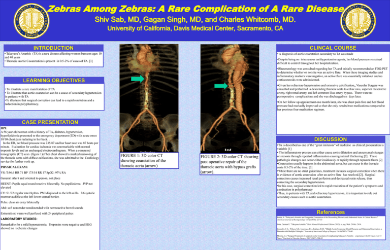
Zebras Among Zebras: A Rare Complication of A Rare Disease Shiv Sab, MD, Gagan Singh, MD, and Charles Whitcomb, MD. University of California, Davis Medical Center, Sacramento, CA CLINCAL COURSE INTRODUCTION • Takayasu’s Arteritis (TA) is a rare disease affecting women between ages 10 and 40 years • Thoracic Aortic Cooarctation is present in 0.5-2% of cases of TA. [3] • A diagnosis of aortic coarctation secondary to TA was made •Despite being on intravenous antihypertensive agents, her blood pressure remained difficult to control throughout her hospitalization. •Rheumatology was consulted regarding her TA and initially recommended an FDG-PET to determine whether or not she was an active flare. When these imaging studies and inflammatory markers were negative, an active flare was essentially ruled out and no corticosteroids were administered. LEARNING OBJECTIVES •Given her refractory hypertension and extensive calcification,, Vascular Surgery was consulted and performed a descending thoracic aorta to celiac axis, superior mesenteric artery, right renal artery, and left common iliac artery bypass. There were no postoperative complications and she was discharged ten days later. • To illustrate a rare manifestation of TA • To illustrate that aortic coarctation can be a cause of secondary hypertension in patients with TA •To illustrate that surgical correction can lead to a rapid resolution and a reduction in polypharmacy. •On her follow up appointment one month later, she was chest pain free and her blood pressure had markedly improved so that she only needed two medications compared to her previous four medication regimen. DISCUSSION CASE PRESENTATION HPI: A 56 year old woman with a history of TA, diabetes, hypertension, hyperlipidemia presented to the emergency department (ED) with acute onset 10/10 chest pain radiating to her back. . In the ED, her blood pressure was 235/87 and her heart rate was 87 beats per minute. Evaluation for cardiac ischemia was unremarkable with normal troponin levels and an unchanged electrocardiogram. When a computed tomographic (CT) scan (figure 1)of her chest showed a marked narrowing of the thoracic aorta with diffuse calfications, she was admitted to the Cardiology service for further work up. PHYSICAL EXAM: VS: T:96.6 HR 71 BP 173/54 RR 17 SpO2: 97% RA General: Aler t and oriented to person, not place HEENT: Pupils equal round reactive bilaterally. No papilledema. JVP not elevated CV: S1/S2 regular rate/rhythm. PMI displaced to the left axilla. 3/6 systolic murmur audible at the left lower sternal border. DISCUSSION FIGURE 1: 3D color CT showing coarctation of the thoracic aorta (arrow) FIGURE 2: 3D color CT showing post operative repair of the thoracic aorta with bypass grafts (arrow). •TA is described as one of the “great imitators” of medicine as clinical presentation is variable [1] • The inflammatory process can either cause aortic dilatation and aneursymal changes or stenosis through repeated inflammation causing intimal thickening [2]. These pathologic changes can occur either insidiously or rapidly through repeated flares [2] •Coarctation usually happens in the abdominal aorta, but can occur in the thoracic aorta 0.5-2% of the time. [3] •While there are no strict guidelines, treatment includes surgical correction when there is evidence of aortic coarctaion after an active flare has resolved.[2]. Surgical correction causes increased renal perfusion and decreased renin release, thus correcting the secondary hypertension. •In this case, surgical correction led to rapid resolution of the patient’s symptoms and a reduction in polypharmacy •Thus, in patients with TA and refractory hypertension, it is important to rule out secondary causes such as aortic coarctation. Pulm: clear air entry bilaterally Abd: soft nontender nondistended with normoactive bowel sounds Extremities: warm well perfused with 2+ peripheral pulses LABORATORY STUDIES: Remarkable for a mild hyponatremia. Troponins were negative and EKG showed no ischemic changes References Lande, A. "Takayasu's Arteritis and Congenital Coarctation of the Descending Thoracic and Abdominal Aorta: A Critical Review." American Journal of Roentgenology 127.2 (1976): 227-33 Gota, Carmen E. "Takayasu Arteritis." Merk Manual Professional Edition (2013): n. pag. Web. 29 Oct. 2014. Connolly, J. E., Wilson, S.E., Lawrence, P.L., Fujitani, R.M.. "Middle Aortic Syndrome: Distal Thoracic and Abdominal Coarctation, a Disorder with Multiple Etiologies." Journal of American College of Surgery 194.6 (2002): 774-81. Moneta, G.l. "Surgical Treatment of Atypical Aortic Coarctation Complicating Takayasu's Arteritis—experience with 33 Cases over 44 Years." Yearbook of Vascular Surgery 2007 (2007): 304-05
