Cavo-tricuspid isthmus ablation triggered cardiac hemangioma
Cavo-tricuspid isthmus ablation triggered cardiac hemangioma
Shaheen T Najafi MD, Gagan D Singh MD, Jeffrey A Southard MD
University of California, Davis Medical Center; Sacramento, CA
Case
A 27-year-old man with a history of paroxysmal atrial flutter treated with CTI ablation two years prior presents with complaints of exertional dyspnea for a month. He denied palpitations and had been exercising thrice weekly since the time of his ablation without incident. His awareness of shortness of breath and exertional fatigue come on with limited effort but are without associated chest pain or lightheadedness.
He denies any sick contacts, recent travel or toxic environmental exposures. He had been on warfarin, flecainide and verapamil around the time of his ablation, but was not taking any medications at the time of presentation. He is 6 feet tall, 192 lbs with a regular pulse at 68 beats per minute. Blood pressure 122/77 mm Hg, oxygen saturation is 100% on room air. He appears fit and is not in distress. Jugular venous pressure is normal. Cardiovascular exam is regular. He has no audible rub, murmur, or gallop. His point of maximal impulse is of normal size and is non-displaced. His lungs are clear. Abdomen and extremities are benign.
Data
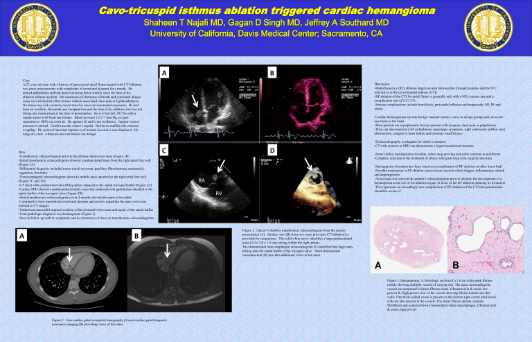
-Transthoracic echocardiogram prior to his ablation showed no mass (Figure 1B)
-Initial transthoracic echocardiogram showed a pedunculated mass from the right atrial free wall
(Figure 1A)
-Differential diagnosis included tumor (atrial myxoma, papillary fibroelastoma, metastasis), vegetation, thrombus
-Transesophageal echocardiogram showed a mobile mass attached to the right atrial free wall
(Figure 1C and 1D)
-CT chest with contrast showed a filling defect adjacent to the septal tricuspid leaflet (Figure 2A)
-Cardiac MRI showed a pedunculated mobile mass that enhanced with gadolinium attached to the septal leaflet of the tricuspid valve (Figure 2B)
-Serial transthoracic echocardiograms over 4 months showed the mass to be stable
-Continued to have intermittent exertional dyspnea and anxiety regarding the mass so he was referred to CT surgery
-Underwent successful surgical excision of the tricuspid valve mass and repair of the septal leaflet
-Final pathologic diagnosis was hemangioma (Figure 3)
-Seen in follow up with no symptoms and no recurrence of mass on transthoracic echocardiograms
Discussion
-Radiofrequency (RF) ablation targets an area between the tricuspid annulus and the IVC referred to as the cavotricupsid isthmus (CTI)
-RF ablation of the CTI for atrial flutter is generally safe with a 90% success rate and a complication rate of 2.5-3.5%
-Serious complications include heart block, pericardial effusion and tamponade, MI, PE and stroke
-Cardiac hemangiomas are rare benign vascular tumors, occur in all age groups and can occur anywhere in the heart
-Most patients are asymptomatic but can present with dyspnea, chest pain or palpitations
-They can also manifest with arrhythmias, neurologic symptoms, right ventricular outflow tract obstructions, congestive heart failure and coronary insufficiency
-Echocardiography is adequate for initial evaluation
-CT with contrast or MRI can demonstrate a hypervascularized structure
-Some cardiac hemangiomas involute, others stop growing and some continue to proliferate
-Complete resection is the treatment of choice with good long term surgical outcomes
-Hemangioma formation has been noted as a complication of RF ablation in other tissue beds
-Possible mechanism is RF ablation causes tissue necrosis which triggers inflammatory stimuli and angiongenesis
-As no mass was seen on the patient’s echocardiogram prior to ablation the development of a hemangioma at the site of the ablation argues in favor of the RF ablation inducing its formation
-This represents an exceedingly rare complication of RF ablation of the CTI that practitioners should be aware of
Figure 1. Apical 4-chamber transthoracic echocardiogram from the current presentation (A). Similar view (B) from two years prior (pre-CTI ablation) is provided for comparison. The solid white arrow identifies a large pedunculated mass (2.0 x 2.0 x 1.5 cm) arising within the right atrium.
Two dimensional trans-esophageal echocardiogram (C) identified the large mass arising near the septal leaflet of the tricuspid valve. Three-dimensional reconstruction (D) provides additional views of the mass.
Figure 3. Hemangioma. A, Histologic section of a 1.0 cm white-pink fibrous nodule showing multiple vessels of varying size. The areas surrounding the vessels are composed of dense fibrous tissue. (Hematoxylin & eosin, low power) B, High-power view of the vessels showing dilated lumina and thin walls. One thick-walled vessel is present on the bottom right corner. Red blood cells are also present in the vessels. The dense fibrous stroma contains fibroblasts and scattered brown hemosiderin-laden macrophages. (Hematoxylin
& eosin, high power)
Figure 2. Non-cardiac gated computed tomography (A) and cardiac gated magnetic resonance imaging (B) providing views of the mass.

