Document 12822778
advertisement
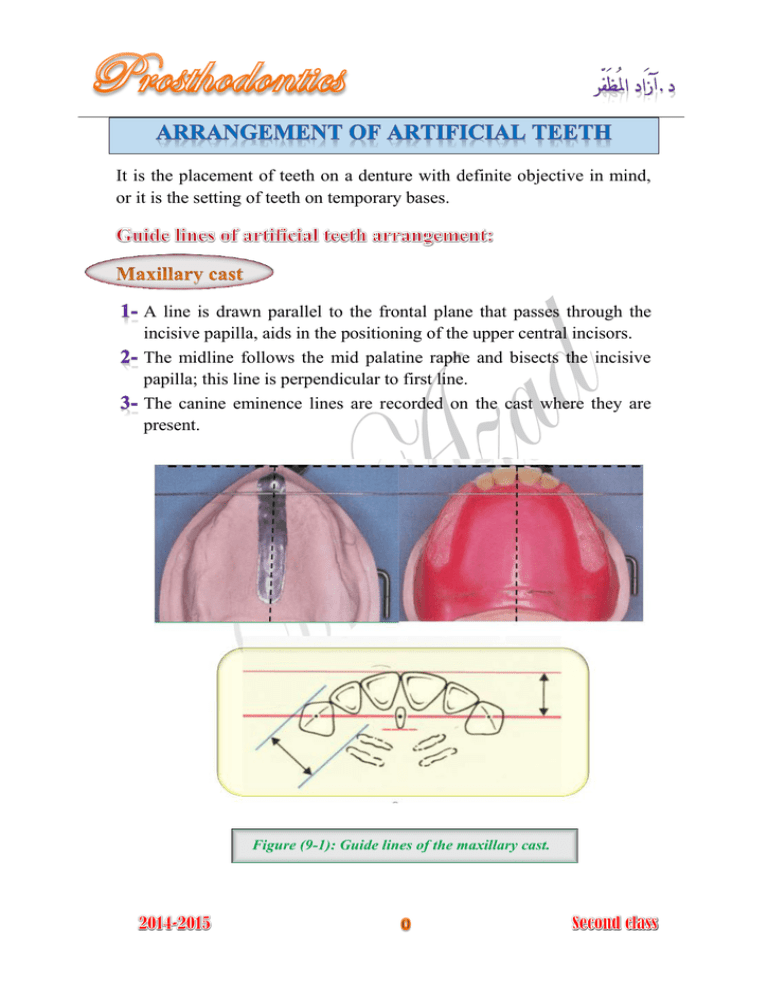
It is the placement of teeth on a denture with definite objective in mind, or it is the setting of teeth on temporary bases. A line is drawn parallel to the frontal plane that passes through the incisive papilla, aids in the positioning of the upper central incisors. The midline follows the mid palatine raphe and bisects the incisive papilla; this line is perpendicular to first line. The canine eminence lines are recorded on the cast where they are present. Figure (9-1): Guide lines of the maxillary cast. A line is drawn parallel to the frontal plane bisecting the residual ridge, aids in positioning of the mandibular central incisors. A point designates the distal of the mandibular canine. A line follows the crest of the residual ridge from the canine point to the middle of retromolar pad, aids in the buccolingual position of the mandibular posterior teeth. A line that bisects the vertical height of the retromolar pad aids in establishing the vertical position of the occlusal surfaces of the posterior teeth. Figure (9-2): Guide lines of the mandibular cast. Maxillary anterior teeth: Following the maxillary occlusion rim. Mandibular anterior teeth: Using the occlusion rims and maxillary teeth as guides. Mandibular posterior teeth: Using the anterior teeth, retromolar pads, and residual ridges as guides. Maxillary posterior teeth: Using the mandibular posterior teeth as guides. The anterior teeth should be arranged to provide: 1234- Proper lip support. Permit satisfactory phonetic. Pleasing esthetic. To set the teeth in place where they grew. The bone loss is upward and backward direction for the maxillary residual ridge; downward and outward for the mandibular residual ridge, therefore the maxillary artificial teeth should be arranged anteriorly and inferiorly to the residual ridge to occupy the space formerly occupied by the natural teeth. In setting the maxillary teeth, make sure the central and lateral incisors are placed so they begin to turn along the curvature of the arch. Figure (9-3) In frontal view The contact point between the right and left central incisors should be coinciding with the midline of cast. The incisal edge of each one should touch the occlusal plane. The long axis is perpendicular to the occlusal plane. Figure (9-4) In sagittal view The central incisors should have slight (5 degrees) labial inclination. Figure (9-5) In horizontal view (occlusal plane) The two central incisors should be placed to give the beginning of curvatures of the arch. Generally the labial surfaces of the two central incisors will be 8-10 mm anterior to the center of the incisive papilla. Figure (9-6) In frontal view The incisal edge of the lateral incisor should be 1 mm above the occlusal plane, and the long axis show little distal inclination. Figure (9-7) In sagittal view The upper lateral incisor should have slight labial inclination (10 degrees); the neck is slightly depressed . Figure (9-8) In horizontal view The cervical area is depressed more than the central incisor, and the distal edge should be rotated lingually to form the arch curvature. Figure (9-9) The maxillary canine represents the corner of the mouth, it is the turning point of the maxillary arch, and also it forms the transition from the anterior teeth to posterior teeth. In frontal view The tip of the canine should touch the occlusal plane, and the long axis is perpendicular to the plane, or tilted slightly to the distal. Figure (9-10) In sagittal view The long axis of canine is vertical. Figure (9-11) In horizontal view The cervical area of canine is prominent. Figure (9-12) In frontal view The long axis is vertical and the midline of the mandibular central incisors, coincide with the maxillary midline. Figure (9-13) Figure (9-14) In sagittal view The mandibular central incisors should have slight labial inclination. The incisal edge should have 1 mm of vertical overlap (overlap), and 1 mm of horizontal overlap (overjet) in respect to maxillary central incisors. Figure (9-15) Overbite (vertical overlap): It is the vertical extension of the maxillary anterior teeth over the mandibular teeth in a vertical direction, when the opposing posterior teeth are in contact in centric occlusion. Overjet (horizontal overlap): It is the projection maxillary anterior teeth beyond their antagonist in a horizontal direction. Figure (9-16) Figure (9-17) Figure (9-19) Figure (9-18) The incisal guide angle denotes the angle by the palatal surface of the maxillary anteriors against the horizontal plane. The incisal guidance can be raised by altering the labial proclination, overjet, and overbite of the maxillary anteriors. In frontal view The long axis is slightly distal inclined to the occlusal plane. Figure (9-20) In sagittal view The lateral incisor is fairly upright, and the incisal edge should be 1 mm of horizontal and vertical overlap in respect with the maxillary central incisor. Figure (9-21) In horizontal view The distal edge rotated lingually to have the arch curvature. Figure (9-22) In frontal view The long axis should have slight distal inclination, and the tip of the mandibular canine should be placed in the embrasure between maxillary lateral and canine. Figure (9-23) In sagittal view The long axis should have slight lingual inclination. Figure (9-24) In horizontal view The cervical area is prominent. Figure (9-25) The arrangement of anterior teeth should follow the form of the arch which is ovoid, tapered, or square. In complete denture fabrication the mandibular incisors should not touch the maxillary incisors in centric relation (the incisal guidance angle as low as possible) to allow free movement of the teeth in eccentric jaw movement without compromising the denture stability. Importance of arrangement of posterior teeth (Significance) Correct placement of posterior teeth is important for the retention and stability of both dentures. Prior to arrangement of the posterior teeth, we must understand some of the definitions which are related to posterior teeth arrangement Curve of Spee: It is an anatomical curvature of the occlusal alignment of teeth, beginning at the tip of mandibular canine and following the buccal cusps of the natural premolars and molars, continuing to the anterior border of the ramus of mandible; figure (). Curve of Wilson: It is a curve extends mediolaterally from one side of the arch to the other side; figure (). Figure (9-27) Figure (9-26) Compensating curve: It is the anteroposterior, and lateral curvature in the alignment of the occluding surfaces and incisal edges of artificial teeth, which is used to develop balanced occlusion. (It compensates the opening that occurs during forward and lateral movement of the mandible). Figure (9-28) Christensen’s phenomenon: This is the posterior opening of the dental arches or occlusion rims during forward movement of the mandible. To compensate for the posterior opening during forward or protrusive movement we incorporate the compensating curve. Figure (9-29) The mandibular posterior teeth will be before the maxillary posterior, because there are more anatomical landmarks to locate the guidelines which are: The line of the crest of the mandibular residual ridge, which extends between the middle of retromolar pad and tip of mandibular canine, the central grooves of the mandibular posterior teeth should coincide with this line. The line extending between the tip of mandibular canine and upper 2/3 of retromolar pad will determine the height of mandibular posterior teeth. Figure (9-30) In buccal view The tooth should be set perpendicular to the occlusal plane. The tip of its buccal cusp should be 1 mm below the line is planed from the tip of canine and the 2/3 of the vertical height of retromolar pad. Figure (9-31) In horizontal view The central groove should be over the crest of residual ridge. Figure (9-32) It should be arranged in the someway as mandibular first premolar. In buccal view The mesiobuccal cusp should be 1 mm below the line, and the distobuccal cusp should be ½ mm below the line. Figure (9-33) In horizontal view The central groove should coincide with the crest of the residual ridge. In buccal view The mesiobuccal cusp is ½ mm below the line, and the distobuccal cusp should touch the line. In horizontal view The central groove should coincide with the crest of the residual ridge. Figure (9-34) In order to get normal molar relation, the mesiobuccal cusp of maxillary first molar should rest in the buccal groove of the mandibular first molar, and the mesiopalatal cusp should seat into the central fossa of mandibular first molar. The palatal cusp should seat into the embrasure formed between the mandibular second premolar and first molar. The palatal cusp should seat into the embrasure between the mandibular first and second premolars. The mesiobuccal cusp should rest in the buccal groove of mandibular second molar, and the mesiopalatal cusp should seat into the central fossa of the mandibular second molar. Figure (9-39) Figure (9-40) Figure (9-41) Maxillary teeth overlap the mandibular teeth horizontally; the overlap must be present posteriorly to prevent cheek biting. The long axis of each maxillary tooth is distal to that of corresponding mandibular tooth. Each tooth in both arches is opposed by two teeth, except the mandibular central incisor and the maxillary second molar. This arrangement of posterior teeth will provide maximum contact between the occlusal surfaces of mandibular and maxillary teeth in centric occlusion. Figure (9-42) Setting mandibular anterior teeth too forward in order to meet maxillary teeth. Failure to make the canine the turning point of the arch. Setting the mandibular first premolars to the buccal side of the canines. Failure to establish the occlusal plane at the proper level and inclination. Establishing the occlusal plane by an arbitrary line on the face. When it is too low or too high, it is not look natural and cause difficulty in the mastication. The posterior teeth should not appear longer than those teeth when the patient smile, the patient will have (reverse smile); figure (9-44). Lack of lingual rotation of anterior teeth to give a narrow effect. Tooth arranged too wide posteriorly, appearance like many teeth in the mouth. Setting the mandibular posterior teeth too far to the lingual side in the second molar region which cause tongue interference and mandibular denture displacement. Teeth arranged too far toward the tongue or palate, there will be dark space between the check and teeth when patient talk or smile (dark buccal corridors); figure (9-43). Figure (9-43) Figure (9-44)
