Anatomy and physiology of Temporomandibular Joint
Anatomy and physiology of Temporomandibular Joint
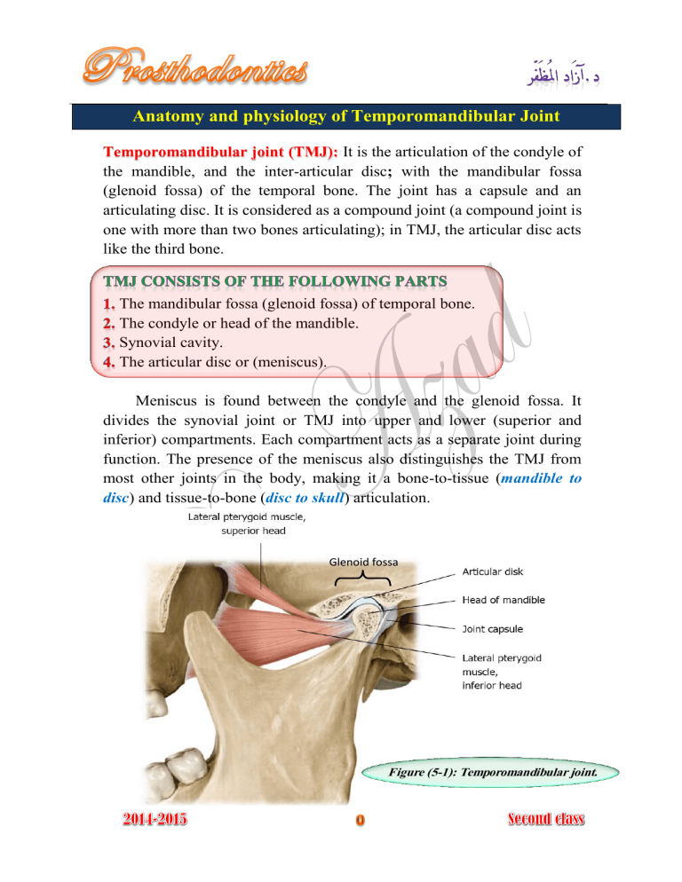
Temporomandibular joint (TMJ): It is the articulation of the condyle of the mandible, and the inter-articular disc ; with the mandibular fossa
(glenoid fossa) of the temporal bone. The joint has a capsule and an articulating disc. It is considered as a compound joint (a compound joint is one with more than two bones articulating); in TMJ, the articular disc acts like the third bone.
The mandibular fossa (glenoid fossa) of temporal bone.
The condyle or head of the mandible.
Synovial cavity.
The articular disc or (meniscus).
Meniscus is found between the condyle and the glenoid fossa. It divides the synovial joint or TMJ into upper and lower (superior and inferior) compartments. Each compartment acts as a separate joint during function. The presence of the meniscus also distinguishes the TMJ from most other joints in the body, making it a bone-to-tissue ( mandible to disc ) and tissue-to-bone ( disc to skull ) articulation.
Glenoid fossa
Figure (5-1): Temporomandibular joint.
There are three groups of muscles:
Closing muscles.
Gliding muscles.
Opening muscles.
The temporalis, masseter and medial pterygoid muscles supply the power for pulling the mandible against the maxilla (elevating and closing the mandible).
Temporalis muscle
Masseter muscle
Medial pterygoid muscle
Figure (5-4): Closing muscles.
Masseter muscle
The lateral pterygoid muscle connects the mandible to the lateral pterygoid plate in such a way as to act as the steering mechanism for the mandible and act to protrude the jaw or to move it laterally.
Lateral pterygoid
(superior part)
Lateral pterygoid
(inferior part)
Figure (5-5): Gliding muscle.
The muscles that depress (open) mandible consist of three groups, suprahyoid muscles, infrahyoid muscles, and platysma.
Suprahyoid
Hyoid bone
Infrahyoid
Figure (5-6): Opening muscles.
Temporomandibular and capsular ligaments.
Sphenomandibular ligament.
Stylomandibulalar ligament.
Platysma
Figure (5-3): TMJ ligaments.
Good prosthodontic treatment bears a direct relation to the structures of the temporomandibular articulation, since occlusion is one of the most important parts of treatment of the patients with complete dentures. The temporomandibular joints affect the dentures and likewise the dentures affect health and function of the joints.
The mandibular bone has specific relationships to the bones of the cranium. The mandible is connected to the cranium at the two temporomandibular joint by the temporomandibular and capsular ligaments. The sphenomandibular and stylomandibular ligaments also connect the bones in such a way as to limit some motions of the mandible.
There are three axes around which the mandibular movements take place, the mandibular movements are related to three planes of skull (sagittal, transverse (horizontal), and coronal (frontal)), figure (5-8).
1- Hinge axis or transverse axis
It is an imaginary line around which the mandible may rotate within the sagittal plane
(during opening and closing movement).
2- Sagittal axis of the mandible
It is an imaginary anteroposterior line around which the mandible may rotate within the frontal plane.
3- Vertical axis of the mandible
It is an imaginary line around which the mandible may rotate through the horizontal plane.
Transverse plane Coronal plane Sagittal plane
Figure (5-7): Body planes. Figure (5-8): Skull planes.
Based on the dimension involved in the movement
1- Rotational aRotation around the transverse or hinge axis. bRotation around the anteroposterior or sagittal axis. cRotation around the vertical axis.
2- Translational or gliding
They are considered as basic movements of the mandible.
Rotation Translation
Figure (5-9): Basic mandibular movements.
The upper compartment shows anteroposterior gliding movement, when this movement takes place, the condyle and the disc move as a single unit against the glenoid fossa.
The lower compartment shows hinge movement, during hinge movement the condyle moves against the articular disc and the glenoid fossa, which together act as a single unit. True condylar rotation is 12° with the maximum incisal separation of 22 mm. See figure (5-14)
Based on the type of movement
1- Hinge movement.
2- Protrusive movement.
3- Retrusive movement.
4- Lateral movement. aLateral rotation or (laterotrusion).
Right.
Left. bLateral translation or (Bennett movement).
Immediate side shift.
Progressive side shift.
Precurrent side shift.
(1) (2) (3)
Figure (5-10): 1- Closed mouth. 2- Hinge movement. 3- Protrusion. 4- Retrusion.
(4)
(L) (R)
Figure (5-11): Laterotrusion (left and right).
O R
Figure (5-12): Bennett movement.
75% of the shift takes place during the first
3 mm of anterior movement of the condyle.
The angle formed between the sagittal plane and the average path of the advancing condyle as viewed in the horizontal plane during lateral mandibular movements
Figure (5-13): Bennett angle: the angle formed between the progressive lateral path and the sagittal plane (Note there is immediate side shift ISS followed by progressive side shift PSS) .
1- Border movement aExtreme movement in the sagittal plane. bExtreme movement in the horizontal plane. cExtreme movement in the frontal plane. dEnvelope of motion.
2- Intra-border movement aFunctional movement.
Chewing cycle.
Swallowing.
Yawing.
Speech. bPara-functional movement.
Clenching.
Bruxism.
Other habitual movements.
12 °
22 mm
Figure (5-14): Extreme movement in the sagittal plane.
C Centric relation.
A Centric occlusion.
G Edge to edge relationship.
B Maximum protrusion.
D Maximum mandibular opening.
C-E Hinge motion.
E-D Gliding.
R Resting position.
38 mm 22 mm
G
9 mm
G
Figure (5-15): Beak tracing
Borders movements recorded in the sagittal plane .
CO Centric occlusion.
RD Right disocclusion.
MRL Maximum right lateral position.
MMO Maximum mouth opening.
MLL Maximum left lateral position.
LD Left disocclusion.
Figure (5-16): Shield tracing
Border movements recorded in the coronal plane.
CR Centric relation.
MRL Maximum right lateral position.
MP Maximum protrusion.
MLL Maximum left lateral position.
Figure (5-17): Diamond tracing
Border movements recorded in the horizontal plane.
When we combine the border movements of all the three planes, we get a three dimensional space within which mandibular movements is possible, this three dimensional limiting space is called the ( envelope of motion ).
Figure (5-18): Envelope of motion
(It is the combination of border movements in all three planes).
