HERPES SIMPLEX VIRUS (HSV)
HERPES SIMPLEX VIRUS (HSV)
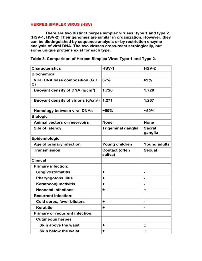
There are two distinct herpes simplex viruses: type 1 and type 2
(HSV-1, HSV-2) Their genomes are similar in organization. However, they can be distinguished by sequence analysis or by restriction enzyme analysis of viral DNA. The two viruses cross-react serologically, but some unique proteins exist for each type.
Table 3. Comparison of Herpes Simplex Virus Type 1 and Type 2.
Characteristics HSV-1
Biochemical
Viral DNA base composition (G +
C)
Buoyant density of DNA (g/cm 3 )
67%
1.726
Buoyant density of virions (g/cm 3 ) 1.271
Homology between viral DNAs
Biologic
Animal vectors or reservoirs
Site of latency
Epidemiologic
Age of primary infection
Transmission
HSV-2
69%
1.728
1.267
~50%
Young children
Contact (often saliva)
~50%
None None
Trigeminal ganglia Sacral ganglia
Young adults
Sexual
Clinical
Primary infection:
Gingivostomatitis
Pharyngotonsillitis
Keratoconjunctivitis
Neonatal infections
Recurrent infection:
Cold sores, fever blisters
Keratitis
Primary or recurrent infection:
Cutaneous herpes
Skin above the waist
Skin below the waist
+
+
+
±
+
+
+
±
-
-
-
+
-
-
±
+
Hands or arms
Herpetic whitlow
Eczema herpeticum
Genital herpes
Herpes encephalitis
Herpes meningitis
+
+
+
±
+
±
+
+
-
+
-
+
Diagnosis of HSV Infections
- Cytopathology
A rapid cytologic method is to stain scrapings obtained from the base of a vesicle (eg, with Giemsa's stain); the presence of multinucleated giant cells indicates that herpesvirus (HSV-1 or HSV-2) and contain Cow dry type An inclusion bodies Formation , In the course of viral multiplication within cells, virus-specific structures called inclusion bodies may be produced. They become far larger than the individual virus particle and often have an affinity for acid dyes (eg, eosin). They may be situated in the nucleus (herpesvirus), in the cytoplasm (poxvirus), or in both (measles virus). In many viral infections, the inclusion bodies are the site of development of the virions (the viral factories). Variations in the appearance of inclusion material depend largely upon the tissue fixative used. The presence of inclusion bodies may be of considerable diagnostic aid. The intracytoplasmic inclusion in nerve cells
—the Negri body
—is pathognomonic for rabies .
- Quantitation of Viruses
The cells can also be stained with specific antibodies in an immunofluorescence test and it is also possible to detect viral DNA by
in situ hybridization. Type-specific antibodies can distinguish between
HSV-1 and HSV-2.
- Polymerase Chain Reaction (PCR)
PCR assays can be used to detect virus and are sensitive and specific. PCR amplification of viral DNA .
- Serology
Antibodies appear in 4
–7 days after infection and reach a peak in 2–4 weeks. They persist with minor fluctuations for the life of the host.
Treatment, Prevention, & Control
Several antiviral drugs have proved effective against HSV infections, including acyclovir, valacyclovir, and vidarabine. Acyclovir is currently
the standard therapy. All are inhibitors of viral DNA synthesis. Acyclovir, a nucleoside analog.
Vaccine
Recombinant HSV-2 glycoprotein ( that found in viral envelope ) can be used .

![Info. Speech Packet [v6.0].cwk (DR)](http://s3.studylib.net/store/data/008110988_1-db39bdd1f22b58bf46d9a39ab146e2e3-300x300.png)
