THE SHIGELLAE Morphology & Identification TYPICAL ORGANISMS : -
advertisement
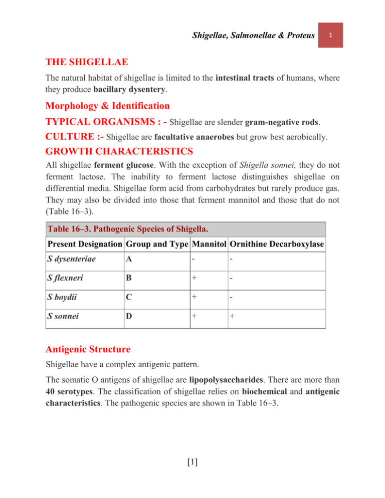
Shigellae, Salmonellae & Proteus 1 THE SHIGELLAE The natural habitat of shigellae is limited to the intestinal tracts of humans, where they produce bacillary dysentery. Morphology & Identification TYPICAL ORGANISMS : - Shigellae are slender gram-negative rods. CULTURE :- Shigellae are facultative anaerobes but grow best aerobically. GROWTH CHARACTERISTICS All shigellae ferment glucose. With the exception of Shigella sonnei, they do not ferment lactose. The inability to ferment lactose distinguishes shigellae on differential media. Shigellae form acid from carbohydrates but rarely produce gas. They may also be divided into those that ferment mannitol and those that do not (Table 16–3). Table 16–3. Pathogenic Species of Shigella. Present Designation Group and Type Mannitol Ornithine Decarboxylase S dysenteriae A - - S flexneri B + - S boydii C + - S sonnei D + + Antigenic Structure Shigellae have a complex antigenic pattern. The somatic O antigens of shigellae are lipopolysaccharides. There are more than 40 serotypes. The classification of shigellae relies on biochemical and antigenic characteristics. The pathogenic species are shown in Table 16–3. [1] Shigellae, Salmonellae & Proteus 2 Pathogenesis & Pathology - Shigella infections are almost always limited to the gastrointestinal tract; bloodstream invasion is rare. - Shigellae are highly communicable; the infective dose is on the order of 103 organisms (whereas it usually is 105–108 for salmonellae and vibrios). - invasion of the mucosal epithelial cells (eg, M cells) by induced phagocytosis, escape from the phagocytic vacuole, multiplication and spread within the epithelial cell cytoplasm, and passage to adjacent cells. - Microabscesses in the wall of the large intestine and terminal ileum lead to necrosis of the mucous membrane. - Superficial ulceration, bleeding, and formation of a "pseudomembrane" on the ulcerated area. This consists of fibrin, leukocytes, cell debris, a necrotic mucous membrane, and bacteria. As the process subsides, granulation tissue fills the ulcers and scar tissue forms. Toxins 1. (endotoxins) Upon autolysis, all shigellae release their toxic lipopolysaccharide. This endotoxin probably contributes to the irritation of the bowel wall. 2. Shigella dysenteriae (exotoxins) - S dysenteriae type 1 (Shiga bacillus) produces a heat-labile exotoxin that affects both the gut and the central nervous system. The exotoxin is a protein that is antigenic (stimulating production of antitoxin) and lethal for experimental animals. - Acting as an enterotoxin, it produces diarrhea. In humans, the exotoxin also inhibits sugar and amino acid absorption in the small intestine. - Acting as a "neurotoxin," this material may contribute to the extreme severity and fatal nature of S dysenteriae infections and to the central nervous system reactions observed in them (ie, meningismus, coma). - The two may act in sequence, the toxin producing an early nonbloody, [2] Shigellae, Salmonellae & Proteus 3 voluminous diarrhea and the invasion of the large intestine resulting in later dysentery with blood and pus in stools. Clinical Findings - After a short incubation period (1–2 days), - there is a sudden onset of abdominal pain, fever, and watery diarrhea. - A day or so later, as the infection involves the ileum and colon, the number of stools increases; they are less liquid but often contain mucus and blood. - Each bowel movement is accompanied by straining and tenesmus (rectal spasms), with resulting lower abdominal pain. - In more than half of adult cases, fever and diarrhea subside spontaneously in 2– 5 days. - in children and the elderly, loss of water and electrolytes may lead to dehydration, acidosis, and even death. - On recovery, most persons shed dysentery bacilli for only a short period, but a few remain chronic intestinal carriers and may have recurrent bouts of the disease. - Upon recovery from the infection, most persons develop circulating antibodies to shigellae, but these do not protect against reinfection. Diagnostic Laboratory Tests SPECIMENS Specimens include fresh stool, mucus flecks, and rectal swabs for culture. Large numbers of leukocytes and red blood cells are seen microscopically. Serum specimens, if desired, must be taken 10 days apart to demonstrate a rise in titer of agglutinating antibodies. CULTURE The materials are streaked on differential media (eg, MacConkey's or EMB agar) and on selective media (Hektoen enteric agar or salmonella-shigella agar). Colorless (lactose-negative) colonies are inoculated into triple sugar iron agar. Organisms that fail to produce H2S, that produce acid but not gas in the butt and an [3] Shigellae, Salmonellae & Proteus 4 alkaline slant in triple sugar iron agar medium, and that are nonmotile should be subjected to slide agglutination by specific shigella antisera. SEROLOGY : - Serology is not used to diagnose shigella infections. Immunity - Infection is followed by a type-specific antibody response. - Injection of killed shigellae stimulates production of antibodies in serum but fails to protect humans against infection. - IgA antibodies in the gut may be important in limiting reinfection; these may be stimulated by live attenuated strains given orally as experimental vaccines. - Serum antibodies to somatic shigella antigens are IgM. Treatment - Ciprofloxacin, ampicillin, doxycycline, and trimethoprim-sulfamethoxazole are most commonly inhibitory for shigella. - They may fail to eradicate the organisms from the intestinal tract. Multiple drug resistance can be transmitted by plasmids, and resistant infections are widespread. - Many cases are self-limited. - Opioids should be avoided in shigella dysentery. Epidemiology, Prevention, & Control - Shigellae are transmitted by "food, fingers, feces, and flies" from person to person. Most cases of shigella infection occur in children under 10 years of age. - Since humans are the main recognized host of pathogenic shigellae, control efforts must be directed at eliminating the organisms from this reservoir by (1) sanitary control of water, food, and milk; sewage disposal; and fly control; (2) isolation of patients and disinfection of excreta; (3) detection of subclinical cases and carriers, particularly food handlers; and (4) antibiotic treatment of infected individuals. [4] Shigellae, Salmonellae & Proteus 5 THE SALMONELLA-ARIZONA GROUP - Salmonellae are often pathogenic for humans or animals when acquired by the oral route. - They are transmitted from animals and animal products to humans, where they cause enteritis, systemic infection, and enteric fever. - Salmonellae isolates are motile. - Salmonellae grow readily on simple media, - They survive freezing in water for long periods. - Salmonellae are resistant to certain chemicals (eg, sodium tetrathionate, sodium deoxycholate). Classification - The members of the genus Salmonella were originally classified on the basis of epidemiology, host range, biochemical reactions, and structures of the O, H, and Vi (when present) antigens. - DNA-DNA hybridization studies have demonstrated that there are seven evolutionary groups. Nearly all of the salmonella serotypes that infect humans are in DNA hybridization group I . (Table 16–4). Table 16–4. Representative Antigenic Formulas of Salmonellae. O Group Serotype Antigenic D S Typhi 9, 12 (Vi):d:— A S Paratyphi A 1, 2, 12:a— C1 S Choleraesuis 6, 7:c:1,5 B S Typhimurium 1, 4, 5, 12:i:1, 2 D S Enteritidis 1, 9, 12:g, m:— 1 O antigens: boldface numerals. [5] Formula1 Shigellae, Salmonellae & Proteus 6 (Vi): Vi antigen if present. Phase 1 H antigen: lower-case letter. Phase 2 H antigen: numeral. Pathogenesis & Clinical Findings - Salmonella Typhi, Salmonella Choleraesuis, and perhaps Salmonella Paratyphi A and Salmonella Paratyphi B are primarily infective for humans, and infection with these organisms implies acquisition from a human source. - The vast majority of salmonellae, however, are chiefly pathogenic in animals that constitute the reservoir for human infection: poultry, pigs, rodents, cattle, pets. - The organisms enter via the oral route, usually with contaminated food or drink. - The mean infective dose to produce clinical or subclinical infection in humans is 105–108 salmonellae. Among the host factors that contribute to resistance to salmonella infection are gastric acidity, normal intestinal microbial flora, and local intestinal immunity. - Salmonellae produce three main types of disease in humans, but mixed forms are frequent. 1. THE "ENTERIC FEVERS" (TYPHOID FEVER) - This syndrome is produced by only Salmonella Typhi (typhoid fever). - salmonellae reach the small intestine, from which they enter the lymphatics and then the bloodstream. They are carried by the blood to many organs. The organisms multiply in intestinal lymphoid tissue and are excreted in stools. - After an incubation period of 10–14 days, fever, malaise, headache, constipation, bradycardia, and myalgia occur. The fever rises to a high plateau, and the spleen and liver become enlarged. Rose spots, usually on the skin of the abdomen or chest, are seen briefly in rare cases. In the preantibiotic era, the chief complications of enteric fever were intestinal hemorrhage and perforation, and the mortality rate was 10–15%. Treatment with antibiotics has reduced the mortality rate to less than 1%. [6] Shigellae, Salmonellae & Proteus 7 - The principal lesions are hyperplasia and necrosis of lymphoid tissue (eg, Peyer's patches), hepatitis, focal necrosis of the liver, and inflammation of the gallbladder, periosteum, lungs, and other organs. 2. BACTEREMIA WITH FOCAL LESIONS - This is associated commonly with S choleraesuis but may be caused by any salmonella serotype. - Following oral infection, there is early invasion of the bloodstream (with possible focal lesions in lungs, bones, meninges, etc), but intestinal manifestations are often absent. Blood cultures are positive. 3. ENTEROCOLITIS - This is the most common manifestation of salmonella infection. - Salmonella Typhimurium and Salmonella Enteritidis are prominent, but enterocolitis can be caused by any of the group I serotypes of salmonellae. Eight to 48 hours after ingestion of salmonellae, there is nausea, headache, vomiting, and profuse diarrhea. Low-grade fever is common. Inflammatory lesions of the small and large intestine are present. Bacteremia is rare (2–4%) except in immunodeficient persons. Blood cultures are negative, but stool cultures are positive. Diagnostic Laboratory Tests SPECIMENS - Blood for culture must be taken repeatedly. In enteric fevers and septicemias, blood cultures are often positive in the first week of the disease. - Urine cultures may be positive after the second week. - Stool specimens also must be taken repeatedly. In enteric fevers, the stools yield positive results from the second or third week on; in enterocolitis, during the first week. ISOLATION OF SALMONELLAE 1. BACTERIOLOGIC METHODS 2. SEROLOGIC METHODS [7] Shigellae, Salmonellae & Proteus 8 Serologic techniques are used to identify unknown cultures with known sera and may also be used to determine antibody titers in patients with unknown illness. Agglutination Test In this test, known sera and unknown culture are mixed on a slide. Clumping, when it occurs can be observed within a few minutes. This test is particularly useful for rapid preliminary identification of cultures. There are commercial kits available to agglutinate and serogroup salmonellae by their O antigens: A, B, C 1, C2, D, and E. Tube Dilution Agglutination Test (Widal Test) Serum agglutinins rise sharply during the second and third weeks of Salmonella Typhi infection. The Widal test to detect these antibodies against the O and H antigens has been in use for decades. At least two serum specimens, obtained at intervals of 7–10 days, are needed to prove a rise in antibody titer. Serial dilutions of unknown sera are tested against antigens from representative salmonellae. False-positive and false-negative results occur. The interpretive criteria when single serum specimens are tested vary, but a titer against the O antigen of > 1:320 and against the H antigen of > 1:640 is considered positive. High titer of antibody to the Vi antigen occurs in some carriers. Results of serologic tests for salmonella infection must be interpreted cautiously because the possible presence of cross-reactive antibodies limits the use of serology. The test is not useful in diagnosis of enteric fevers caused by salmonella other than Salmonella Typhi. Immunity - Infections with Salmonella Typhi or Salmonella Paratyphi usually confer a certain degree of immunity. - Reinfection may occur. - Circulating antibodies to O and Vi are related to resistance to infection and disease. - Secretory IgA antibodies may prevent attachment of salmonellae to intestinal epithelium. Treatment - In severe diarrhea, replacement of fluids and electrolytes is essential. [8] Shigellae, Salmonellae & Proteus 9 - Antimicrobial therapy of invasive salmonella infections is with ampicillin, trimethoprim-sulfamethoxazole, or a third-generation cephalosporin. - Multiple drug resistance transmitted genetically by plasmids among enteric bacteria is a problem in salmonella infections. Susceptibility testing is an important adjunct to selecting a proper antibiotic. - In most carriers, the organisms persist in the gallbladder (particularly if gallstones are present) and in the biliary tract. - Some chronic carriers have been cured by ampicillin alone, but in most cases cholecystectomy must be combined with drug treatment. Prevention & Control - Sanitary measures . - Infected poultry, meats, and eggs must be thoroughly cooked. - Carriers must not be allowed to work as food handlers. - Two injections of acetone-killed bacterial suspensions of Salmonella Typhi, followed by a booster injection some months later, give partial resistance to small infectious inocula of typhoid bacilli but not to large ones. - Oral administration of a live avirulent mutant strain of Salmonella Typhi has given significant protection in areas of high endemicity. - Vaccines against other salmonellae give less protection and are not recommended. Epidemiology - The feces of persons who have unsuspected subclinical disease or are carriers are a more important source of contamination. - Many animals, including cattle, rodents, and fowl, are naturally infected with a variety of salmonellae and have the bacteria in their tissues (meat), excreta, or eggs. - The high incidence of salmonellae in commercially prepared chickens has been widely publicized. - The problem probably is aggravated by the widespread use of animal feeds containing antimicrobial drugs that favor the proliferation of drug-resistant salmonellae and their potential transmission to humans. [9] Shigellae, Salmonellae & Proteus 10 CARRIERS After manifest or subclinical infection, some individuals continue to harbor salmonellae in their tissues for variable lengths of time .Three percent of survivors of typhoid become permanent carriers, harboring the organisms in the gallbladder, biliary tract, or, rarely, the intestine or urinary tract. SOURCES OF INFECTION Water, Milk and Dairy Products (Ice Cream, Cheese, Custard),Shellfish and Dried or Frozen Eggs. Proteus is a genus of Enterobacteriacae, widely distributed in nature as saprophytes, being found in decomposing animal matter, in sewage, in manure soil, and in human and animal feces. They are opportunistic pathogens, commonly responsible for urinary and septic infections, often nosocomial. Clinical significance Three species—P. vulgaris, P. mirabilis, and P. penneri—are opportunistic human pathogens. Proteus includes pathogens responsible for many human urinary tract infections. P. mirabilis is often found as a free-living organism in soil and water.causes wound and urinary tract infections. Most strains of P. mirabilis are sensitive to ampicillin and cephalosporins. P. vulgaris is isolated less often in the laboratory and usually only targets immunosuppressed individuals. P. vulgaris occurs naturally in the intestines of humans and a wide variety of animals, and in manure, soil, and polluted waters. P. mirabilis, once attached to the urinary tract, infects the kidney more commonly than E. coli. About 10-15% of kidney stones are struvite stones, caused by alkalinization of the urine by the action of the urease enzyme (which splits urea into ammonia and carbon dioxide) of Proteus (and other) bacterial spp. [10]
