International Research Journal of Medical Sciences ____________________________________ 1(3),
advertisement
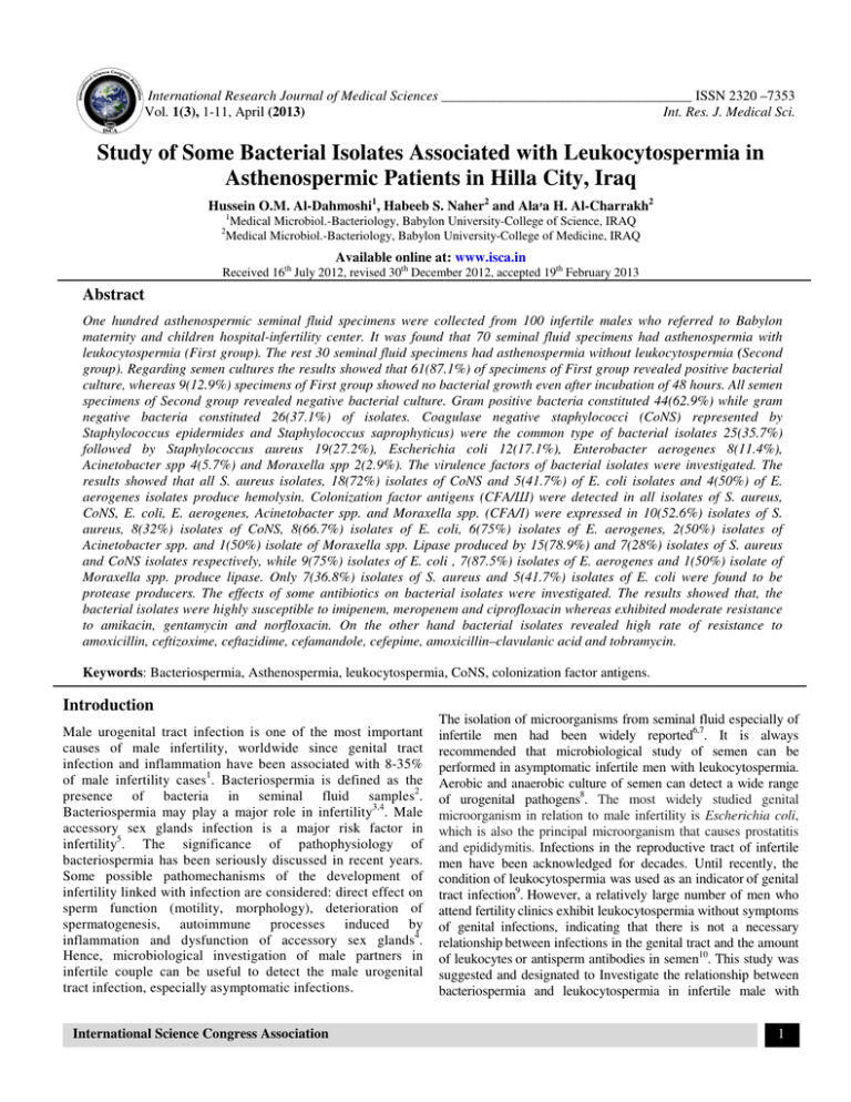
International Research Journal of Medical Sciences ____________________________________ ISSN 2320 –7353 Vol. 1(3), 1-11, April (2013) Int. Res. J. Medical Sci. Study of Some Bacterial Isolates Associated with Leukocytospermia in Asthenospermic Patients in Hilla City, Iraq Hussein O.M. Al-Dahmoshi1, Habeeb S. Naher2 and Ala׳a H. Al-Charrakh2 1 2 Medical Microbiol.-Bacteriology, Babylon University-College of Science, IRAQ Medical Microbiol.-Bacteriology, Babylon University-College of Medicine, IRAQ Available online at: www.isca.in Received 16th July 2012, revised 30th December 2012, accepted 19th February 2013 Abstract One hundred asthenospermic seminal fluid specimens were collected from 100 infertile males who referred to Babylon maternity and children hospital-infertility center. It was found that 70 seminal fluid specimens had asthenospermia with leukocytospermia (First group). The rest 30 seminal fluid specimens had asthenospermia without leukocytospermia (Second group). Regarding semen cultures the results showed that 61(87.1%) of specimens of First group revealed positive bacterial culture, whereas 9(12.9%) specimens of First group showed no bacterial growth even after incubation of 48 hours. All semen specimens of Second group revealed negative bacterial culture. Gram positive bacteria constituted 44(62.9%) while gram negative bacteria constituted 26(37.1%) of isolates. Coagulase negative staphylococci (CoNS) represented by Staphylococcus epidermides and Staphylococcus saprophyticus) were the common type of bacterial isolates 25(35.7%) followed by Staphylococcus aureus 19(27.2%), Escherichia coli 12(17.1%), Enterobacter aerogenes 8(11.4%), Acinetobacter spp 4(5.7%) and Moraxella spp 2(2.9%). The virulence factors of bacterial isolates were investigated. The results showed that all S. aureus isolates, 18(72%) isolates of CoNS and 5(41.7%) of E. coli isolates and 4(50%) of E. aerogenes isolates produce hemolysin. Colonization factor antigens (CFA/Ш) were detected in all isolates of S. aureus, CoNS, E. coli, E. aerogenes, Acinetobacter spp. and Moraxella spp. (CFA/Ι) were expressed in 10(52.6%) isolates of S. aureus, 8(32%) isolates of CoNS, 8(66.7%) isolates of E. coli, 6(75%) isolates of E. aerogenes, 2(50%) isolates of Acinetobacter spp. and 1(50%) isolate of Moraxella spp. Lipase produced by 15(78.9%) and 7(28%) isolates of S. aureus and CoNS isolates respectively, while 9(75%) isolates of E. coli , 7(87.5%) isolates of E. aerogenes and 1(50%) isolate of Moraxella spp. produce lipase. Only 7(36.8%) isolates of S. aureus and 5(41.7%) isolates of E. coli were found to be protease producers. The effects of some antibiotics on bacterial isolates were investigated. The results showed that, the bacterial isolates were highly susceptible to imipenem, meropenem and ciprofloxacin whereas exhibited moderate resistance to amikacin, gentamycin and norfloxacin. On the other hand bacterial isolates revealed high rate of resistance to amoxicillin, ceftizoxime, ceftazidime, cefamandole, cefepime, amoxicillin–clavulanic acid and tobramycin. Keywords: Bacteriospermia, Asthenospermia, leukocytospermia, CoNS, colonization factor antigens. Introduction Male urogenital tract infection is one of the most important causes of male infertility, worldwide since genital tract infection and inflammation have been associated with 8-35% of male infertility cases1. Bacteriospermia is defined as the presence of bacteria in seminal fluid samples2. Bacteriospermia may play a major role in infertility3,4. Male accessory sex glands infection is a major risk factor in infertility5. The significance of pathophysiology of bacteriospermia has been seriously discussed in recent years. Some possible pathomechanisms of the development of infertility linked with infection are considered: direct effect on sperm function (motility, morphology), deterioration of spermatogenesis, autoimmune processes induced by inflammation and dysfunction of accessory sex glands4. Hence, microbiological investigation of male partners in infertile couple can be useful to detect the male urogenital tract infection, especially asymptomatic infections. International Science Congress Association The isolation of microorganisms from seminal fluid especially of infertile men had been widely reported6,7. It is always recommended that microbiological study of semen can be performed in asymptomatic infertile men with leukocytospermia. Aerobic and anaerobic culture of semen can detect a wide range of urogenital pathogens8. The most widely studied genital microorganism in relation to male infertility is Escherichia coli, which is also the principal microorganism that causes prostatitis and epididymitis. Infections in the reproductive tract of infertile men have been acknowledged for decades. Until recently, the condition of leukocytospermia was used as an indicator of genital tract infection9. However, a relatively large number of men who attend fertility clinics exhibit leukocytospermia without symptoms of genital infections, indicating that there is not a necessary relationship between infections in the genital tract and the amount of leukocytes or antisperm antibodies in semen10. This study was suggested and designated to Investigate the relationship between bacteriospermia and leukocytospermia in infertile male with 1 International Research Journal of Medical Sciences ________________________________________________ ISSN 2320 –7353 Vol. 1(3), 1-11, April (2013) Int. Res. J. Medical Sci. Asthenospermia and Studying some of the virulence factors and antimicrobial susceptibility patterns of the isolated bacteria. Material and Methods Patients: Asthenospermic seminal fluid specimens were collected from (100) infertile males. The asthenospermic specimens were divided into two group according to the presence of leukocytes in their specimens (leukocytospermia): First group: this group included 70 asthenospermic specimens with leukocytospermia (>1×10⁶ pus cell/ml of seminal fluid). Second group: this group included 30 asthenospermic specimens without leukocytospermia (<1×10⁶ pus cell/ml of seminal fluid). Infertile male age rang from (25-44) years with mean age of (32.11) years. Abstinence time range from (72-120 hrs.). The specimens of patients who treated with antibiotic were excluded. Methods: Seminal fluid specimens were collected from infertile patients by masturbation, under aseptically conditions. They were also asked to pass urine first and then wash and rinse hands and penis before the specimens were collected11. The specimens were collected into clean wide-mouthed 15ml sterile plastic vials and incubated at 37ºC for 30 minutes for liquefaction and then seminal fluid analysis (SFA) was done to diagnose asthenospermia and leukocytospermia. Swabs were inserted into the specimens and then directly inoculated on blood agar, chocolate agar and MacConkey agar. All plates were incubated aerobically at 37ºC for 24-48 hrs. Seminal fluid analysis (SFA): In this experiment SFA method was used to investigate leukocytospermia and asthenospermia . According to World Health Organization criteria asthenospermia defined as less than 50% of spermatozoa with forward progression or less than 25% of spermatozoa with rapid progression within 60 min after semen collection. Leukocytospermia was defined as more than 1×10⁶ pus cell/ml of seminal fluid11. According to the diagnostic procedures recommended by Collee and his colleagues (1996)12; MacFaddin (2000)13 and Forbes and his colleagues (2007)14, the isolation and identification of G+ve and G-ve bacteria associated with bacteriospermia in asthenospermic patients were done. Virulence factors tests: Blood agar medium was streaked with a pure culture of bacterial isolate to be tested and incubated at 37ºC for 24-48 hrs. The appearance of a clear zone surrounding the colony is an indicator of β- hemolysin while the greenish zone is an indicator of α- hemolysin14. Haemagglutination test (HA) was performed to show the ability of bacterial isolates to produce colonization factors antigen (CFA). Lipase test was carried out in egg-yolk agar medium to determine the ability of microorganisms to produce lipase enzyme. After inoculation of the medium agar, International Science Congress Association plates were incubated for overnight at 37ºC. The appearance of opaque pearly layer around the colonies indicated for a positive result12. Antimicrobial susceptibility test was performed according to CLSI (2010)15. Statistical analysis: The χ2 (Chi-square) test was used for statistical analysis. P<0.01 was considered to be statistically significant. Results and Discussion Asthenospermia and leukocytospermia: One hundred asthenospermic seminal fluid specimens were diagnosed using seminal fluid analysis (SFA). Motile spermatozoa in all specimens were ranged 10-40% with mean motile spermatozoa (25%) and this result revealed asthenospermia according to world health organization criteria. Asthenospermic seminal fluid specimens were divided into two groups according to leukocytospermia, 70 specimens, first group, who had leukocytospermia and 30 specimens, second group, who had no leukocytospermia. White blood cells (WBCs) in seminal fluid specimens were counted and the results showed that, all specimens of first group had more than 1×10⁶ pus cell/ml of seminal fluid revealed to leukocytospermia which indicates an infection11, while all specimens of second group had no leukocytospermia as shown in table.1. Bacterial isolates from asthenospermic specimens: The results of this experiment showed that 61(87.1%) specimens of first group revealed positive bacterial culture as shown in table.1 whereas 9(12.9%) specimens of first group showed no bacterial growth even after 48 hours, which may be due to the presence of another type of causative agents that might need special technique for their detection such as viruses, Chlamydia or Mycoplasma. These results were corresponding to those results being reported by Shefi and Turek16. However the results were higher than those reported by Jiao and his colleagues17, who found that (5-15%) of samples, gave positive culture. All specimens of second group gave negative bacterial culture. The results in table.1 were statistically analyzed by using χ2 test showed that there was a strong relationship between the bacteriospermia and asthenospermia (P<0.01). This result agreed with that result being reported by Golshani and his colleagues18 who declared that semen specimens of infertile men, especially those contain high number of E. coli and Enterococci isolates, had high rate of nonmotile and morphologically abnormal sperms. Philip and Folstad19 confirmed that there was a significant positive effect of antibiotic treatment for the following sperm parameters: sperm volume, sperm concentration, sperm motility, and sperm morphology. Antibiotic treatment also significantly reduced the number of leukocytes in ejaculates of male infertility patients. Thus, in general, males treated with antibiotics were relieved from leukocytospermia and produced ejaculates of high quality. Also there was a strong relationship between bacteriospermia and leukocytospermia (P<0.01). 2 International Research Journal of Medical Sciences ________________________________________________ ISSN 2320 –7353 Vol. 1(3), 1-11, April (2013) Int. Res. J. Medical Sci. Asthenospermia Leukocytospermia Bacteriospermia Table-1 Illustration of asthenospermia, leukocytospermia and bacteriospermia Specimens Cases First group n(%) n=70 Second group n(%) n=30 70(100%) 30(100%) Positive 70(100%) 0.0 Negative 0.0 30(100%) Positive 61(87.1%) 0.0 Negative 9(12.9%) 30(100%) Table-2 Distribution of bacterial isolates from patients with asthenospermia according to the isolates Bacterial species Single isolates n Mixed isolates n Total isolates n (%) Total n (%) S. saprophyticus 14 *4 CoNS 25 (35.7) S. epidermides 7 0 44(62.9) S. aureus 14 5 19(27.2) Escherichia coli 9 **3 12(17.1) Enterobacter aerogenes 6 2 8(11.4) 26(37.1) Acinetobacter spp. 4 0 4(5.7) Moraxella spp. 2 0 2(2.9) Total 56 14 70 (100) 100% *Four isolates of S. saprophyticus were mixed with Four isolated of S. aureus. **Three isolates of E. coli were mixed with one isolate of S. aureus and two isolates of E. aerogenes A total of (70) bacterial isolates were obtained from the (61) seminal fluid specimens in which gram positive bacteria constituted 44(62.9%) of the total isolates and were considered as the largest etiological agent of bacteriospermia compared with gram negative bacteria which constituted 26(37.1%) as indicated in table-2 and this might be due to the fact that grams positive bacteria are commensals of mucosal surfaces of urogenital tract and these results were similar to those results being reported by Chimura and Saito20 who found that G+ve bacterial strains constituted (78.4%), while G-ve bacterial strains constituted (21.6%). Pathogenicity of bacteria in asthenospermic patients: The present study showed that asthenospermia were caused by 70 bacterial isolates Table-2. Coagulase negative staphylococci (CoNS) represented by S. epidermides and S. saprophyticus which constituted 25(35.7%), S. aureus constituted 19(27.2%) were predominant in causative microorganism of bacteriospermia followed by E. coli 12(17.1%). However, each of the following bacteria E. aerogenes, Acinetobacter spp. and Moraxella spp. constituted 8(11.4); 4(5.7) and 2(2.9) respectively. CoNS organisms were the most common bacterial group isolated from seminal fluid infections (35.7%); CoNS infections in the present study were less than those reported by other researchers21 who found that these infections constituted (5089%), but they were more than those reported by Virecoulon F. International Science Congress Association et al22, who reported that seminal fluid infections caused by CoNS were constituted (15.7%). The high percentage of CoNS infections may be due to that they are common contaminant of skin and urethral meatus, and also their ability to resist antibiotics commonly used in medical therapy. These commensals bacteria may have a role as opportunistic pathogens in the presence of weakened local tissue defense when immunosuppressive agents were used, and the antibiotics had been associated with emergence of opportunistic infection by microorganisms not previously regarded as pathogenic bacteria23. S. aureus was the second in occurrence in seminal fluid specimens, which constituted 19(27.2%). This was in line with reports from other studies24,25. S. aureus had detrimental effect of spermatozoa resulted from damage of sperm membrane lipids26. The pathogenesis of S. aureus was attributed to the combined effects of extracellular factors and toxins, together with invasive properties such as adherence, biofilm formation, and resistance to phagocytosis27. S. aureus may inherent nature of developing resistant strains for antibiotics. S. aureus also contains teichoic acid and lipoteichoic acid, capsular material which facilitated the adherence of these bacteria to epithelium of urogenital tract28. The detection of staphylococci from seminal fluid specimens was documented. It was found that staphylococci involved in the pathogenesis of chronic pelvic pain syndrome (CPPS)29. They were identified in focal colonies adherent to the prostatic duct walls30. 3 International Research Journal of Medical Sciences ________________________________________________ ISSN 2320 –7353 Vol. 1(3), 1-11, April (2013) Int. Res. J. Medical Sci. Bacteria S. aureus CoNS E. coli Enterobacter aerogenes Acinetobacter spp. Moraxella spp. Table-3 Virulence factor of bacterial isolate Virulence factor Hemolysin production Lipase production *CFA Ι 19 (100%) 15 (78.9%) 10 (52.6%) 18 (72%) 7 (28%) 8 (32%) 5 (41.7) 9 (75) 8 (66.7) 4 (50) 7 (87.5) 6 (75) 0 (0.0) 0 (0.0) 2 (50) 0 (0.0) 1 (50 ) 1 (50) Results of this study also found that (37.1%) of bacteriospermia were caused by gram negative bacteria. E. coli represented the common gram negative bacteria isolated from seminal fluid specimens. They accounted for (17.1%) of total bacterial isolates of asthenospermic patients. This result was close to the finding by other researchers24,31. In other studies E. coli isolates were found to be less than 10%21,25. Immobilizing effect of certain bacteria, particularly E. coli on spermatozoa had been demonstrated, and this was the mechanism responsible for the asthenospermia resulted from bacteriospermia. Also, E. coli has the ability to cause sperm membrane lipid damage26. The other group of gram negative bacteria isolated from seminal fluid specimens were E. aerogenes (11.4%), Acinetobacter spp. (5.7) and Moraxella spp. (2.9%). This result was the highest of those reported by other studies as in Alwash (2006)32. E. aerogenes posses many factor that facilitate their pathogenicity as endotoxin, which have deleterious effect on seminal fluid; capsules and adhesion proteins that support their attachment to mucosal surfaces of urogenital and also have the ability of resistance to multiple antimicrobial agents14. Virulence factors of the bacterial isolates: The factors that determine the initiation, development, and outcome of an infection involve a series of complex and shifting interaction between the host and the parasite, which can vary with different infecting microorganisms. Virulence factors of the bacterial isolates demonstrated in this work included coagulase, hemolysin, capsule, siderophore, bacteriocin, lipase and extracellular protease production as well as colonization factor antigens (CFA/I, and CFA/III). Microorganisms evolve a number of mechanisms for the acquisition of iron from their environments. One of them is the production of hemolysins, which acts to release iron complexed to intracellular heme and hemoglobin. Another mechanism for iron acquisition is to produce siderophores which chelate iron with a very high affinity and which compete effectively with transferrin and lactoferrin to mobilize iron for microbial use33.The results of this study revealed that all isolates of S. aureus were able to expressed β-hemolytic mode on blood agar. Among CoNS isolates only 18(72%) exhibited α-hemolytic pattern, while the rest CoNS isolates were γ-hemolytic (non hemolytic) pattern, which no color change around the bacterial International Science Congress Association **CFA Ш 19 (100%) 25 (100%) 12 (100) 8 (100) 4 (100) 2 (100) colonies Table-3. This agreed with the result mentioned by Dinges and his colleagues (2000)34. The production of hemolysin by S. aureus is well known and considered as a main virulence factor for these bacteria and it associated with increased severity of infections35. In G-ve, bacterial isolates five isolates of E. coli and four isolates of E. aerogenes displayed βhemolytic pattern. The other G-ve isolates demonstrated γhemolytic pattern (table 3-6). Iron can increase disease risk by functioning as a readily available essential nutrient for invading microbial and neoplastic cell. To survive and replicate in hosts, microbial pathogens must acquire host iron. Highly virulent strains possess exceptionally powerful mechanisms for obtaining host iron from health hosts35. Production of lipase were detected among bacterial isolates and the results showed that 15(78.9%) of S. aureus and 7(28%) of CoNS isolates were capable of lipase production (table 3-5). Results of lipase production test in G-ve bacterial isolates revealed that 9(75%) of E. coli, 7(87.5%) of E. aerogenes and 1(50%) isolate of Moraxella spp. were lipase producer (table 36). Host cell membranes contain lipids in their components; lipase enzyme will destroy these elements and aids the pathogen to penetrate the host tissue to develop the infections36. All isolates were tested for their ability to produce colonization factor antigens type (CFA/I) and (CFA/III). The results revealed that all G+ve isolates were able to produce (CFA/III) and 10(52.6%) of S. aureus, 8(32%) of CoNS isolates were capable to produce (CFA/I) as shown in table (3-5). These factors are considered primary factors, which cause adhesion of bacteria to the target host cell, and their presence indicates that the bacteria contain cell surface fimbrial antigens. Detection of CFA in G-ve bacterial isolates were done and the results indicated presence of (CFA/III) in all G-ve isolates, while (CFA/I) were found in 8(66.7%) of E. coli, 6(75%) of E. aerogenes, 2(50%) of Acinetobacter spp. and 1(50%) of Moraxella spp. isolates (table 3-6). The (CFA/I) contributed and aided the bacteria to adhere and multiply within eukaryotic cells. Bacterial adherence to host tissues is a complex process that, in many cases, involves the participation of several distinct adhesions, all of which may act at the same time or at different stages during infection. Many pathogenic bacteria displayed polymeric adhesive fibers termed "pili" or "fimbriae" that facilitated the initial attachment to epithelial cells and subsequent successful colonization of the 4 International Research Journal of Medical Sciences ________________________________________________ ISSN 2320 –7353 Vol. 1(3), 1-11, April (2013) Int. Res. J. Medical Sci. host37. Pili are virulence factors that mediate interbacterial aggregation and biofilm formation, or mediate specific recognition of host-cell receptors (Jonson et al., 2005). It is clear that pili play similar biological roles for commensals bacteria because they also have to colonize specific niches and overcome the host's natural clearing mechanisms. It is thought that commensal and some pathogenic Escherichia coli strains use type I pili or curli to colonize human and animal tissues38. Effect of some antibiotics on bacterial isolates: figure (3-1) displays the resistance of all G+ve and G-ve bacterial isolates to amoxicillin and amoxicillin-clavulanic acid .The results revealed that all bacterial isolates showed high resistance (75% 100%) to amoxicillin, but less resistance to amoxicillinclavulanic acid (47.4% - 75%). Among G+ve bacterial isolates the resistance of S. aureus and CoNS isolates to amoxicillin were (100%) for both. These results are agreeable with results obtained by Dan39 who confirmed that the resistance of CoNS isolates to β-lactams was mediated by β-lactamase enzymes production under chromosomal control. Both S. aureus and CoNS isolates exhibited low level of resistance toward amoxicillin-clavulanic acid 9(47.4%), 13(52%) respectively. Addition of clavulanic acid can inhibit the action of βlactamases enzyme40. These results matched those obtained by Romolo and his colleagues41 who pointed out that the uropathogens resistance to amoxicillin was as high as to amoxicillin-clavulanic acid. The use of clavulanic acid decreased the resistance of bacteria to β-lactame antibiotics .The mechanism of this resistance is mostly due to either production of β-lactamases that hydrolyze β-lactame ring which was controlled by plasmid or chromosomal regulation, or lack of penicillin receptors on cell wall and/or alteration in their permeability to β-lactam antibiotics and preventing the uptaking of antibiotics42. Among G-ve bacterial isolates the resistance of E. coli to amoxicillin was 12(100%) which was higher than to amoxicillin-clavulanic acid 8(66.7%). This result was in line with other results reported by Dulawa and his group43 who observed an upward trend in the resistance of E. coli to amoxillin/ampcillin and this resistance is predominantly caused by plasmid-encoded β-lactamase TEM-1; these enzymes preferentially hydrolyze penicillin, which was sensitive to βlactamase inhibitors such as clavulanic acid. So, addition of clavulanic acid can inhibit the action of these enzymes and only 70% were resistant to amoxicillin-clavulanic acid40. Generally, resistance to beta-lactam antibiotics in G-ve bacteria can be due to four mechanisms: Decreased permeability of the drug into the cell, hydrolysis of the drug by ß-lactamase, decreased affinity of the target penicillin-binding proteins (PBPs), or by pumpmediated resistance14. The resistance of Acinetobacter to amoxicillin was (100%) and this result was higher than those reported by Alwash B.H.32 and Al-Shukri M.S.44 who clarified that the resistance rate of uropathogenic Acinetobacter to amoxicillin was (63.6%) and (80%) respectively .Enzyme resistance was resulted from the ability of Acinetobacter to produce β-lactamase14, 44. Only three isolates of E. aerogenes were resisted to amoxicillin and this results in agreement with those results being reported by other researcher45. Also Dumarche and his colleagues46 reported that all E. aerogenes isolates which produce (ESBL) had one or more of plasmids which carry multiresistance genes. Two isolates of Moraxella spp. were resistant to amoxicillin and amoxicillin-clavulanic acid. Mechanism of resistance exhibited by Moraxella was similar to those of Acinetobacter. Varon and his researchers (2000)47 found that M. catarrhalis were fully sensitive to amoxillin. Percentage (%) of resistant isolates 120 100 AM 100 AM 100 AM 100 80 60 AMC 52 AMC 47.4 AM 100 AM 100 AMC 75 AMC 66.7 AM 75 AMC 50 AMC 50 40 20 0 S. aureus CoNS E. coli Acinetobacter spp E. aerogenes M oraxella spp Antibiotics AMC AM Figure-1 Resistance of bacterial isolates to amoxicillin and amoxicillin clavulanic acid. AM: amoxillin, AMC: amoxicillin–clavulanic acid International Science Congress Association 5 International Research Journal of Medical Sciences ________________________________________________ ISSN 2320 –7353 Vol. 1(3), 1-11, April (2013) Int. Res. J. Medical Sci. Resistance of bacterial isolates to the cephalosporins was studied. Figure (3-2) reveals variable levels of resistance to different generations of cephalosporins. S. aureus resistance to cefamandole (2nd generation), ceftizoxime, ceftazidime (3rd generation) and cefepime (4th generation) were 73.7% , 84.2% , 100% and 68.4% respectively .This result revealed that S. aureus exhibited low level of resistance to 4th generation cephalosporin than other cephalosporins. This result agreed with Brooks and his colleagues48. CoNS isolates displayed low level of resistance to cephalosporins (56%-80%) than those exhibited by S. aureus. Resistance to cephalosporins mediated by cephalosporinase production14. All G-ve bacterial isolates were fully (100%) resistance to cefamandole (second-generation cephalosporin) except E. coli and E. aerogenes (91.7%, 75%) respectively. S. aureus and CoNS isolates exhibited less level of resistance to cefamandole than G-ve isolates. All isolates of Gve bacteria exhibited nearly similar levels of resistance to cephalosporins. Acinetobacter spp. isolates were fully resistance to cephalosporins, also six isolates of E. aerogenes were resistant to all cephalosporins. This resistance may be resulted from combination of unusually restricted outer membrane permeability and chromosomally encoded β-lactamase. This agreed with results mentioned by Bisiklis and his workers49. Figure (3-3) showed that all bacterial isolates exhibited high sensitivity to imipenem and meropenem (carbapenems) except in Moraxella spp. which displayed resistance to both of these antibiotics which might be due to the low number of Moraxella isolates in the present study. However, the result was in accordance with those reported by Watanabe and his colleagues (2000)50 and Nomura and Nagayama51. Imipenem and meropenem are broad-spectrum carbapenems antibiotics. Betalactam rings of these antibiotics are resistant to hydrolysis by most beta-lactamases and the activity of meropenem against most clinical isolates was comparable with imipenem. These antibiotics pass through the outer membrane of G-ve bacteria via the water filled porin channels to reach their targets, penicillin binding proteins14. Deletion or diminished production of these outer membrane proteins (porins) decreases outer membrane permeability of some G-ve bacteria for diffusion of these antibiotics and decreases susceptibility to imipenem and meropenem48. Generally a distinct difference was present between β-lactamase production by G+ve and G-ve bacterial isolates, for example β-lactamase produced by staphylococci were excreted into the surrounding environment where the hydrolysis of β-lactams takes place before the drug can bind to PBPs in the cell membrane. In contrast, β-lactamase produced by G-ve bacteria remained intracellular in the periplasmic space where they were strategically positioned to hydrolyze β-lactams as they transverse the outer membrane through water filled, protein lined porin channels14. 120 P e r c e n ta g e ( % ) o f r e s is ta n t is o la t e s 100 100 91.7 84.2 80 68.4 73.7 80 83.3 80 100 100 100100 100 100 83.3 75 75 75 75 72 56 60 100 66.7 50 40 20 0 S. aureus CoNS MA ZOX FEP CAZ E. coli Acinetobacter spp E. aerogenes Moraxella spp Antibiotics Figure-2 Resistance of bacterial isolates (gram positive and gram negative) to cephalosporins. MA: cefamandole, ZOX: Ceftizoxime, FEP: Cefepime, CAZ: ceftazidime International Science Congress Association 6 Percentage (%) of resistant isolates International Research Journal of Medical Sciences ________________________________________________ ISSN 2320 –7353 Vol. 1(3), 1-11, April (2013) Int. Res. J. Medical Sci. 120 110 100 90 80 70 60 50 40 30 20 10 0 100 50 10.5 5.3 4 0 S. aureus MEM 8.3 8 CoNS 0 E. coli 0 0 0 Acinetobacter E. aerogenes spp Moraxella spp Antibiotics IPM Figure-3 Resistance of bacterial isolates to carbapenems. MEM: meropenem, IPM: imipenem Percentage(%) of resistant isolates 100 90 80 87.6 75 7575 70 60 57.9 50 50 36.8 40 36 30 50 37.5 32 25 20 10 0 TOB 91.7 88 84.2 AK 0 S. aureus CoNS E. coli Acinetobacter spp 0 0 E. aerogenes Moraxella spp Antibiotics CN Figure-4 Resistance of bacterial isolates to aminoglycosides. TOB: tobramycin, AK: amikacin, CN: gentamycin. Resistance of the bacterial isolates to aminoglycosides were established in figure -4. The results revealed that S. aureus and CoNS isolates showed similar status of resistance to gentamycin (84.2%, 88%) respectively. The mechanism of aminoglycosides resistance by staphylococcal isolates is enzymatic modification, in which modifying enzymes alter various sites on the aminoglycosides molecule so that the ability of drug to bind the ribosome and halt protein synthesis was greatly diminished or lost. This result was agreed with Alwash B.H.32, who found that (80%) of Staphylococcus spp. isolates were exhibited resistance International Science Congress Association to gentamycin. However, Khorshed (2005)52 reported that Staphylococcus spp. isolated from UTI were very sensitive to gentamycin (low level of resistance 15%). S. aureus and CoNS gave low level of resistance to amikacin, (36.3%, and 32% respectively) and also to tobramycin (57.9%, 36% respectively) when compared with their resistance to gentamycin. Resistance to gentamycin had been identified in CoNS isolates. Moreover, CoNS may function as a reservoir for antibiotic resistant genes to S. aureus. Among G-ve bacterial isolates, 7 International Research Journal of Medical Sciences ________________________________________________ ISSN 2320 –7353 Vol. 1(3), 1-11, April (2013) Int. Res. J. Medical Sci. 91.7% of E. coli isolates were resistant to tobramycin. Only (75%) of E. coli isolates were resistant to amikacin and gentamycin. These results agreed with those reported with AlMuhanna53 and Al-Nuaimi54, who found that E. coli was fully resistant to amikacin. However, this result disagreed with other local studies as given by Alwash B.H.32 who found that E. coli isolated from patients with urinary tract infections (UTI) and from those with prostatitis exhibited low level of resistance to amikacin (7.7%-25%). This resistance could be interpreted depending on the fact that many strains of E. coli have acquired plasmids conferring resistance to one or more than one type of antibiotics, therefore antimicrobial therapy should be guided by laboratory result test of sensitivity55. Acinetobacter spp. isolates were fully sensitive to tobramycin, but they showed low resistance to amikacin (1/4) and (2/2) of them were resist gentamycin. This result was in line with those documented by Al-Shukri M.S.44 and Al-Hamawandi J.A.55 who observed that Acinetobacter was resistant to gentamycin and this resistance was produced through alteration of the ribosomal target site, and production of aminoglyside-modifying enzyme. Moreover, Hpa established that resistance of uropathogenic Acinetobacter to gentamycin and amikacin were 43% and 5% respectively. Concerning E. aerogenes resistance of aminoglycosides, the results revealed that (6/8) of E. aerogenes isolates were resistant to gentamycin (7/8) were resistant to amikacin and (3/8) of them were resisted tobramycin. confer high level resistance to all aminoglycosides except streptomycin in G-ve human pathogens including E. aerogenes56. Moraxella spp. isolates were fully sensitive to gentamycin and amikacin. Only (1/2) of Moraxella spp. isolates were resist to tobramycin. In the present study the results of fluoroquinolones (ciprofloxacin and norfloxacin) resistance are displayed in figure (3-5). G+ve isolates exhibited low resistance to both ciprofloxacin and norfloxacin, (42.1%) of S. aureus and (12%) of CoNS isolates were resist to ciprofloxacin, while resistance to norfloxacin was (36.8%, 32%) respectively. This result agreed with other local studies as given by Khorshed P.A.52 who found that only (20%) of staphylococcus spp. isolated from patients with UTI were resistant to ciprofloxacin. Also, Alwash32 found that (33.3%) of S. aureus and (11.1%) of CoNS isolates were resisted ciprofloxacin. Similarly, Rachid and his group (2000)57 observed that there were an increased number of strains resistant to ofloxacin and ciprofloxacin. Kurt and Naber (2001)58 document that the ciprofloxacin was the first choice for seminal fluid tract infection. Moroever, Donnell and Gelone, (2000)59 reported that the resistance to flouroquinolones was through chromosomal mutations or alterations affecting the ability of fluoroquinolones to permeate the bacterial cell wall. Fortunately, separate isomerases were required to produce this form of resistance41. Forbes and his colleagues14 stated that staphylococci had two mechanisms to resist flouroquinolones; the first one was efflux mechanism in which an activation of efflux pump that removes flouroquinolones before intracellular concentration sufficient for inhibiting DNA metabolism can be achieved. The other mechanism (target alteration) included changes in DNA gyrase subunits decrease ability of flouroquinolones to bind this enzyme and interfere with DNA processes. percentage(%) of resistant isolates Enterobacter spp. resistance to gentamycin was (75%). Park and his colleagues had stated that the resistance rate of Enterobacter spp. to gentamycin was (33.3%) while it was (54%) for amikacin and that differ from the results in the present study. The mechanism of E. aerogenes resistance to aminoglycosides was mediated by the production of more than one type of aminoglycosidases located on the R plasmid.Other mechanism was post transcriptional modification of 16S rRNA which can CIP 120 110 100 90 80 70 60 50 40 30 20 10 0 100 100 75 50 36.8 42.1 32 25 25 12 12.5 0 S. aureus NOR CoNS E. coli Acinetobacter spp E. aerogenes Moraxella spp Antibiotics Figure-5 Resistance of bacterial isolates to Fluoroquinolones. CIP: Ciprofloxacin, NOR: norfloxacin International Science Congress Association 8 International Research Journal of Medical Sciences ________________________________________________ ISSN 2320 –7353 Vol. 1(3), 1-11, April (2013) Int. Res. J. Medical Sci. Flouroquinolones resistance among G-ve bacterial isolates were also studied. (25%) of E. coli isolates were resistant to both ciprofloxacin and norfloxacin. This result was in line with results obtained by (32,52) who found that, the resistance rate of E. coli to ciprofloxacin was (36.4%), and differed from Klligore and his colleagues60 who demonstrated that the resistance rate of uropathogenic E. coli to ciprofloxacin was (0.4%, 13%) respectively. (4/4) and (2/4) of Acinetobacter spp. isolates were resistant to norfloxacin respectively. Resistance of G-ve isolates to flouroquinolones occurred by one of the two strategies, either by alteration in the outer membrane led to diminishes uptake of drug, or by changes in DNA gyrase subunits which decreases ability of flouroquinolones to bind this enzyme and interfere with DNA processes14. In addition to that, Jacoby and his collageus (2006)61 stated that Enterobacter had plasmidmediated quinolones resistance gene which confer their resistance to the flouroquinolones. From the data gathered above we can conclude that, There is a significant relationship between asthenospermia and bacteriospermia. Staphylococcus aureus (CoNS) represented by Staphylococcus epidermidis and Staphylococcus saprophyticus, Escherichia coli, Enterobacter aerogenes, Acinetobacter spp. and Moraxella spp. seem to be the most common bacteria associated with bacteriospermia. There is a significant relationship between leukocytospermia and bacteriospermia and leukocytospermia can be used as predictor of bacteriospermia.The bacterial isolates associated with bacteriospermia showed resistance to many antibiotics but they were highly susceptible to imipenem, meropenem and ciprofloxacin.All bacterial isolates in this study have the ability to possess more than one virulence factors such as coagulase, capsule, siderophore, hemolysin, extracellular protease, lipase and adherence factors to produce asthenospermia. Conclusion In conclusion, there is a significant relationship between asthenospermia and bacteriospermia. The most common bacteria closely associated with bacteriospermia are Staphylococcus spp., Acinetobacter spp., Moraxella spp., E. coli, and Enterobacter aerogenes. Most of these bacterial types are resistant to antibiotics but in general, they are highly susceptible to Imipenem, Meropenem, and Ciprofloxacin References 1. Elbhar A., Male genital tract infection: the point of view of the bacteriologist, Gynecol. Obstet. Fertil., 33(9), 691-697 (2005) 2. Onemu S.O. and Ibeh I.N., Studies on the significance of positive bacterial semen cultures in male fertility in Nigeria, Int. J. Fertil. Women Med., 46(4), 210-214 (2001) International Science Congress Association 3. Li H.Y. and Liu J.H., Influence of male genital bacterial infection on sperm function, Zhonghoa. Nan. Ke. Xue., 8(6), 442-444 (2005) 4. Bukharin O.V., Kuzimin M.D. and Ivanov I.B., The role of the microbial factor in the pathogenesis of male in fertility, Zh. Microbial. Epidemiol. Immunobiol., 2, 106-110 (2003) 5. Diemer T., Ludwig M., Huwe P., Haler D.B. and Weidner W., Influence of genital urogenital infection on sperm function, Curr. Opin. Urol., 1(1), 39-44 (2000) 6. Orji I., Ezeifeka G., Amadi E.S. and Okafor F., Role of enriched media in bacterial isolation from semen and effect of microbial infection on semen quality: A study on 100 infertile men, Pak. J. Med. Sci., 23(6), 885-888 (2007) 7. Gdoura R., Kchaou W., Znazen A., Chakroun N., Fourati M., Ammar-Keskes L. and Hammami A., Screening for bacterial pathogens in semen samples from infertile men with and without leukocytospermia., 40(4), 209-218 (2008) 8. Palayekar V.V., Joshi J.V., Hazari K.T., Shah R.S. and Chitlange S. M., Comparison of four nonculture diagnostic tests for Chlamydia trachomatis infection, J. Assoc. Physicians, India, 48, 481-483 (2000) 9. Behre H.M., Yeung C.H. and Nieschlag E., Diagnosis of male infertility and hypogonadism. In: Andrology: male reproductive health and dysfunction (Nieschlag E., Behre H. M., eds.), Berlin: Springer-Verlag, 87, 110 (1997) 10. Trum J.W., Mol B.W.J., Pannekoek Y., Spanjaard L., Wertheim P., Bleker O.P. and Veen van der F., Value of detecting leukocytospermia in the diagnosis of genital tract infection in subfertile men, Fertil. Steril., 70, 315-319 (1998) 11. World Health Organization (WHO), WHO laboratory manual for the examination of human semen and spermcervical mucus interaction, 4th edition; Cambridge, Cambridge University Press. UK (1999) 12. Collee J.G., Fraser A.G., Marmino B.P. and Simons A., Mackin and McCartney Practical Medical Microbiology, 14th ed., The Churchill Livingstone, Inc. USA (1996) 13. Mac Faddin J.F., Biochemical tests for the identification of medical bacteria. 3rd ed., The Williams and WilkinsBaltimor, USA (2000) 14. Forbes B.A., Daniel F.S. and Alice S.W, Bailey and Scott's Diagnostic microbiology, 12th ed., Mosby Elsevier company, USA (2007) 15. Clinical and Laboratory Standards Institute (CLSI), Performance Standards for Antimicrobial Susceptibility Testing; Twentieth Informational Supplement M100-S20; 30(1), Replaces M100-S19, 29(3), 1-157 (2010) 9 International Research Journal of Medical Sciences ________________________________________________ ISSN 2320 –7353 Vol. 1(3), 1-11, April (2013) Int. Res. J. Medical Sci. 16. Shefi S. and Turek P.J., Definition and current evaluation of subfertile men, Int. Braz. J. Urol. Rio de Janeiro, 23(4), 212-222 (2006) 29. Stimac G.D., Agreb A.N. and Gorica M.A., New prospect for chronic prostatitis, J. Acta. Clin. Croat. Urol., 40(2), 109-116 (2001) 17. Jiao Y., Long S.D. and Grange W.X., The observation of chronic prostatitis using national institutes of health, J. Hosp. Chin. Acad. Trad. Med., 8(2),127-129 (2002) 30. Lee J.C., Microbiology of prostate, J. Clin. Dis., 1, 159163 (2000) 18. Golshani M., Taheri S., Eslami G., Suleimani A.A., Fallah F. and Goudarzi H., Genital tract infection in asymptomatic infertile men and its effect on semen quality, Iranian J. Publ. Health, 35(3), 81-84 (2006) 19. Philip A.S. and Folstad I., Do bacterial infections cause reduced ejaculate quality? A meta-analysis of antibiotic treatment of male infertility, Behav. Ecolo., 14(1), 40-47 (2003) 20. Chimura T. and Saito H., A clinical study of asymptomatic bacteriospermia, Jpn. J. Antibiot., 43(1), 139-146 (1990) 21. Riegel P., Ruimy R., Briel D., Prevost G., Jehl F., Bimet F., Christen R. and Monteil H., Corynebacterium seminali sp. Nov., new species associated with genital infection in male patients, J. Clinic. Microbiol., 33(9), 2244-2249 (1995) 22. Virecoulon F., Wallet F., Fruchart A.F., Rigot J.M., Peers M.C., Mitchell V. and Courcol R.J., Bacterial flora of the low male genital tract in patients consulting for infertility, Andrologia, 37(5), 160-165 (2005) 23. El-Shamy H.A., Bacteriology of chronic secretory otitis media in children. J. Egypt public health association, Lxv III, 495-505 (1993) 24. Rodin D.M., Larone D. and Goldstein M., Relationship between semen culture, leukospermia and semen analysis in men undergoing fertility evaluation, Fertil. Steril., 79(3), 1555-1558 (2003) 25. Ikechukwu O., George E., Sabinus A.E. and Florence O., Role of enriched media in bacterial isolation from semen and effect of microbial infection on semen quality, Pak. J. Medic. Sci., 23(6), 885-888 (2007) 26. Fraczek M., Szumala A.K., Jedrzejczak P., Kamieniczna M. and Kurpisz M., Bacteria trigger oxygen radical release and sperm lipid peroxidation in vitro model of semen inflammation, Fertil. Steril., 88(40), 1076-1085 (2007) 27. Eiichi A., Monden K., Mitsuhata R., Kariyama R. and Kumon H., Biofilm formation among methicillin resistant staphylococcus aureus isolates from patients with urinary tract infection, Acta. Medica. Okayama., 58(4), 207-214 (2004) 28. Yassin H., Chronic otitis media microbiological and epidemiological study, Msc. thesis. College of medicine, University of Basrah , Iraq (1990) International Science Congress Association 31. Merino G., Carranza L.S., Murrieta S., Rodriguez L., Cnevas E. and Moran C., Bacterial infection and semen characteristics in infertile men, Arch. Androl., 35(1), 43-47 (1995) 32. Alwash B.H., A bacteriological and clinical study of patients with benign prostatic hyperplasia and or chronic prostatitis, Msc. thesis, College of medicine, Babylon university Iraq., (2006) 33. Neilands J.B., Siderophores: Structure and function of microbial Iron transport compounds, J. Biolog. Chem., 270(45), 26723-26726 (1995) 34. Dinges M.M., Orwin P.M. and Schlievert P.M., Exotoxins of Staphylococcus aureus, J. Clin. Microbiol, 13(1), 16-34 (2000) 35. Vergis E.N., Shankar N., Chow J.W. and Hayden M.K., Association between the presence of enterococcal virulence factors gelatinase, hemolysin and enterococcal surface protein and mortality among patients with bacteremia due to Enterococcus faecalis, J. Clin. Infect. Dis., 35(5), 570-575 (2002) 36. Bartels E.D., Nielsen J.E., Lindgaard M.L. and Hulten L.M., Endothelial lipase is highly expressed in macrophages in advanced human atherosclerotic lesions Atherosclerosis, J. Appl. Microbiol., 12, 65-73 (2007) 37. Ofek I., Hasty D.L. and Doly R.J., Bacterial Adhesion. Am. Soc. Microbiol. Washington, DC, 63-69 (2002) 38. Maria A.R., Zeus S., Aysen L.E. and Valerio M.N., Commensal and pathogenic Escherichia coli use a common pilus adherence factor for epithelial cell colonization. Proc. National Acad. Sci. USA (2007) 39. Dan P.N., Treatment of acute and chronic bacterial prostatitis caused by Staphylococcus epidermidis. J. Clin. Infect. Dis., 43, 147 (2005) 40. Aggarwal A., Kanna S. and Arova U., Characterization biotyping antibiogram and klebocin typing of Klebsiella, Indian J. Med., 57(2), 68-70 (2003) 41. Romolo G., Facep L., Warcester M.A. and Gideon B., Complicated urinary tract infection: Risk stratification, clinical evaluation and evidence-based antibiotic therapy for out management. Update. J. American Health Consultants, 68(30), 230-274 (2004) 42. Ang J.Y., Ezike E. and Asmar B., Antibacterial resistant, Indian, J. pediatrics, 71(3), 229-239 (2004) 10 International Research Journal of Medical Sciences ________________________________________________ ISSN 2320 –7353 Vol. 1(3), 1-11, April (2013) Int. Res. J. Medical Sci. 43. Dulawa J., Poland K. and Katowice J., Urinary tract infection and metabolic diseases, Ann. Academia Medicae Bialotocensis., 10(3), 45-50 (2003) 44. Al-Shukri M.S., A study of some bacteriological and genital aspects of Acinetobacter isolated from patients in Hilla-city. Msc. thesis. College of Science, Babylon University, Iraq (2003) 45. Tzelepi E., Giakkoupi P., Ofianou D., Loukova S.V., kemeroglou A. and Tsakris A., Detection of extendedspectrum β-lactamases in clinical isolates of Enterobacter cloacae and Enterobacter aerogenes, J. clin. Micorbiol., 38(3), 542-546 (2000) 46. Dumarche P., Dechamps C., Sirot D., Chanal C., Bonnet R. and Sirot J., TEM derivative–producing Enterobacter aerogenes Strains: dissemination of a prevalent clone, Antimicrobiol. Agent Chemother, 46(4), 1128-1131(2000) 47. Varon E., Levy C., Dela R. and Boucherat M., Impact of antimicrobial therapy on nasopharyngeal carriage of Streptococcus pneumoniae, Haemophilus influenzae and Branhamella catarrhalis in children with respiratory tract infections, J. Clin. Infect. Dis., 31, 477-481 (2000) 48. Brooks G.F., Butel J.S. and Morse S.A., Medical microbiology. 23rd ed. Lange Medical books, McGraw-Hill Companies.USA (2004) 49. Bisiklis A., Tsiakiri E. and Alexiou-Danieel S., Phenotypic detection of Pseudomonas aeruginosa producing metalloβ- lactamases, European Society of clinical microbiology and infectious disease, 15th European congress of clinical microbiology and infectious disease, 1134, 1-11 (2005) 50. Watanabe A., Takahashi H., Kikuchi T., Kobayashi T., Gomi K., Fujimura S., Tokue Y. and Nukiwa T., Comparative in vitro activity of S-4661, a new parenteral carbapenems and other antimicrobial agents against respiratory pathogens, J. Antimicrob. Agents Chemother, 46, 184-187 (2000) 51. Nomura S. and Nagayama A., In vitro antibacterial activity of S-4661, a new parenteral carbapenems, against urological pathogens isolated from patients with International Science Congress Association complicated urinary tract infections, J. Chemother, 14, 155-160 (2002) 52. Khorshed P.A., Bacteriological study of some pathogens causing urinary tract infection in pateints attending Azadi general hospital in Kirkuk city.Msc thesis, College of Education – Tikret Univercity.Iraq (2005) 53. Al-Muhanna A.S., Identification of bacteria causing pneumonia in three medical centers in Najaf. M.Sc. thesis. Kufa University, Iraq (2001) 54. Al-Nuaimi E.M., Urinary tract infection in pregnant women .M.Sc. thesis. College of science. Al-Mustansiriya university, Iraq (2002) 55. Al-Hamawandi J.A., Bacteriological and immunological study on infants pneumonia at Babylon governorate, M.Sc. thesis. College of Science, Al-Mustansiriya University, Iraq (2005) 56. Galimand M., Lambert T. and Courvalin P., Emergence and dissemination of a new mechanism of resistance to aminoglycosides in gram- negative bacteria: 16s rRNA methylation, J. Biol. Chem., 10(1), 243-331(2005) 57. Rachid S.A., Witte V.M. and Hacker J.Z., Effect of subinhbitory antibiotic concentration on Intracellular adhesion expression in biofilm-forming Staphylococcus epidermidis, J. Antimicrob. Agents Chemother, 32, 755760 (2000) 58. Kurt G. and Naber I., Which fluoroquinolones are suitable for the treatment of urinary tract infection, Intern, J. Antimicrob. Agent., 17, 331-341 (2001) 59. Donnell J.A. and Gelone S.P., Antimicrobial therapy: fluoroquinolones, J. Clin. Infect. Dis., 4, 488-513 (2000) 60. Klligore D., March K.L. and Blance B.J., Risk factors for community acquired ciprofloxacin resistant Escherichia coli urinary tract infection, J. Ann. Pharmacol., 38(7), 1148-1152 (2004) 61. Jacoby G.A., Walsh K.E., Mills D.M., Walker J., Oh H., Robicsek A. and Hooper D.C., A qnrB, another plasmid mediated gene for quinolone resistance, Antimicrob. Agents and Chemother, 50(4), 1178-1182 (2006) 11

