Toll Receptor-Mediated Hippo Signaling Controls Drosophila Innate Immunity in Article
advertisement
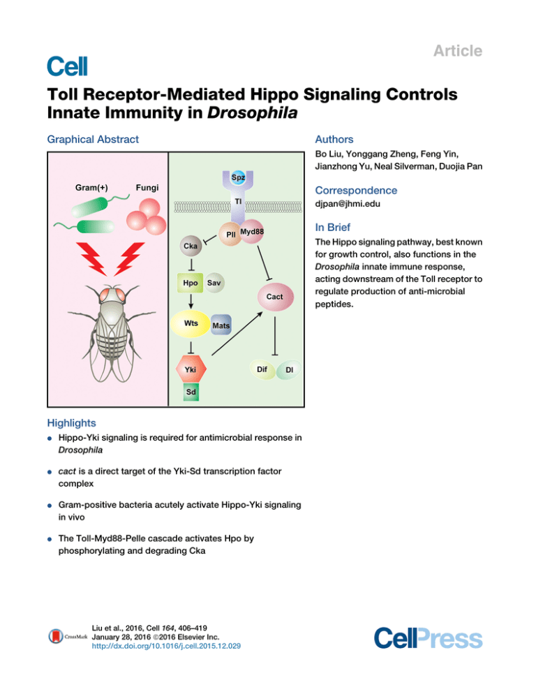
Article Toll Receptor-Mediated Hippo Signaling Controls Innate Immunity in Drosophila Graphical Abstract Authors Bo Liu, Yonggang Zheng, Feng Yin, Jianzhong Yu, Neal Silverman, Duojia Pan Correspondence djpan@jhmi.edu In Brief The Hippo signaling pathway, best known for growth control, also functions in the Drosophila innate immune response, acting downstream of the Toll receptor to regulate production of anti-microbial peptides. Highlights d d Hippo-Yki signaling is required for antimicrobial response in Drosophila cact is a direct target of the Yki-Sd transcription factor complex d Gram-positive bacteria acutely activate Hippo-Yki signaling in vivo d The Toll-Myd88-Pelle cascade activates Hpo by phosphorylating and degrading Cka Liu et al., 2016, Cell 164, 406–419 January 28, 2016 ª2016 Elsevier Inc. http://dx.doi.org/10.1016/j.cell.2015.12.029 Article Toll Receptor-Mediated Hippo Signaling Controls Innate Immunity in Drosophila Bo Liu,1 Yonggang Zheng,1 Feng Yin,1 Jianzhong Yu,1 Neal Silverman,2 and Duojia Pan1,* 1Department of Molecular Biology and Genetics, Howard Hughes Medical Institute, Johns Hopkins University School of Medicine, Baltimore, MD 21205, USA 2Division of Infectious Diseases, Department of Medicine, University of Massachusetts Medical School, Worcester, MA 01605, USA *Correspondence: djpan@jhmi.edu http://dx.doi.org/10.1016/j.cell.2015.12.029 SUMMARY The Hippo signaling pathway functions through Yorkie to control tissue growth and homeostasis. How this pathway regulates non-developmental processes remains largely unexplored. Here, we report an essential role for Hippo signaling in innate immunity whereby Yorkie directly regulates the transcription of the Drosophila IkB homolog, Cactus, in Toll receptor-mediated antimicrobial response. Loss of Hippo pathway tumor suppressors or activation of Yorkie in fat bodies, the Drosophila immune organ, leads to elevated cactus mRNA levels, decreased expression of antimicrobial peptides, and vulnerability to infection by Gram-positive bacteria. Furthermore, Grampositive bacteria acutely activate Hippo-Yorkie signaling in fat bodies via the Toll-Myd88-Pelle cascade through Pelle-mediated phosphorylation and degradation of the Cka subunit of the Hippoinhibitory STRIPAK PP2A complex. Our studies elucidate a Toll-mediated Hippo signaling pathway in antimicrobial response, highlight the importance of regulating IkB/Cactus transcription in innate immunity, and identify Gram-positive bacteria as extracellular stimuli of Hippo signaling under physiological settings. INTRODUCTION The Hippo signaling pathway is a central regulator of organ size in a diverse range of animals, from Drosophila to mammals (Harvey and Tapon, 2007; Johnson and Halder, 2014; Pan, 2010). In Drosophila, this pathway comprises a core kinase cascade linking two kinase complexes—Hippo (Hpo)-Salvador (Sav) and Warts (Wts)-Mob as tumor suppressor (Mats)—to the phosphorylation of the oncoprotein Yorkie (Yki). This phosphorylation event excludes Yki from the nucleus, where it normally functions as a transcriptional coactivator for the expression of target genes involved in cell growth, proliferation, and survival. Studies in mul406 Cell 164, 406–419, January 28, 2016 ª2016 Elsevier Inc. tiple model systems suggest that the Hippo kinase cascade represents a signaling module that integrates multiple biological inputs such as cell polarity, adhesion, mechanical forces, and secreted ligands (Boggiano and Fehon, 2012; Piccolo et al., 2014; Yu and Guan, 2013). Accentuating its importance in growth control, dysregulation of Hippo signaling has been linked to a variety of human cancers, including the hereditary cancer syndrome neurofibromatosis type II (Hamaratoglu et al., 2006; Zhang et al., 2010). Much of the studies to date on Hippo signaling have focused on its role in developing or regenerating tissues. While these studies have firmly established a critical role for Hippo signaling in tissue growth, differentiation, regeneration, and homeostasis, whether and how the Hippo pathway participates in non-developmental and non-growth-related processes remains largely unexplored. Of particular interest to this study is innate immunity. As in other metazoans, antimicrobial defense in Drosophila includes cellular reactions, which involve direct attack/engulfment of microbes by circulating blood, and humoral reactions, which involve the production of antimicrobial peptides by fat bodies (Hultmark, 2003). Pioneering studies in Drosophila have delineated two signaling pathways, namely Toll and Imd, that govern the humoral reactions (Georgel et al., 2001; Lemaitre et al., 1996). Fungi or Gram-positive bacterial infection initiates a proteolytic cascade that activates Spatzle (Spz), a ligand for the transmembrane receptor Toll (Morisato and Anderson, 1994). Activated Toll signals through the adaptor proteins Myd88 and the protein kinase Pelle (Pll) to promote the phosphorylation and degradation of Cactus (Cact), a cytoplasmic ankyrin-repeat-containing protein that normally inhibits the NF-kB family transcription factors Dorsal (Dl) and Dorsalrelated immune factor (Dif) by retaining the latter in the cytoplasm. As a result, Toll signaling allows Dl and Dif to translocate into the nucleus, where they can induce the expression of antimicrobial peptides such as Drosomycin and Metchnikowin (Brennan and Anderson, 2004; Hoffmann, 2003). In contrast to the Toll pathway, the Imd pathway is primarily activated by Gram-negative bacteria. The NF-kB protein in the Imd pathway, Relish, carries its own inhibitory ankyrin-repeat sequence in the carboxy terminus. Activation of the Imd pathway leads to the cleavage of Relish, which removes its ankyrin repeat and releases the amino terminus of Relish into the nucleus to induce the expression of a distinct set of A A’ S.aureus 120 A’’ E.faecalis 120 100 100 80 80 80 Survival (%) 100 ** ** ** ** ** 60 40 0 60 60 ** ** ** ** ** 40 20 20 0 1 2 3 4 Days post infection 0 5 ** 20 ** ** ** ** 2 3 4 Days post infection Hpo RNAi Wts RNAi 5 Yki OE 80 60 60 60 40 ** 40 ** 40 20 ** ** ** ** 20 ** ** ** ** 20 0 5 GFP RNAi C Wts RNAi 80 80 60 60 40 40 20 20 ** 3 4 1 2 5 Days post infection GFP RNAi Hpo RNAi 0 0 5 Yki OE 1 2 3 4 Days post infection Wts RNAi Yki OE 0 1 2 3 4 Days post infection 5 Tl RNAi F ** 0 ** ** ** ** Spz RNAi E.coli 120 100 0 2 3 4 Days post infection Hpo RNAi 100 0 1 C’ Ecc15 120 0 ** S.aureus Drs expression (fold) 2 3 4 Days post infection A.fumigatus 1.5 1.0 ** ** ** ** ** ** ** 0.5 0.0 G F H PR p W o RNAi ts N R Ai N SpYki Ai z OE M T RN yd l R A 88 N i Pl RN Ai lR A N i Ai Survival (%) 100 80 5 Tl RNAi 120 100 1 2 3 4 Days post infection B’’ 80 0 1 Spz RNAi E.faecalis 120 0 100 0 Survival (%) 1 B’ S.aureus 120 40 0 0 GFP RNAi B A.fumigatus 120 5 TAK1 RNAi Ecc15 S.aureus 0 ** ** 1 3 4 5 2 Days post infection WT matse235/A33 spzrm7 120 100 80 60 40 20 0 G Ecc15 0 ** 1 4 3 5 2 Days post infection WT matse235/A33 Tak12 Dpt expression (fold) 120 100 80 60 40 20 0 E 1.5 N.S. N.S. N.S. N.S. N.S. N.S. N.S. 1.0 0.5 0.0 G FP H R po N W R Ai ts NA R i N SpYki Ai z OE M T RN yd l A 88 R N i A Pl RN i lR A N i Ai Survival (%) D Figure 1. Hippo Signaling Is Required for Antimicrobial Response 3- to 6-days-old adult flies were used in all analyses. All data shown are mean ± SD; **p < 0.01; N.S., non-significant. (A–A0 0 ) Adult males expressing the indicated UAS constructs by r4-Gal4 were infected by various pathogens. Fly survival was monitored daily. (B–B0 0 ) Adult virgin females expressing the indicated UAS constructs by YP1-Gal4 were infected and monitored for survival. (C–C0 ) Adult males expressing the indicated UAS constructs by r4-Gal4 were infected with Ecc15 or E. coli. Note the increased sensitivity of TAK1 RNAi flies and the normal sensitivity of Hpo RNAi, Wts RNAi, or Yki overexpression flies. (D) Adult males of wild-type, mats (matse235/matsA33), or spz mutants (spzrm7/spzrm7) were infected with S. aureus and monitored for survival. Note the higher sensitivity of mats mutants and spz mutants than wild-type flies. (legend continued on next page) Cell 164, 406–419, January 28, 2016 ª2016 Elsevier Inc. 407 antimicrobial peptides, such as Diptericin, Attacin, Cecropin, and Drosocin (Brennan and Anderson, 2004; Hoffmann, 2003; Kleino and Silverman, 2014). Here, we report the identification and characterization of the Hippo pathway as an essential regulator of innate immunity in Drosophila. We show that the Hippo signaling pathway functions through Yki and its cognate DNA-binding partner Scalloped to regulate the transcription of Cact, which in turn regulates the expression of antimicrobial peptides and vulnerability to infection by Gram-positive bacteria. We further show that Gram-positive bacteria activate Hippo signaling in fat bodies through Toll-dependent, Pelle-mediated phosphorylation and degradation of Cka, an essential subunit of the STRIPAK PP2A complex, which normally associates with Hpo and antagonizes its kinase activity. Our studies elucidate a Toll-mediated Hippo signaling pathway in innate immunity and implicate bacteria as extracellular stimuli of Hippo signaling under physiological settings. RESULTS Hippo Signaling Activity in the Fat Bodies Is Required for Antimicrobial Response in Adult Flies In an effort to investigate Hippo signaling in adult physiology, we examined the effect of depleting Hpo and Wts or the overexpression of Yki on antimicrobial response. Given the fact that the null mutants of Hpo or Wts are larval lethal, we used the r4-Gal4 driver to generate adult flies with fat-body-specific RNAi of Hpo or Wts or with overexpression of Yki. These flies were injected with Staphylococcus aureus (S. aureus), a Gram-positive bacterium, and monitored daily for survival. Similar to flies with depletion of the Toll pathway component Spz or Toll (Tl), flies with knockdown of Hpo or Wts or with overexpression of Yki were significantly more sensitive to infection, succumbing to death at earlier time than control flies (Figure 1A). Adult flies with Hpo/Wts depletion or with Yki overexpression also showed higher sensitivity to another Gram-positive bacterium Enterococcus faecalis (E. faecalis) as well as the fungus Aspergillus fumigatus (A. fumigatus) (Figures 1A0 –1A00 ). To exclude the possibility that these immunity defects are due to developmental defects, we made use of another driver, YP1-Gal4, which is only expressed in the fat bodies of adult females (Georgel et al., 2001). Similar to the r4-Gal4 driver, adult females with YP1Gal4-directed Hpo or Wts knockdown or with Yki overexpression showed higher sensitivity to Gram-positive bacteria S. aureus or E. faecalis and to fungus A. fumigatus (Figures 1B–1B00 ). We noted that in these survival experiments, flies with Hpo RNAi generally showed less severe phenotypes than flies with Wts RNAi. This is consistent with the less severe overgrowth phenotypes induced by Hpo RNAi in the eye (Figures S1A–S1A00 ). To further probe the specificity of the immune phenotypes we have observed in adult flies with defective Hippo signaling, we infected these animals with the Gram-negative bacteria Erwinia carotovora carotovora 15 (Ecc15) or Escherichi coli (E. coli). In contrast to their high sensitivity to Gram-positive bacteria and fungi, adult flies with Hpo/Wts knockdown or with Yki overexpression were not sensitive to these Gram-negative bacteria (Figures 1C and 1C0 ). In contrast, adult flies with knockdown of TAK1, a key mediator of the Imd pathway (Vidal et al., 2001), were highly sensitive to infection by both Gram-negative bacteria (Figures 1C and 1C0 ). To further corroborate a functional requirement for the Hippo pathway in immunity, we took advantage of matsA33, a newly isolated hypomorphic allele of mats carrying a Gly137Asp missense mutation. Unlike the larval lethality that results from null alleles for any of the core components of the Hippo kinase cascade (hpo, sav, wts, and mats) (Wu et al., 2003; Xu et al., 1995; Tapon et al., 2002; Lai et al., 2005), transheterozygotes of matsA33 over the null allele matse235 produced viable survivors with no visible morphological defects. We found that the matse235/matsA33 flies were extremely sensitive to S. aureus infection, displaying a survival rate comparable to the null mutants of spz (Figure 1D). In contrast, the matse235/matsA33 transheterozygotes were not sensitive to Ecc15 infection (Figure 1E). Taken together, these findings suggest that defective Hippo signaling confers preferential sensitivity to the Toll-mediated response to Gram-positive bacteria and fungi, but not the Imdmediated response to Gram-negative bacteria. Consistent with this view, the mRNA level of the Toll pathway target gene Drosomycin (Drs) upon S. aureus infection was significantly decreased in adult flies with Hpo knockdown, Wts knockdown, or Yki overexpression than in control flies, similar to flies with RNAi depletion of the major Toll pathway components Spz, Tl, Myd88, or Pll (Figure 1F). In contrast, the mRNA level of the Imd pathway target Diptericin (Dpt) was not affected in these flies upon Ecc15 infection (Figure 1G). As a more quantitative assessment of the selective vulnerability of the Hippo-defective flies to infection by Gram-positive bacteria, we measured post-infection bacterial load in adult flies with fat-body-specific Wts knockdown or Yki overexpression. Bacterial load (as measured by colony-forming units, or CFUs) was significantly higher than that in the control flies 24 hr after infecting these flies with Gram-positive bacteria S. aureus, similar to flies with RNAi knockdown of the major Toll pathway components Spz, Tl, Myd88, and Pll (Figure S1B). In contrast, knockdown of Hpo or Wts had no effect on the load of the Gramnegative bacteria Ecc15 (Figure S1C), consistent with the normal survival rate of these animals upon Ecc15 infection. Taken together, the differential sensitivity to Gram-positive versus Gram-negative bacteria suggests a potential link between Hippo signaling and the Toll-mediated innate immunity pathway. (E) Adult males of wild-type, mats (matse235/matsA33), or Tak1 mutants (Tak12/Tak12) were infected with Ecc15 and monitored for survival. Note the comparable sensitivity of mats mutant and wild-type flies. Also note the higher sensitivity of the Tak1 mutant flies. (F–G) Adult males expressing the indicated UAS constructs by r4-Gal4 were infected with S. aureus (F) or Ecc15 (G). Drs (F) or Dpt (G) mRNA was monitored by qRT-PCR 6 hr post infection and normalized by the expression of ribosomal protein rp49. See also Figure S1 408 Cell 164, 406–419, January 28, 2016 ª2016 Elsevier Inc. A S.aureus B E.coli C wtsX1 A’ lacZ wtsX1 B’ B” Dpt-GFP lacZ hpo42-47 C’ sav3 Merge C’’ Cact D’ Merge D” GFP wtsX1 E’ Cact E GFP matse235 F’ Cact F GFP Merge Drs-GFP GFP D A” Merge E” Merge F” Cact Merge Figure 2. The Hippo Kinase Cascade Cell-Autonomously Regulates Antimicrobial Gene Expression and Cact Protein Levels in Larval Fat Bodies Third instar larval fat bodies containing the indicated mutant clones were analyzed. (A–B0 0 ) Drs-GFP or Dpt-GFP (green) was examined 6 hr after S. aureus (A–A0 0 ) or E. coli (B–B0 0 ) infection. wts mutant clones (arrowheads) were marked by the lack of the ubiquitously expressed Arm-lacZ marker (red). Note the decreased expression of Drs-GFP but not Dpt-GFP in wts mutant cells. (C–F0 0 ) GFP-negative mutant clones of the indicated genotypes were stained for Cact protein (red). The merged images also include the nuclear dye DAPI (blue). Note the strong upregulation of Cact expression in hpo (C–C0 0 ), sav (D– D0 0 ), wts (E–E0 0 ), or mats (F–F0 0 ) mutant cells. Representative mutant cells are outlined with dashed lines. See also Figure S2 and Table S1 The Hippo Kinase Cascade Cell-Autonomously Regulates Toll Signaling Outputs in Larval Fat Bodies, Including Target Gene Expression and Cact Protein Levels To investigate the molecular mechanism by which Hippo signaling intersects the Toll-mediated immunity pathway, we turned to the larval fat bodies. A major advantage of the larval fat bodies over the adult fat bodies is the ease of immunostaining and mosaic analysis, which allows a robust genetic analysis at single-cell resolution. Consistent with the downregulation of the Toll pathway target gene Drs after S. aureus infection in Wts-depleted adult flies, we found that loss-of-function (LOF) mutant clones of wts in larval fat bodies showed a cell-autonomous decrease of Drs expression after S. aureus infection (see Table S1 for quantification of all clonal phenotypes reported in this paper), as revealed by a Drs-GFP reporter line (Ferrandon et al., 1998) (Figures 2A–2A00 ). On the other hand, the expression of the Imd pathway reporter Dpt-GFP (Tzou et al., 2000) was not affected in wts LOF mutant clones in the larval fat bodies after infection by the Gram-negative bacteria E. coli (Figures 2B–2B00 ). Taken together, these findings implicate a cell-autonomous requirement for Hippo signaling in conferring normal levels of Toll pathway target gene expression. Degradation of Cactus represents a direct biochemical output of the Toll signaling pathway (Belvin et al., 1995; Nicolas et al., 1998; Reach et al., 1996). In order to establish a functional link between Hippo signaling and the Toll pathway, we investigated the effect of Hippo signaling on Cact protein levels. For this purpose, we examined Cact protein in larval fat bodies containing LOF mutant clones of hpo, sav, wts, and mats, key components of the Hippo kinase cascade. LOF mutant clones for any of the four tumor suppressor genes showed a robust increase of Cact protein levels in a cell-autonomous manner (Figures 2C– 2F00 ). Besides the fat bodies, a similar upregulation of Cact protein levels was observed in mutant clones of the respective Hippo pathway components in the imaginal discs (Figure S2), suggesting that Hippo signaling is a general regulator of Cact expression. Taken together, the cell-autonomous requirement of Hippo signaling for Drs and Cact expression further supports a functional link between the Hippo and the Toll pathways. The Hippo Kinase Cascade Regulates Cact through a Yki-Mediated Transcriptional Mechanism Since the best understood mode of regulating Cact in innate immunity is through Toll pathway-mediated phosphorylation and degradation (Nicolas et al., 1998), we first considered the simplest possibility that the Hippo kinase cascade may directly or indirectly promote the phosphorylation and degradation of Cact. However, in transfection experiments in Drosophila S2R+ cells, co-expression of Hpo and Wts failed to induce mobility shift or degradation of Cact (Figure S3), suggesting that Hippo signaling downregulates Cact through a previously undescribed mechanism. Since the Hippo kinase cascade functions almost exclusively through the transcriptional coactivator Yki in diverse biological processes, we considered the alternative possibility that the regulation of Cact by the Hippo kinase cascade may be mediated by Yki through a transcriptional mechanism. Consistent Cell 164, 406–419, January 28, 2016 ª2016 Elsevier Inc. 409 A >Yki A’ GFP B ykiB5 Cact B’ GFP C >Yki sd47M >Yki Cact wtsX1 GFP Merge C” Cact D’ Merge D” cact-lacZ GFP E Merge B” C’ GFP D A” E’ Merge E” cact-lacZ Merge Figure 3. The Hippo Kinase Cascade Regulates Cact through a YkiMediated Transcriptional Mechanism Third instar larval fat bodies containing the indicated mutant clones were analyzed unless otherwise indicated. (A–B0 0 ) GFP-positive Yki overexpressing clones (A–A0 0 ) or GFP-negative ykiB5 mutant clones (B–B0 0 ) were stained for Cact protein (red), showing increased or decreased expression of Cact, respectively. (C–C0 0 ) GFP-positive sd47M mutant clones with Yki overexpression were stained for Cact protein. Note the unaltered Cact expression in the GFPpositive cells (outlined in C0 ). (D–D0 0 ) Similar to A–A0 0 except that lacZ expression (red) from the cact-lacZ enhancer trap line cactK17027 was examined. Note the increased lacZ staining in Yki-overexpressing cells. (E–E0 0 ) A third instar wing imaginal disc containing GFP-negative wtsX1 mutant clones and the cact-lacZ enhancer trap line cactK17027 stained for lacZ expression (red), showing increased lacZ staining in the wts mutant clones (outlined in E0 ). See also Figures S3, S4, and Table S1 with this hypothesis, Yki overexpression clones in fat bodies showed a dramatic increase of Cact protein levels in a cellautonomous manner (Figures 3A–3A00 ), mimicking the LOF 410 Cell 164, 406–419, January 28, 2016 ª2016 Elsevier Inc. mutant clones for hpo, sav, wts, and mats. A similar upregulation of Cact protein levels was observed in Yki overexpression clones in many other tissues, such as the imaginal disc (Figures S4A– S4A00 ), the gut (Figures S4B–S4B00 ), and the salivary gland (Figures S4C–S4C00 ), suggesting that it represents a general mode of Cact regulation. Conversely, LOF mutant clones of yki in fat bodies showed a decrease of Cact protein levels (Figures 3B–3B00 ). Additional evidence further supports our model of transcriptional regulation of Cact by Hippo signaling. As a transcriptional coactivator, Yki functions together with its major DNA-binding transcription factor partner Scalloped (Sd) to control target gene expression (Wu et al., 2008; Zhang et al., 2008). One would thus expect Yki-induced Cact expression to be Sd-dependent. To test this prediction, we used the MARCM (mosaic analysis with a repressible cell marker) technique (Lee and Luo, 1999) to generate sd mutant clones with Yki overexpression. Indeed, in both fat bodies and imaginal discs, sd mutant clones with Yki overexpression showed normal Cact expression (Figures 3C–3C00 and Figures S4D–S4D00 ), demonstrating that Ykiinduced upregulation of Cact expression is Sd-dependent. To more directly assay cact transcription, we took advantage of cactK17027, a P[lacZ] enhancer trap reporter inserted into the cact locus. Indeed, lacZ expression from cactK17027 was strongly induced in Yki-overexpression clones in fat bodies (Figures 3D– 3D00 ) or wts LOF mutant clones in imaginal discs (Figures 3E– 3E00 ), further demonstrating that cact is regulated by Hippo signaling through a transcription mechanism. cact Is a Direct Target of the Yki-Sd Transcription Factor Complex After demonstrating that Cact is regulated by the Hippo signaling through a transcriptional mechanism, we wished to investigate whether cact is a direct target of the Yki-Sd transcription factor complex. Through systematic dissection of Hippo-responsive element (HRE) in the classic Hippo target gene diap1, we have previously identified a minimal HRE that contains an Sd-binding site (Wu et al., 2008). Examination of the cact locus revealed nine potential Sd-binding motifs as defined by the consensus Sd-binding sequence CATTCNN (Figure 4A). We first used a cell-based luciferase reporter assay to test their potential function as HREs. For this purpose, each Sd-binding motif and 50 bp flanking sequence on each side was cloned upstream of the hsp70 basal promoter in the pGL3 luciferase reporter vector, with the exception of #4, which contains two Sd-binding motifs only 21 bp apart (Figure 4A). Among all the constructs, only #4 could be activated by co-expression of Sd and Yki (Figure 4B), suggesting that it may function as HRE. Since #4 contains two potential Sd-binding motifs (CATTCCA and CATTCCC), we mutated each site individually and found that mutation of CATTCCA (#4M1), but not CATTCCC (#4M2), abolished the Yki-Sd-induced luciferase activity (Figure 4B), suggesting that the former motif is required for HRE activity. Strikingly, this motif is identical to the Sd-binding sequence in the HRE of diap1 (Wu et al., 2008). To validate the physiological relevance of our cell-based assay, we cloned #4 or a mutant version of #4 with mutation of the CATTCCA motif (#4M1) into the enhancer detector vector CPLZN, which contains the hsp70 basal promoter and lacZ #3 A #1 CATTCGC CATTCTT #4 CATTCCA Relative Luc Activity (fold) B CATTCTG #6 CATTCCC CATTCCT #2 #7 CATTCAT #5 CATTCAC CATTCAT #8 Yki+Sd pAC 8 7 6 5 4 3 2 1 GFP F GFP >Yki F’ Merge D” #4M1-lacZ G 2 1 M E” #4-lacZ #4M1-lacZ H 2 3 4 Days post infection 5 7 U AS -Y ki 1 8 ki 0 9 -Y 20 0 11 10 AS ** 12 N 40 ** R 60 N.S. 13 G FP Survival (%) 80 S.aureus Ai GFP RNAi UAS-Yki UAS-Yki;Cact RNAi 100 Merge U 120 Colony-forming units/ml (log10) S.aureus Merge F” GFP Merge #4 #4 M #8 #7 #6 #5 #4 #3 #2 #1 ap #4-lacZ >Yki E’ Ai GFP >Yki D’ E N D C” ac tR >Yki C’ ;C C di G L3 1 0 Figure 4. cact Is a Transcriptional Target of the Yki-Sd Complex (A) Schematic representation of the cactus locus. Nine potential Sd-binding motifs and the DNA constructs tested for HRE activity are indicated. Also indicated are coding exons (yellow boxes) and non-coding exons (gray boxes). (B) Luciferase reporter assay in Drosophila S2R+ cells. Reporter constructs containing individual Sd-binding motifs were co-transfected with Sd- and Ykiexpressing plasmids. Note the induction of luciferase activity by Yki-Sd for reporter #4. Also included are the empty luciferase reporter (GL3: negative control) and a luciferase reporter containing the minimal diap1 HRE (diap1: positive control) (Wu et al., 2008). Data are mean ± SD; **p < 0.01. (legend continued on next page) Cell 164, 406–419, January 28, 2016 ª2016 Elsevier Inc. 411 A Yki B Merge Ctrl Ctrl S.aureus 0.5 h E.coli 0.5 h S.aureus 2h E.coli 2h S.aureus 4h E.coli 4h Yki Merge (A–B) Third instar larvae injected with S. aureus (A) or E. coli (B) were immunostained with a-Yki antibody (red) at the indicated time points postinfection. Nuclear staining (DAPI) is shown in green in the merged channel. Note the gradual accumulation of cytoplasmic Yki signal upon S. aureus (A), but not E. coli infection (B). (C–C0 0 ) Third instar fat bodies containing GFPnegative hpo mutant clones were stained with a-Yki antibody (red) 2 hr after S. aureus infection. The merged channel also includes the nuclear dye DAPI (blue). Reduced cytoplasmic Yki staining was observed in most hpo mutant cells (C0 ). (D) Drosophila Mbn2 cells were treated with a mixture containing heat inactivated S. aureus (5 ml/ml) and E. faecalis (5 ml/ml) or with the peptidoglycan derived from M. luteus (20 mg/ml) for the indicated time. Cell lysates were probed with antibodies against the indicated endogenous proteins. Note the induction of Hpo phosphorylation and Yki phosphorylation by either treatment (compare lanes 2–5 with lane 1). See also Table S1 D Bacteria PGN Ctrl 0.5h 3h 0.5h 3h p-Hpo C C” hpo42-47 C’ Figure 5. Gram-Positive Bacteria Activate Hippo Signaling in Drosophila Fat Bodies Hpo p-Yki that knockdown of Cact rescued both the higher sensitivity and the higher bacterial load upon S. aureus infection in Yki-overexpressing flies (Figures 4G–4H), further supporting our molecular model implicating Cact as a transcriptional target of the Hippo-Yki pathway. Yki S.aureus GFP Yki Merge Tubulin reporter (Wharton and Crews, 1993). LacZ reporter expression was then examined in imaginal discs and fat bodies containing Yki-overexpression clones. Consistent with our in vitro assay, the lacZ reporter containing #4, but not #4M1, was strongly induced in Yki-overexpression clones in both imaginal discs (Figures 4C–4D00 ) and fat bodies (Figures 4E–4F00 ). Taken together, these results pinpoint an essential HRE in the cact locus and identify cact as a direct target of the Sd-Yki transcription factor complex. If cact is a physiological target of Yki in innate immunity, one would expect the immune phenotypes of the Hippo-defective flies to be critically dependent on Cact activity. Indeed, we found Bacterial Infection Activates Hippo Signaling In Vivo In the course of our studies, we noted that infection by the Gram-positive bacteria S. aureus (Figure 5A), but not the Gram-negative bacteria E. coli (Figure 5B), induced a rapid nuclear-to-cytoplasmic translocation of Yki in the larval fat body cells. Yki is highly enriched in fat body nuclei in unchallenged animals (Figure 5A). A shift of Yki to cytoplasmic localization was detectable within 30 min of S. aureus infection. By 4 hr post-infection, Yki was diffusely distributed in most fat body cells and no longer enriched in the cell nuclei (Figure 5A). S. aureus-induced cytoplasmic translocation of Yki in the fat bodies is reminiscent of Hippo-dependent cytoplasmic retention of phosphorylated Yki in the imaginal discs (Dong et al., 2007), suggesting that S. aureus infection may activate Hippo signaling (C–F0 0 ) Analysis of the cact HRE activity in vivo. lacZ reporter containing #4 or #4M1 (red) was examined in third instar wing discs (C–D0 0 ) or fat bodies (E–F0 0 ) containing GFP-positive Yki overexpression clones. Note the induction of #4-lacZ (C and E), but not #4M1-lacZ, in Yki-overexpression clones in both tissues. (G) 3- to 6-days-old adult males expressing the indicated UAS constructs under the control of r4-Gal4 were infected with S. aureus and monitored for survival. Data shown are mean ± SD; **p < 0.01. Note the higher sensitivity of the Yki-overexpressing flies to S. aureus, which was rescued by RNAi of Cact. (H) Adult flies of the indicated genotypes were infected with S. aureus. Bacterial CFU in individual flies was measured at 24 hr post-infection. Data are mean ± SD; **p < 0.01; N.S., non-significant. Note the significant increase of bacterial load in Yki-overexpressing flies, which was rescued by RNAi of Cact. See also Table S1 412 Cell 164, 406–419, January 28, 2016 ª2016 Elsevier Inc. in vivo. Consistent with this hypothesis, loss of hpo abolished S. aureus-induced cytoplasmic translocation of Yki in the fat bodies (Figures 5C–5C00 ). In agreement with this in vivo finding, treating Drosophila Mbn2 cells (a macrophage-like cell line) with heat-inactivated Gram-positive bacterial cocktail containing S. aureus and E. faecalis induced rapid and robust phosphorylation of endogenous Hpo and Yki (Figure 5D). Treating Mbn2 cells with peptidoglycan (PGN) derived from another Gram-positive bacterial strain M. luteus (Micrococcus luteus) had a similar effect (Figure 5D). Interestingly, in both treatment conditions, Hpo phosphorylation peaked at 0.5 hr while elevated Yki phosphorylation persisted at 3 hr, a time course that agrees with the cytoplasmic translocation of Yki observed in vivo. These results demonstrate that bacterial infection can rapidly and potently activate the Hippo-Yorkie pathway, thus implicating Gram-positive bacteria as a class of signals that induce Hippo signaling in vivo. We further infer from these data that Gram-positive bacteria activate the Hippo signaling cascade at the level of Hpo or upstream of Hpo. The Toll-Myd88-Pelle Cascade Mediates BacteriaInduced Hippo Signaling in Drosophila Fat Bodies To investigate how Hippo signaling is rapidly activated by bacterial infection, we considered the possibility that the Toll receptor, which is rapidly activated by Gram-positive bacteria, may itself mediate the activation of Hippo signaling by S. aureus infection. We conducted several experiments to test this hypothesis. One prediction of this model is that Tl itself may be required for S. aureus-induced Hippo signaling in the fat bodies. Consistent with this prediction, we found that S. aureus-induced Yki cytoplasmic translocation was blocked in Tl mutant animals (Figure 6B). To investigate which additional components of the canonical Tl signaling pathway are required for S. aureusinduced Hippo signaling, we examined Yki localization after S. aureus infection in LOF mutants of positive signaling components Spz, Myd88, and Pll and gain-of-function overexpression clones of the negative component Cactus. We also included LOF mutants of Dredd and Tak1, two key components of the Imd pathway (Leulier et al., 2000; Vidal et al., 2001), as control. Most of these LOF mutants are homozygous or transheterozygous viable except for pll, for which LOF mutant clones were examined. Similar to the Tl mutants, S. aureus-induced Yki cytoplasmic localization was blocked in the absence of spz (Figure 6C), Myd88 (Figure 6D), and pll (Figure 6F). In contrast, S. aureus-induced Yki cytoplasmic localization was not affected in LOF mutants of Dredd (Figure 6E), Tak1 (Figure S5), or cells with Cact overexpression (Figure 6G). These results suggest that Toll signaling components upstream of Cactus are required for S. aureus-induced Hippo signaling. Another prediction of our model is that artificial activation of Toll signaling may be sufficient to activate Hippo signaling, even in the absence of bacterial infection. We first tested this prediction by generating mosaic fat bodies containing Tl-overexpression clones. Strikingly, in unchallenged flies, Tl-overexpression clones in the fat bodies showed robust cytoplasmic localization of Yki, in sharp contrast to the nuclear localization of Yki in the neighboring wild-type cells (Figure 6H). A similar cytoplasmic translocation of Yki was observed in fat body clones overexpressing the Toll signaling component Pelle (Figure 6I). These results indicate that artificial activation of Tl or Pll is sufficient to activate Hippo signaling in Drosophila fat bodies in a cell-autonomous manner. Consistent with genetic analysis in the fat bodies, overexpression of Pelle or a constitutively activated mutant of Tl (Tl10b) (Schneider et al., 1991) promoted the phosphorylation of Yki in Drosophila S2 cells (Figure 6J). Furthermore, Pll-induced Yki phosphorylation was greatly attenuated by knockdown of Hpo or Wts (Figure 6J), consistent with the requirement of the Hippo kinase cascade in Yki phosphorylation. Pll and Tl10b also induced the phosphorylation of Hpo (Figure 6K), in agreement with our results showing increased Hpo phosphorylation by Gram-positive bacteria (Figure 5D). These results further support our genetic analysis demonstrating the requirement of Tl and Pll in S. aureus-induced Hippo signaling in Drosophila fat bodies. Pelle Activates Hpo by Phosphorylating and Inducing the Degradation of Cka, an Essential Subunit of the Hpo-Inhibitory STRIPAK PP2A Complex The results presented so far suggest a model whereby Grampositive bacteria, through a pathway involving Tl and its downstream signaling components Myd88 and Pll, promote the phosphorylation of Hpo. Since Pll encodes a protein kinase, the simplest model is that Pll may directly phosphorylate Hpo. We tested this possibility by conducting an in vitro kinase assay using bacterially purified glutathione-S-transferase (GST) fusion protein with a kinase-dead form of Hpo (HpoKD) to avoid autophosphorylation of Hpo, as described in previous studies (Boggiano et al., 2011). We failed to detect phosphorylation of GSTHpoKD by Pll in this assay (Figure S6A). As a control, GST-HpoKD was robustly phosphorylated by Tao1, a bona fide upstream activating kinase of Hpo (Boggiano et al., 2011; Poon et al., 2011) (Figure S6A). Thus, it is unlikely that Pll functions as a direct Hpo kinase. After excluding Pll as a direct Hpo kinase, we explored whether Pll activates Hpo indirectly by phosphorylating a Hippo pathway component upstream of Hpo. For this purpose, we systematically conducted co-immunoprecipitation assays between Pll and proteins that have been implicated upstream of Hpo, including Tao1, Ex, Mer, and Kibra, as well as different subunits of the STRIPAK (striatin-interacting phosphatase and kinase)PP2A (protein phosphatase 2A) complex. The STRIPAK-PP2A complex is known to associate with Hpo and inhibit Hpo activity by dephosphorylating Hpo at its activation loop (Thr195) (Ribeiro et al., 2010). Among all of these proteins, the only protein that interacted with Pll was Cka, a subunit of the STRIPAK complex (Figures 7A and S6B–S6D). We confirmed that Cka was required to inhibit the phosphorylation of Hpo as reported (Ribeiro et al., 2010), as RNAi of Cka induced robust Hpo phosphorylation in S2R+ cells (Figure S6E). Furthermore, LOF mutant clones of Cka in larval fat bodies showed robust cytoplasmic localization of Yki, consistent with activation of Hippo signaling upon loss of Cka in fat body cells (Figures 7B–7B00 ). While examining the physical interactions between Pll and Cka, we noticed that cotransfection of Pll induced mobility retardation of Cka on SDSPAGE as well as greatly decreased Cka protein levels (Figure 7C), Cell 164, 406–419, January 28, 2016 ª2016 Elsevier Inc. 413 D WT B Merge Yki Merge Yki Merge Tlr3/Tlr4 S.aureus C Yki Yki spzrm7 S.aureus Yki Merge Merge Yki DreddB118 Yki Ctrl Merge S.aureus Merge Merge S.aureus GFP H >Tl H’ Yki HA-Pll Tl10b-Flag HA-Yki + + + + + + + + + Merge G” Merge H” GFP Merge Yki > Pll I’ I” GFP Merge J F” Yki I Yki pll2 F’ S.aureus GFP G >Cact G’ Merge DreddB118 spzrm7 Ctrl Yki Myd88KG S.aureus E Tlr3/Tlr4 Ctrl Ctrl FP d p o sR N W ds A ts R N ds A R N A S.aureus Yki WT F H Ctrl Myd88KG G A Merge Yki K + + HA-Pll Tl10b-Flag Myc-HpoKD + p-Yki p-Hpo Yki Hpo Wts HA (Pll) Flag (Tl10b) Myc (HpoKD) + + + + HA (Pll) Flag (Tl10b) Tubulin Tubulin Figure 6. The Toll-Myd88-Pelle Cascade Mediates Bacteria-Induced Hippo Signaling in Drosophila Fat Bodies (A–E) Third instar fat bodies with the indicated genotypes were stained for Yki protein (red) and counterstained with DAPI (green) before (Ctrl, upper panels) or 2 hr after S. aureus infection (lower panels). Note the cytoplasmic translocation of Yki induced by S. aureus in wild-type (A) or DreddB11 mutant flies (E) and the absence of such translocation in Tlr3/Tlr4 (B), spzrm7 (C) or Myd88KG03447 (D) mutant fat bodies. (F) Third instar fat bodies containing pll mutant clones (GFP-negative) were stained for Yki protein (red) and counterstained with DAPI (blue) 2 hr after S. aureus infection. Note the absence of Yki cytoplasmic translocation in pll2 mutant cells (outlined in F0 ), as compared to the neighboring wild-type cells. (G) Similar to (F) except that Cact-overexpression clones (GFP-positive) were examined 2 hr after S. aureus infection. Note the normal cytoplasmic translocation of endogenous Yki in Cact-overexpression cells (outlined in G0 ), as compared to the neighboring wild-type cells. (H–I) Fat bodies from non-infected third instar larvae containing GFP-positive clones with overexpression of Tl (H) or Pll (I) were stained for Yki (red) and counterstained with DAPI (blue). Note the cytoplasmic enrichment of Yki in Tl- or Pll-overexpression cells (outlined in H0 and I0 ). (J) S2 cells were transfected with indicated plasmids. Note the induced Yki phosphorylation by Tl10b (compare lanes 2 and 1) or Pll (compare lanes 3 and 1), which was inhibited by depleting Hpo (compare lanes 5 and 3) or Wts (compare lanes 6 and 3). (K) S2 cells were transfected with indicated plasmids. A kinase dead form of Hpo (HpoKD) was used to avoid auto-phosphorylation of Hpo, as reported in previous studies (Boggiano et al., 2011). Note the induction of Hpo phosphorylation by Tl10b (compare lanes 2 and 1) and Pll (compare lanes 3 and 1). See also Figure S5 and Table S1 suggesting that Pll may induce phosphorylation and degradation of Cka. This hypothesis was supported by additional biochemical evidence. First, the mobility shift of Cka induced by Pll was abrogated by phosphatase treatment, demonstrating that this mobility shift is due to protein phosphorylation (Figure S6F). Sec414 Cell 164, 406–419, January 28, 2016 ª2016 Elsevier Inc. ond, in contrast to wild-type Pll, a kinase-dead mutant form of Pll (Pll-K240R) (Daigneault et al., 2013) failed to induce mobility shift or degradation of Cka (Figure 7D). Lastly, we performed in vitro kinase assay to examine whether Pll can directly phosphorylate bacterially purified GST-Cka fusion protein. In this assay, A C Fl a Fl g - C ag ka Fl -S a lm Fl g-F ap ag go Fl -S p2 ag tri -M p ob 4 B Ckas1883 B’ HA-Pll + + + + + + IB: α-HA GFP IP: Flag ed 16 14 12 10 8 6 4 2 0 > Pll H’ Ctrl I GFP > Pll I’ Flag (Cka) S.aureus GFP Flag (Cka) H” au Ai N FP A R i C N ka Ai R N Ai * R R G ka FP G G K 5h pll RNAi K’ Tl Crb K” 5h 0. Myd88 0. ia N Pll PG er trl Ba ct C Merge Spz Cka Cact Cka p-Hpo Hpo Merge I” M J + + + re H + + N FP C RN ka A G RN i FP A i C RN ka A R i N Ai 0.0 - + + + - + + PNBM HA-Pll-K240R HA-Pll GST-Cka Thiophosphate-ester (Cka) Thiophosphate-ester (Pll) HA (Pll-K240R & Pll) GST-Cka + + ** Drs expression (fold) (fold) * 0.5 us ct fe -in on N 1.5 C N S. on au -in re fe us G * + + 0.2 0.4 + + + + S. ed ct F 1.0 E + HA-Pll-K240R HA-Pll Flag-Cka Flag (Cka) HA (Pll) HA (Pll-K240R) Tubulin lysates α-HA cact expression Yki D IB: α-Flag HA-Pll (μg) GFP + Flag-Cka + Flag (Cka) HA (Pll) GFP Tubulin Merge B” Ctrl L GFP pll RNAi L’ Flag (Cka) Merge L” p-Yki Yki Tubulin Sav Hpo Dif Mats Wts Dl Yki S.aureus GFP Flag (Cka) Merge Antimicrobial peptides Dif Dl Yki Sd cact Figure 7. Pelle Activates Hpo by Phosphorylating and Inducing the Degradation of Cka (A) S2 cells expressing HA-Pll with FLAG-tagged constructs encoding different subunits of the STRIPAK complex were immunoprecipitated with anti-FLAG beads. Note the interactions between Pll and Cka, but not the other subunits of the STRIPAK complex (Slmap, Fgop2, Strip, or Mob4). (legend continued on next page) Cell 164, 406–419, January 28, 2016 ª2016 Elsevier Inc. 415 ATP-gS was used as phosphate donor to generate thiophosphorylated substrate, which further reacts with p-nitrobenzyl mesylate (PNBM) to form a thiophosphate ester that can be detected by thiophosphate-ester-specific antibody (Allen et al., 2007). As shown in Figure 7E, Cka was phosphorylated in vitro by wild-type Pll, but not the kinase-dead mutant Pll-K240R, supporting our model implicating Cka as a direct substrate of Pll. Taken together, the above data suggest a working model in which Gram-positive bacteria activate Hippo signaling through Pll-mediated phosphorylation and degradation of Cka, ultimately leading to decreased Yki activity, decreased Cact transcription, and increased Drs expression (Figure 7M). This model predicts that Cka should positively regulate the transcription of cact and negatively regulate the transcription of Drs. Indeed, fatbody-specific RNAi knockdown of Cka resulted in decreased cact mRNA levels and increased Drs mRNA levels, both in S. aureus-infected and non-infected adult flies (Figures 7F–7G). To further corroborate our model, we used clonal analysis in larval fat bodies to examine the regulation of Cka by Pll at single-cell resolution. Due to the lack of anti-Cka antibodies suitable for immunostaining, we monitored Cka protein levels in vivo using a FLAG-tagged Cka transgene driven by the tubulin promoter (tub-FLAG-Cka) that fully rescued the lethality of Cka mutant flies. Consistent with our model, Pll-overexpression clones in the larval fat bodies showed a cell-autonomous decrease of FLAG-Cka protein levels compared to the neighboring wildtype cells, both in S. aureus-infected and non-infected flies (Figures 7H–7I00 ). Of note, the Cka protein levels in wild-type fat body cells were significantly lower in S. aureus-infected flies than noninfected flies, consistent with bacteria inducing degradation of Cka (compare the staining intensity of FLAG-Cka in wild-type fat body cells in Figure 7I0 versus Figure 7H0 or in Figure 7L0 versus Figure 7K0 ). A similar decrease of Cka protein levels was observed in Mbn2 cells treated with heat-inactivated Gram-positive bacteria or peptidoglycan derived from Gram- positive bacteria (Figure 7J). Complementary to the effect of Pll overexpression on Cka protein levels, larval fat body clones with Pll knockdown showed a modest, cell-autonomous increase of Cka protein levels compared to the neighboring wildtype cells in the absence of S. aureus infection (Figures 7K– 7K00 ). The difference in Cka protein levels between Pll-depleted and wild-type cells was even more obvious when the larvae were infected with S. aureus (Figures 7L–7L00 ). These results further support our model linking Toll to Hippo signaling through Pll-mediated degradation of Cka. DISCUSSION An emerging theme of biological systems is that a limited number of signaling pathways are used reiteratively to control a myriad of biological processes. In this study, we have characterized an essential role for Hippo signaling, a developmental signaling pathway, in innate immune response. It is interesting to note that organ size control and innate immunity are two ancient features of metazoans (Leulier and Lemaitre, 2008; Sebé-Pedrós et al., 2012). Our findings that the signaling pathways underlying these seemingly disparate processes (Hippo and Toll) are functionally intertwined may therefore have a deep evolutionary origin. Interestingly, Toll receptor and Hippo signaling have both been implicated in other biological processes such as cell competition in the wing disc (Meyer et al., 2014; Neto-Silva et al., 2010) and polarized cell rearrangements in Drosophila embryos (Marcinkevicius and Zallen, 2013; Paré et al., 2014), although their molecular relationship in these processes has not been determined. The Hippo signaling pathway was initially defined as an intracellular signaling cascade linking tumor suppressor kinases to transcriptional regulation, and there has been longstanding interest in the field to understand how Hippo signaling activity is regulated in normal development and physiology. An emerging (B–B0 0 ) Non-infected third instar fat bodies containing Cka mutant clones (GFP-negative) were stained for Yki protein (red) and counterstained with DAPI (blue). Note the cytoplasmic enrichment of Yki in Cka mutant cells (outlined in B0 ), as compared to the neighboring wild-type cells. (C) S2R+ cells were transfected with FLAG-Cka and GFP constructs together with different amount of HA-Pll. Note the mobility shift (compare white and black dots next to protein band) and reduced protein level of Cka when Pll was co-transfected. Also note the greater reduction of Cka protein level with increased expression of Pll. In contrast, the protein level of the co-transfected GFP was not affected by the expression of Pll. (D) S2R+ cells were transfected with indicated plasmids. Note the mobility shift and reduced protein level of Cka caused by wild-type Pll but not the Pll-K240R mutant. (E) Direct phosphorylation of Cka by Pll in vitro. Kinase assay was performed using bacterially purified GST-Cka and Pll or Pll-K240R immunoprecipitated from S2R+ cells. Note the thiophosphate-ester signals on Cka as well as Pll (due to autophosphorylation) after incubating with Pll, but not with Pll-K240R. (F–G) 3- to 6-days-old adult males expressing the indicated UAS-RNAi constructs under the control of r4-Gal4 were uninfected or infected with S. aureus. cact (F) or Drs (G) mRNA was monitored by qRT-PCR 6 hr post infection and normalized by rp49 level. Data shown are mean ± SD; *p < 0.05; **p < 0.01. Note the lower cact level and higher Drs level in Cka knockdown flies than in control flies in both uninfected and infected conditions. (H–I) Fat bodies from non-infected control (H) or S. aureus-infected (I) third instar larvae containing Pll-overexpressing clones (GFP-positive) and tub-FLAG-Cka transgene were stained for FLAG-Cka (red) and counterstained with DAPI (blue). Note the reduced level of Cka in Pll-overexpressing cells (outlined in H0 and I0 ), as compared to the neighboring wild-type cells. Also note the overall reduction of Cka signal (red) in the wild-type cells (GFP-negative) upon infection (compare I–I0 with H–H0 , confocal images taken under identical parameters). (J) Mbn2 cells were treated as in Figure 5D. Note the reduced Cka protein level upon bacterial or PGN treatment. Also note the increased p-Hpo and p-Yki levels upon bacterial or PGN treatment. (K–L) Similar to (H–I) except that Pll-knockdown clones (GFP-positive) were analyzed. Note the higher level of Cka in Pll-knockdown cells (outlined in K0 and L0 ), as compared to the neighboring wild-type cells. (M) A schematic model depicting the coordinate regulation of Cact by Toll and Hippo signaling. Activation of the Toll receptor by Gram-positive bacteria not only induces Cact degradation but also suppresses cact transcription through the Hippo-Yki pathway. Activation of Hippo signaling requires Spz, Tl, Myd88, and Pelle, which phosphorylates and degrades Cka, a negative regulator of Hpo. See also Figure S6 and Table S1 416 Cell 164, 406–419, January 28, 2016 ª2016 Elsevier Inc. theme from studies in both Drosophila and mammals indicates that the Hippo kinase cascade functions as a signaling module that integrates multiple upstream inputs such as cell polarity, adhesion, mechanical forces, and secreted ligands (Boggiano and Fehon, 2012; Piccolo et al., 2014; Yu and Guan, 2013). In this regard, we note that, while many conditions have been reported to modulate YAP (the mammalian counterpart of Yki) in cell cultures such as cell density (Zhao et al., 2007), cell detachment (Zhao et al., 2012), certain GPCR ligands (Yu et al., 2012), cell shape (Dupont et al., 2011; Wada et al., 2011), oxygen availability (Ma et al., 2015), and energy stress (DeRan et al., 2014), it remains a challenge to define the exact physiological contexts in which these biological inputs are employed to regulate Hippo signaling in intact tissues. Thus, our findings demonstrating rapid cytoplasmic translocation of Yki in Drosophila fat bodies upon bacterial infection implicate Gram-positive bacteria as a unique class of signals capable of activating Hippo signaling under physiological settings. It is interesting to note that before the recent attention to the Hippo signaling pathway due to its critical role in growth control, the mammalian counterparts of Hpo were biochemically purified as kinases responsive to stress (thus the acronym Krs1/2) (Taylor et al., 1996). In cultured NIH 3T3 cells, Krs1/2 (corresponding to Mst2/1) was activated by extreme forms of stress such as okadaic acid, staurosporine, sodium arsenite, or extreme heat shock at 55 C (Taylor et al., 1996). The physiological relevance of these observations, however, has been unclear. Our findings that bacterial infection rapidly activates Hippo signaling in fat bodies provide a robust system to study the regulation of Hippo signaling by a non-developmental signal. Our genetic and molecular interrogation led us to elucidate a Toll-mediated pathway that activates Hpo through Pll-mediated phosphorylation and degradation of Cka. These findings uncover the STRIPAK PP2A complex as an entry point by which regulatory inputs converge on the Hippo signaling pathway. In this regard, it is worth noting that among the diverse conditions that have been previously reported to modulate YAP phosphorylation in cell cultures—ranging from cell-cell contacts to GPCR ligands—none of them affects Hpo phosphorylation, suggesting that they converge on Hippo signaling at a level downstream of Hpo. Thus, Gram-positive bacteria represent a unique external signal that activates Hippo signaling upstream of Hpo. While much of the research on Toll receptor signaling has focused on the phosphorylation and degradation of Cact at the protein level (Brennan and Anderson, 2004; Hoffmann, 2003), our studies highlight the physiological importance of regulating Cact at transcriptional level. We suggest that regulation of Cact at both the transcriptional and posttranscriptional level is required for proper antimicrobial responses by the Toll receptor (Figure 7M). Since the innate immune pathway is at least partially conserved (Beutler, 2009; Medzhitov and Janeway, 2000), it will be interesting to investigate whether the functional link between Hippo and TLR-NF-kB/IkB signaling is conserved in innate immunity in mammals. We further speculate that such a link may potentially contribute to the oncogenic activity of YAP/TAZ, whereby YAP/TAZ may protect the tumor cells from immune attack through the induction of IkB expression and inhibition of inflammation (Ben-Neriah and Karin, 2011). EXPERIMENTAL PROCEDURES Drosophila Genetics The hypomorphic matsA33 allele was isolated in an EMS mutagenesis screen for mutations on chromosome 3R that result in increased clone size in mosaic eyes using the eyeless-Flp system (Newsome et al., 2000). Mutant clones of matsA33 were larger than wild-type clones but much less overgrown than null alleles of mats. Sequencing of genomic DNA revealed a Gly137Asp missense mutation in the matsA33 allele. Transheterozygous matse235/matsA33 adult survivors were used in this study. Other flies used in the study have been described before: hpo4247 (Wu et al., 2003), sav3 (Tapon et al., 2002), wtsX1 (Xu et al., 1995), matse235 (Lai et al., 2005), and ykiB5 (Huang et al., 2005). r4-Gal4, YP1-Gal4, spzrm7, Tlr3, Tlr4, Myd88KG03447, pll2, Tak12, DreddB118, and Ckas1883 were obtained from Bloomington stock center. All RNAi lines were collected from Vienna Drosophila Resource Center (VDRC) except for Cact RNAi line, which was obtained from Bloomington Stock Center. P [lacW]Cactk17027 enhancer reporter line was obtained from Kyoto Drosophila Genetic Resource Center. Drs-GFP and Dpt-GFP reporter lines have been reported previously (Ferrandon et al., 1998; Tzou et al., 2000) (gift from Louisa Wu). Flp-out was used to generate Yki overexpression clones (Pignoni and Zipursky, 1997). MARCM strain was used to generate sd mutant clones with Yki overexpression (Lee and Luo, 1999). To generate the flies carrying a tub-FLAG-Cka transgene, the UAS-hsp70 promoter of pUASTattB was replaced by the a-tubulin promoter to generate the vector pTubattB. DNA sequence of FLAG-tagged Cka was inserted into pTubattB and the resulting construct pTubattB-FLAG-Cka was used to generate the transgenic line Tub-FLAG-Cka-86Fa. Two P element insertion lines of Cka, Ckas1883, and Cka05836 were obtained from Bloomington Drosophila Stock Center. Transheterozygous Ckas1883/ Cka05836 flies died at pupal stage, but 100% of Ckas1883/Cka05836; tub-FLAG-Cka flies survived to adults with normal morphology. Microbial Strains and Culture S. aureus (ATCC 25923), E. faecalis (ATCC 29212), and E. coli (1106) were purchased from ATCC. S. aureus was cultured in tryptic soy broth. E. faecalis was cultured in brain heart infusion broth. Ecc15 and E. coli were cultured in lysogeny broth medium. A. fumigatus spores were harvested as previously described (Tzou et al., 2002). Septic Injuries and Survival Experiments Overnight cultures of bacteria were suspended in working solutions containing S. aureus (OD600 [optical density] = 0.2), E. faecalis (OD600 = 0.2), A. fumigatus (109 spores/ml), Ecc15 (OD600 = 200), or E. coli (OD600 = 200). Adult male or female flies, aged 3–6 days, were anaesthetized with CO2 and infected by being pricked in the thorax with a thin needle dipped previously into the bacterial working solution. Survival experiments were done at 29 C with 20–30 flies for each tested group. Surviving flies were transferred daily into fresh vials and counted. Bacterial Burden Estimation Adult male flies (3- to 6-days-old) were injected thoracically with bacterial suspension (S. aureus OD600 = 0.2 or Ecc15 OD600 = 200) and cultured at 29 C. After 24 hr, the flies were dipped in 70% EtOH and air-dried on the fly pad. Individual flies were mashed in 100 ml sterile bacterial culture medium and followed by spinning to remove the debris. The supernatant from each fly was serially diluted and plated. The plates were incubated at 37 C for 12–16 hr before counting the number of colonies. Larval Infection Third instar larvae were washed three times with sterile ddH2O and injected using a sharp capillary in the posterolateral body with 100 nl bacterial suspension of S. aureus (OD600 = 20) or E. coli (OD600 = 200). Treated larvae were Cell 164, 406–419, January 28, 2016 ª2016 Elsevier Inc. 417 placed in a vial containing regular food and a piece of wet Kimwipes paper for further analysis. SUPPLEMENTAL INFORMATION Supplemental Information includes Supplemental Experimental Procedures and six figures and can be found with this article online at http://dx.doi.org/ 10.1016/j.cell.2015.12.029. AUTHOR CONTRIBUTIONS B.L. and D.P. conceived the project; B.L. and Y.Z. designed experiments; B.L., Y.Z., F.Y., and J.Y. performed the experiments; N.S. and D.P. supervised the study; B.L. and D.P. wrote the manuscript. ACKNOWLEDGMENTS We would like to thank Dr. Louisa Wu (University of Maryland) for Drs-GFP and Dpt-GFP reporter lines and Dr. Steven X. Hou (National Cancer Institute) for Cka antibody. We thank Bloomington Drosophila Stock Center, Kyoto Drosophila Genetic Resource Center, Vienna Drosophila Resource Center, Drosophila Genomics Resource Center, and Developmental Studies Hybridoma Bank for fly strains and reagents. This study was supported in part by grants from the NIH (EY015708 to D.P. and AI060025 to N.S.). D.P. is an investigator of the Howard Hughes Medical Institute. Received: February 23, 2015 Revised: October 18, 2015 Accepted: December 8, 2015 Published: January 28, 2016 REFERENCES Allen, J.J., Li, M., Brinkworth, C.S., Paulson, J.L., Wang, D., Hübner, A., Chou, W.H., Davis, R.J., Burlingame, A.L., Messing, R.O., et al. (2007). A semisynthetic epitope for kinase substrates. Nat. Methods 4, 511–516. Belvin, M.P., Jin, Y., and Anderson, K.V. (1995). Cactus protein degradation mediates Drosophila dorsal-ventral signaling. Genes Dev. 9, 783–793. Ben-Neriah, Y., and Karin, M. (2011). Inflammation meets cancer, with NF-kB as the matchmaker. Nat. Immunol. 12, 715–723. Beutler, B.A. (2009). TLRs and innate immunity. Blood 113, 1399–1407. Boggiano, J.C., and Fehon, R.G. (2012). Growth control by committee: intercellular junctions, cell polarity, and the cytoskeleton regulate Hippo signaling. Dev. Cell 22, 695–702. Boggiano, J.C., Vanderzalm, P.J., and Fehon, R.G. (2011). Tao-1 phosphorylates Hippo/MST kinases to regulate the Hippo-Salvador-Warts tumor suppressor pathway. Dev. Cell 21, 888–895. Brennan, C.A., and Anderson, K.V. (2004). Drosophila: the genetics of innate immune recognition and response. Annu. Rev. Immunol. 22, 457–483. Daigneault, J., Klemetsaune, L., and Wasserman, S.A. (2013). The IRAK homolog Pelle is the functional counterpart of IkB kinase in the Drosophila Toll pathway. PLoS ONE 8, e75150. DeRan, M., Yang, J., Shen, C.H., Peters, E.C., Fitamant, J., Chan, P., Hsieh, M., Zhu, S., Asara, J.M., Zheng, B., et al. (2014). Energy stress regulates hippo-YAP signaling involving AMPK-mediated regulation of angiomotin-like 1 protein. Cell Rep. 9, 495–503. Dong, J., Feldmann, G., Huang, J., Wu, S., Zhang, N., Comerford, S.A., Gayyed, M.F., Anders, R.A., Maitra, A., and Pan, D. (2007). Elucidation of a universal size-control mechanism in Drosophila and mammals. Cell 130, 1120–1133. Dupont, S., Morsut, L., Aragona, M., Enzo, E., Giulitti, S., Cordenonsi, M., Zanconato, F., Le Digabel, J., Forcato, M., Bicciato, S., et al. (2011). Role of YAP/ TAZ in mechanotransduction. Nature 474, 179–183. 418 Cell 164, 406–419, January 28, 2016 ª2016 Elsevier Inc. Ferrandon, D., Jung, A.C., Criqui, M., Lemaitre, B., Uttenweiler-Joseph, S., Michaut, L., Reichhart, J., and Hoffmann, J.A. (1998). A drosomycin-GFP reporter transgene reveals a local immune response in Drosophila that is not dependent on the Toll pathway. EMBO J. 17, 1217–1227. Georgel, P., Naitza, S., Kappler, C., Ferrandon, D., Zachary, D., Swimmer, C., Kopczynski, C., Duyk, G., Reichhart, J.M., and Hoffmann, J.A. (2001). Drosophila immune deficiency (IMD) is a death domain protein that activates antibacterial defense and can promote apoptosis. Dev. Cell 1, 503–514. Hamaratoglu, F., Willecke, M., Kango-Singh, M., Nolo, R., Hyun, E., Tao, C., Jafar-Nejad, H., and Halder, G. (2006). The tumour-suppressor genes NF2/ Merlin and Expanded act through Hippo signalling to regulate cell proliferation and apoptosis. Nat. Cell Biol. 8, 27–36. Harvey, K., and Tapon, N. (2007). The Salvador-Warts-Hippo pathway - an emerging tumour-suppressor network. Nat. Rev. Cancer 7, 182–191. Hoffmann, J.A. (2003). The immune response of Drosophila. Nature 426, 33–38. Huang, J., Wu, S., Barrera, J., Matthews, K., and Pan, D. (2005). The Hippo signaling pathway coordinately regulates cell proliferation and apoptosis by inactivating Yorkie, the Drosophila Homolog of YAP. Cell 122, 421–434. Hultmark, D. (2003). Drosophila immunity: paths and patterns. Curr. Opin. Immunol. 15, 12–19. Johnson, R., and Halder, G. (2014). The two faces of Hippo: targeting the Hippo pathway for regenerative medicine and cancer treatment. Nat. Rev. Drug Discov. 13, 63–79. Kleino, A., and Silverman, N. (2014). The Drosophila IMD pathway in the activation of the humoral immune response. Dev. Comp. Immunol. 42, 25–35. Lai, Z.C., Wei, X., Shimizu, T., Ramos, E., Rohrbaugh, M., Nikolaidis, N., Ho, L.L., and Li, Y. (2005). Control of cell proliferation and apoptosis by mob as tumor suppressor, mats. Cell 120, 675–685. Lee, T., and Luo, L. (1999). Mosaic analysis with a repressible cell marker for studies of gene function in neuronal morphogenesis. Neuron 22, 451–461. Lemaitre, B., Nicolas, E., Michaut, L., Reichhart, J.M., and Hoffmann, J.A. (1996). The dorsoventral regulatory gene cassette spätzle/Toll/cactus controls the potent antifungal response in Drosophila adults. Cell 86, 973–983. Leulier, F., and Lemaitre, B. (2008). Toll-like receptors–taking an evolutionary approach. Nat. Rev. Genet. 9, 165–178. Leulier, F., Rodriguez, A., Khush, R.S., Abrams, J.M., and Lemaitre, B. (2000). The Drosophila caspase Dredd is required to resist gram-negative bacterial infection. EMBO Rep. 1, 353–358. Ma, B., Chen, Y., Chen, L., Cheng, H., Mu, C., Li, J., Gao, R., Zhou, C., Cao, L., Liu, J., et al. (2015). Hypoxia regulates Hippo signalling through the SIAH2 ubiquitin E3 ligase. Nat. Cell Biol. 17, 95–103. Marcinkevicius, E., and Zallen, J.A. (2013). Regulation of cytoskeletal organization and junctional remodeling by the atypical cadherin Fat. Development 140, 433–443. Medzhitov, R., and Janeway, C., Jr. (2000). Innate immunity. N. Engl. J. Med. 343, 338–344. Meyer, S.N., Amoyel, M., Bergantiños, C., de la Cova, C., Schertel, C., Basler, K., and Johnston, L.A. (2014). An ancient defense system eliminates unfit cells from developing tissues during cell competition. Science 346, 1258236. Morisato, D., and Anderson, K.V. (1994). The spätzle gene encodes a component of the extracellular signaling pathway establishing the dorsal-ventral pattern of the Drosophila embryo. Cell 76, 677–688. Neto-Silva, R.M., de Beco, S., and Johnston, L.A. (2010). Evidence for a growth-stabilizing regulatory feedback mechanism between Myc and Yorkie, the Drosophila homolog of Yap. Dev. Cell 19, 507–520. Newsome, T.P., Asling, B., and Dickson, B.J. (2000). Analysis of Drosophila photoreceptor axon guidance in eye-specific mosaics. Development 127, 851–860. Nicolas, E., Reichhart, J.M., Hoffmann, J.A., and Lemaitre, B. (1998). In vivo regulation of the IkappaB homologue cactus during the immune response of Drosophila. J. Biol. Chem. 273, 10463–10469. Pan, D. (2010). The hippo signaling pathway in development and cancer. Dev. Cell 19, 491–505. Paré, A.C., Vichas, A., Fincher, C.T., Mirman, Z., Farrell, D.L., Mainieri, A., and Zallen, J.A. (2014). A positional Toll receptor code directs convergent extension in Drosophila. Nature 515, 523–527. Vidal, S., Khush, R.S., Leulier, F., Tzou, P., Nakamura, M., and Lemaitre, B. (2001). Mutations in the Drosophila dTAK1 gene reveal a conserved function for MAPKKKs in the control of rel/NF-kappaB-dependent innate immune responses. Genes Dev. 15, 1900–1912. Piccolo, S., Dupont, S., and Cordenonsi, M. (2014). The biology of YAP/TAZ: hippo signaling and beyond. Physiol. Rev. 94, 1287–1312. Wada, K., Itoga, K., Okano, T., Yonemura, S., and Sasaki, H. (2011). Hippo pathway regulation by cell morphology and stress fibers. Development 138, 3907–3914. Pignoni, F., and Zipursky, S.L. (1997). Induction of Drosophila eye development by decapentaplegic. Development 124, 271–278. Wharton, K.A., Jr., and Crews, S.T. (1993). CNS midline enhancers of the Drosophila slit and Toll genes. Mech. Dev. 40, 141–154. Poon, C.L., Lin, J.I., Zhang, X., and Harvey, K.F. (2011). The sterile 20-like kinase Tao-1 controls tissue growth by regulating the Salvador-Warts-Hippo pathway. Dev. Cell 21, 896–906. Reach, M., Galindo, R.L., Towb, P., Allen, J.L., Karin, M., and Wasserman, S.A. (1996). A gradient of cactus protein degradation establishes dorsoventral polarity in the Drosophila embryo. Dev. Biol. 180, 353–364. Ribeiro, P.S., Josué, F., Wepf, A., Wehr, M.C., Rinner, O., Kelly, G., Tapon, N., and Gstaiger, M. (2010). Combined functional genomic and proteomic approaches identify a PP2A complex as a negative regulator of Hippo signaling. Mol. Cell 39, 521–534. Schneider, D.S., Hudson, K.L., Lin, T.Y., and Anderson, K.V. (1991). Dominant and recessive mutations define functional domains of Toll, a transmembrane protein required for dorsal-ventral polarity in the Drosophila embryo. Genes Dev. 5, 797–807. Sebé-Pedrós, A., Zheng, Y., Ruiz-Trillo, I., and Pan, D. (2012). Premetazoan origin of the hippo signaling pathway. Cell Rep. 1, 13–20. Tapon, N., Harvey, K.F., Bell, D.W., Wahrer, D.C., Schiripo, T.A., Haber, D., and Hariharan, I.K. (2002). salvador Promotes both cell cycle exit and apoptosis in Drosophila and is mutated in human cancer cell lines. Cell 110, 467–478. Taylor, L.K., Wang, H.C., and Erikson, R.L. (1996). Newly identified stressresponsive protein kinases, Krs-1 and Krs-2. Proc. Natl. Acad. Sci. USA 93, 10099–10104. Wu, S., Huang, J., Dong, J., and Pan, D. (2003). hippo encodes a Ste-20 family protein kinase that restricts cell proliferation and promotes apoptosis in conjunction with salvador and warts. Cell 114, 445–456. Wu, S., Liu, Y., Zheng, Y., Dong, J., and Pan, D. (2008). The TEAD/TEF family protein Scalloped mediates transcriptional output of the Hippo growth-regulatory pathway. Dev. Cell 14, 388–398. Xu, T., Wang, W., Zhang, S., Stewart, R.A., and Yu, W. (1995). Identifying tumor suppressors in genetic mosaics: the Drosophila lats gene encodes a putative protein kinase. Development 121, 1053–1063. Yu, F.X., and Guan, K.L. (2013). The Hippo pathway: regulators and regulations. Genes Dev. 27, 355–371. Yu, F.X., Zhao, B., Panupinthu, N., Jewell, J.L., Lian, I., Wang, L.H., Zhao, J., Yuan, H., Tumaneng, K., Li, H., et al. (2012). Regulation of the Hippo-YAP pathway by G-protein-coupled receptor signaling. Cell 150, 780–791. Zhang, L., Ren, F., Zhang, Q., Chen, Y., Wang, B., and Jiang, J. (2008). The TEAD/TEF family of transcription factor Scalloped mediates Hippo signaling in organ size control. Dev. Cell 14, 377–387. Zhang, N., Bai, H., David, K.K., Dong, J., Zheng, Y., Cai, J., Giovannini, M., Liu, P., Anders, R.A., and Pan, D. (2010). The Merlin/NF2 tumor suppressor functions through the YAP oncoprotein to regulate tissue homeostasis in mammals. Dev. Cell 19, 27–38. Tzou, P., Ohresser, S., Ferrandon, D., Capovilla, M., Reichhart, J.M., Lemaitre, B., Hoffmann, J.A., and Imler, J.L. (2000). Tissue-specific inducible expression of antimicrobial peptide genes in Drosophila surface epithelia. Immunity 13, 737–748. Zhao, B., Wei, X., Li, W., Udan, R.S., Yang, Q., Kim, J., Xie, J., Ikenoue, T., Yu, J., Li, L., et al. (2007). Inactivation of YAP oncoprotein by the Hippo pathway is involved in cell contact inhibition and tissue growth control. Genes Dev. 21, 2747–2761. Tzou, P., Meister, M., and Lemaitre, B. (2002). Methods for studying infection and immunity in Drosophila. Molecular Cellular Microbiology 31, 507–529, (Academic Press). Zhao, B., Li, L., Wang, L., Wang, C.Y., Yu, J., and Guan, K.L. (2012). Cell detachment activates the Hippo pathway via cytoskeleton reorganization to induce anoikis. Genes Dev. 26, 54–68. Cell 164, 406–419, January 28, 2016 ª2016 Elsevier Inc. 419
