The Digestive and Endocrine Systems What You’ll Learn
advertisement
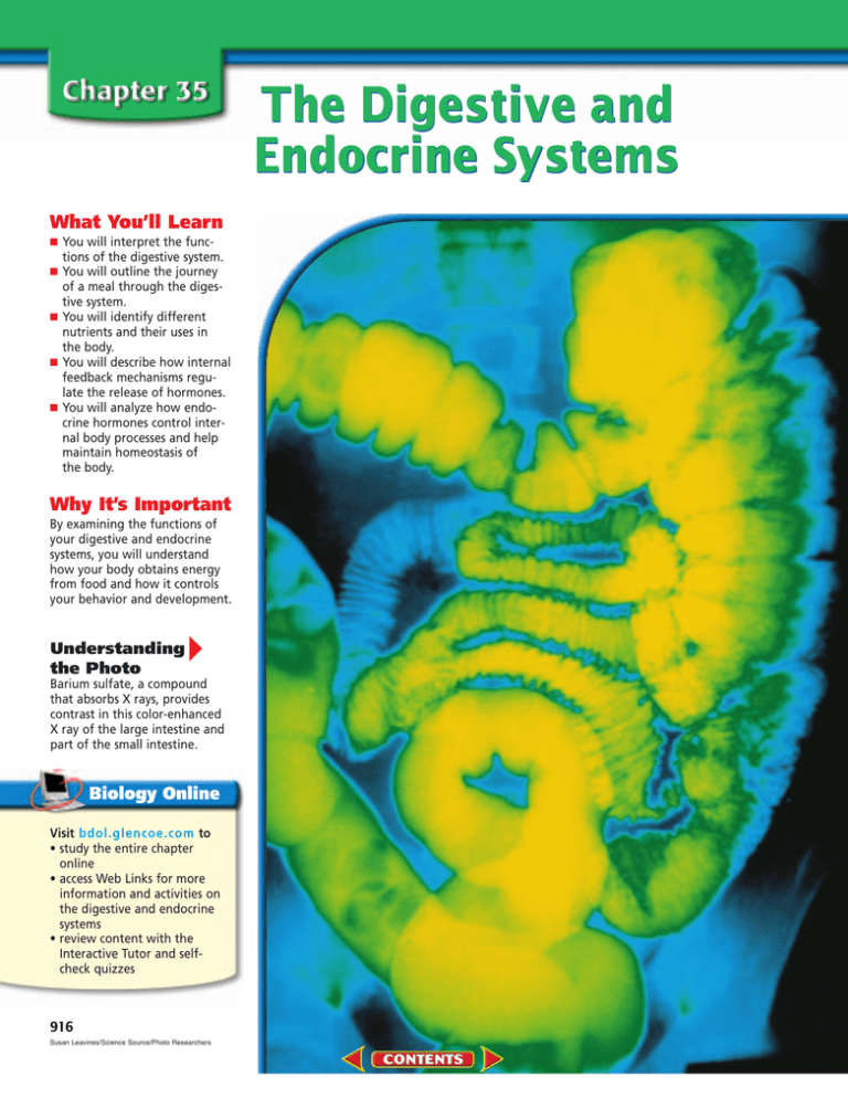
The Digestive and Endocrine Systems What You’ll Learn ■ ■ ■ ■ ■ You will interpret the functions of the digestive system. You will outline the journey of a meal through the digestive system. You will identify different nutrients and their uses in the body. You will describe how internal feedback mechanisms regulate the release of hormones. You will analyze how endocrine hormones control internal body processes and help maintain homeostasis of the body. Why It’s Important By examining the functions of your digestive and endocrine systems, you will understand how your body obtains energy from food and how it controls your behavior and development. Understanding the Photo Barium sulfate, a compound that absorbs X rays, provides contrast in this color-enhanced X ray of the large intestine and part of the small intestine. Visit bdol.glencoe.com to • study the entire chapter online • access Web Links for more information and activities on the digestive and endocrine systems • review content with the Interactive Tutor and selfcheck quizzes 916 Susan Leavines/Science Source/Photo Researchers 35.1 SECTION PREVIEW Following Digestion of a Meal Objectives Interpret the different functions of the digestive system organs. Outline the pathway food follows through the digestive tract. Identify the role of enzymes in chemical digestion. Digestive System Make the following Foldable to help you learn more about the structures and functions of the digestive system. STEP 1 Draw a mark at the midpoint of a sheet of paper along the side edge. Then fold the top and bottom edges in to touch the midpoint. STEP 2 Fold in half from side to side. STEP 3 Open and cut along the inside fold lines to form four tabs. STEP 4 Label each tab as shown. Review Vocabulary enzyme: type of protein found in all living things that increases the rate of chemical reactions (p. 161) New Vocabulary amylase esophagus peristalsis epiglottis stomach pepsin small intestine pancreas liver bile gallbladder villus large intestine rectum Mouth Small Intestine Stomach Large Intestine Identify As you read Chapter 35, list the functions of each of these digestive system structures beneath the appropriate tab. Functions of the Digestive System The main function of the digestive system is to disassemble the food you eat into its component molecules so that it can be used as energy for your body. In this sense, your digestive system can be thought of as a sort of disassembly line. Digestion is accomplished through a number of steps. First, the system takes ingested food and begins moving it through the digestive tract. As it does so, it digests—or breaks down mechanically and chemically—the complex food molecules. Then, the system absorbs the digested food and distributes it to your cells. Finally, it eliminates undigested materials from your body. As you read about each digestive organ, use Figure 35.1 on the next page to locate its position within the system. The Mouth The first stop along the digestive disassembly line is your mouth. Suppose it’s lunchtime and you have just prepared a bacon, lettuce, and tomato sandwich. The first thing you do is bite off a piece and chew it. 35.1 FOLLOWING DIGESTION OF A MEAL 917 action of enzymes. What purpose do the different structures inside your mouth serve? Find out by examining Figure 35.2. Figure 35.1 All the digestive organs work together to break down food into simpler compounds that can be absorbed by the body. Describe Interpret the functions of the digestive system. Salivary glands Teeth Mouth Tongue Pharynx Esophagus Diaphragm Liver Stomach Gallbladder Pancreas Large intestine Small intestine Appendix Rectum Anus Physical Science Connection Physical and chemical changes in matter Digestion involves both physical and chemical changes in matter. Describe the digestive processes that occur in the mouth. Classify each as a physical or a chemical change. What happens as you chew? As you chew, your tongue moves the food around and helps position it between your teeth. Chewing is a form of mechanical digestion, the physical process of breaking food into smaller pieces. Mechanical digestion prepares food particles for chemical digestion. Chemical digestion is the process of changing food on a molecular level through the Swallowing your food Once you’ve thoroughly chewed your bite of sandwich, your tongue shapes it into a ball and moves it to the back of your mouth to be swallowed. Swallowing forces food from your mouth into your throat and from there into your esophagus, a muscular tube that connects your mouth to your stomach. Food moves down the esophagus by way of peristalsis. Peristalsis (per uh STAHL sus) is a series of involuntary smooth muscle contractions along the walls of the digestive tract. Table 35.1 Digestive Enzymes Organ Enzyme Molecules Digested Product Salivary glands Salivary amylase Starch Disaccharide Stomach Pancreas Pepsin Pancreatic amylase Trypsin Pancreatic lipase Nucleases Maltase Sucrase Lactase Peptidase Nuclease Proteins Starch Proteins Fats Nucleic acids Disaccharide Disaccharide Disaccharide Peptides Nucleotides Peptides Disaccharide Peptides Fatty acids and glycerol Nucleotides Monosaccharide Monosaccharide Monosaccharide Amino acids Sugar and nitrogen bases Small intestine 918 Chemical digestion begins in the mouth Some of the nutrients in your sandwich are starches, large molecules known as polysaccharides. As you chew your bite of sandwich, salivary glands in your mouth secrete saliva. Saliva contains a digestive enzyme, called amylase, which breaks down starch into smaller molecules such as di- or monosaccharides. In the stomach, amylase continues to digest starch in the swallowed food for about 30 minutes. Table 35.1 lists some digestive enzymes that act to break food molecules apart. THE DIGESTIVE AND ENDOCRINE SYSTEMS Your Mouth Figure 35.2 Your mouth houses many structures involved in other functions besides digestion. Some of these structures protect against foreign materials invading your body; others help you taste the food you eat. Critical Thinking Why is it important that the tongue is composed of skeletal muscles? Incisors A Teeth The incisors are adapted Gums for cutting food. The cuspids, or canines, tear or shred food. The three sets of molars can crush and grind food. Often, there is not enough room Hard palate for the third set of molars, called wisdom teeth, Uvula which then must Soft palate B Tonsils A pair of tonsils is located at the back of the mouth. They play a role in preventing infections in the nose and mouth by helping to eliminate foreign bacteria. be removed. C Tongue The tongue is attached to the floor of the mouth. It is made of numerous skeletal muscles covered with a mucous membrane. Molars Color-enhanced SEM Magnification: 245 Bicuspids or premolars Cuspids or canines Enamel CROWN Opening of salivary gland duct Dentin Pulp NECK The tongue is covered by projections that contain numerous taste receptor cells like the ones shown here. Nerve D Structure of a tooth Teeth are made mainly of dentin, a bonelike ROOT Bone Root canal Vascular supply substance that gives a tooth its shape and strength. The dentin encloses a space filled with pulp, a tissue that contains blood vessels and nerves. The dentin of the crown is covered with an enamel that consists mostly of calcium salts. Tooth enamel is the hardest substance in the body. 35.1 FOLLOWING DIGESTION OF A MEAL 919 Omikron/Photo Researchers Figure 35.3 Smooth muscle contractions are responsible for moving food through the digestive system. From mouth To stomach Longitudinal muscle Food mass Circular muscle Contraction Contraction of circular muscles behind food mass Contraction of longitudinal muscles ahead of food mass Figure 35.3 shows how the food is moved along from the mouth to the stomach. The contractions occur in waves: first, circular muscles relax and longitudinal muscles contract; then circular muscles contract and longitudinal muscles relax. When you swallow, the food enters the esophagus. Usually, a flap of cartilage called the epiglottis (ep uh GLAH tus) closes over the opening to the respiratory tract as you swallow, preventing food from entering. After the food passes into your esophagus, the epiglottis opens again. But if you talk or laugh as you swallow, the epiglottis may open, allowing food to enter the upper portion of the respiratory tract. Your response, a reflex, is to choke and cough, forcing the food out of the respiratory tube. The Stomach Contraction in circular muscle layer forces food mass forward Esophagus Figure 35.4 Smooth muscle contractions churn the food in the stomach until it becomes a thin liquid. Stomach Small intestine 920 THE DIGESTIVE AND ENDOCRINE SYSTEMS When the chewed food reaches the end of your esophagus, it enters the stomach. The stomach is a muscular, pouchlike enlargement of the digestive tract. Both physical and chemical digestion take place in the stomach. Muscular churning Three layers of involuntary muscles, lying across one another, are located within the wall of the stomach. When these muscles contract, as shown in Figure 35.4, they work to physically break down the swallowed food, creating smaller pieces. As the muscles continue to work the food pieces, they mix them with digestive juices produced by the stomach. Chemical digestion in the stomach The inner lining of the stomach contains millions of glands that secrete a mixture of chemicals called gastric juice. Gastric juice contains pepsin and hydrochloric acid. Pepsin is an enzyme that begins the chemical digestion of proteins in food. Pepsin works best in the acidic environment provided by hydrochloric acid, which increases the acidity of the stomach contents to pH 2. How is the stomach lining protected from powerful digestive enzymes and strong acids? The stomach lining secretes mucus that forms a protective layer between it and the acidic environment of the stomach. Food remains in your stomach for approximately two to four hours. When food is ready to leave the stomach, it is about the consistency of tomato soup. Peristaltic waves gradually become more vigorous and begin to force small amounts of liquid out of the lower end of the stomach and into the small intestine. The Small Intestine From your stomach, the liquid food moves into your small intestine, a muscular tube about 6 m long. This section of the intestine is called small not because of its length, but because of its narrow diameter— only 2.5 cm. Digestion of your meal is completed within the small intestine. Muscle contractions contribute to further mechanical breakdown of the food. At the same time, carbohydrates and proteins undergo further chemical digestion with the help of enzymes produced and secreted by the pancreas and liver. Explain which types of digestion occur in the small intestine. Chemical action The first 25 cm of the small intestine is called the duodenum (doo ah DEE num). Most of the enzymes and Diaphragm Esophagus Liver Stomach Gallbladder Bile duct Pancreatic duct Duodenum Pancreas Small intestine Figure 35.5 chemicals that function in the duodenum enter it through ducts that collect juices from the pancreas, liver, and gallbladder. These organs, shown in Figure 35.5, play important roles in digestion, even though food does not pass directly through them. Both the pancreas and the liver produce chemicals needed for digestion in the small intestine. Secretions of the pancreas The pancreas is a soft, flattened gland that secretes both digestive enzymes and hormones, which you will learn more about in the last section of this chapter. The mixture of enzymes it secretes breaks down carbohydrates, proteins, and fats. Alkaline pancreatic juices also help to neutralize the acidity of the liquid food, stopping any further action of pepsin. Secretions of the liver The liver is a large, complex organ that has many functions. One of its functions is to produce bile. Bile is a chemical substance that helps break down fats. Once made in the liver, bile is stored in a small organ called the gallbladder. 35.1 FOLLOWING DIGESTION OF A MEAL 921 Sequence How is digestion affected if the gallbladder is removed? Many people have had their gallbladders surgically removed. What changes take place in digestion if the gallbladder is removed? Solve the Problem The following diagrams show the appearance of a normal liver and gallbladder (diagram A) and the appearance when the gallbladder has been removed (diagram B). B A Hepatic duct Bile duct Hepatic duct Bile duct Thinking Critically 1. Identify Where is bile produced? Where is bile stored? 2. Explain How does bile affect fat? 3. Sequence Identify the pathway for bile from the liver to the duodenum in a person with a gallbladder and compare it to the sequence in a person with no gallbladder. 4. Infer The gallbladder is a muscular sac. It squeezes and discharges a large quantity of bile when fats are present in the duodenum. Explain why a person without a gallbladder is unable to digest fats as efficiently as someone who has a gallbladder. Figure 35.6 Gallstones, seen in this color-enhanced X ray, can form in the gallbladder or bile duct. They consist mainly of crystallized bile salts. Gallstones 922 THE DIGESTIVE AND ENDOCRINE SYSTEMS Department of Clinical Radiology, Salisbury District Hospital/Science Photo Library/Photo Researchers From the gallbladder, bile passes into the duodenum. Bile causes further mechanical digestion by breaking apart large drops of fat into smaller droplets. If bile becomes too concentrated due to high levels of cholesterol in the diet, or if the gallbladder becomes inflamed, gallstones can form, as seen in Figure 35.6. Can a person live without a gallbladder? Find out in the Problem-Solving Lab on this page. Absorption of food Liquid food stays in your small intestine for three to five hours and is slowly moved along its length by peristalsis. As digested food moves through the intestine, it passes over thousands of tiny fingerlike structures called villi. A villus (plural, villi) is a single projection on the lining of the small intestine that functions in the absorption of digested food. The villi greatly increase the surface area of the small intestine, allowing for a greater absorption rate. Because the digested food is now in the form of small molecules, it can be absorbed directly into the cells of the villi, as shown in Figure 35.7. The food molecules then diffuse into the blood vessels of the villus and enter the bloodstream. The villi are the link between the digestive system and the circulatory system. What happens to indigestible materials that remain in the digestive tract? The Large Intestine The indigestible material from your meal now passes into your large intestine, a muscular tube that is also called the colon. Although the large intestine is only about 1.5 m long, it is much wider than the small intestine—about 6.5 cm in diameter. The appendix, a tubelike extension off the large intestine thought to be an evolutionary remnant from our herbivorous ancestors, seems to serve no function in human digestion. Water absorption As the indigestible mixture passes through the large intestine, water and salts are absorbed by the intestinal walls, leaving behind a more solid material. In this way, the water is not wasted. A secondary function of the large intestine is vitamin synthesis. Anaerobic bacteria in the large intestine synthesize some B vitamins and vitamin K, which are absorbed as needed by the body. The presence of certain bacteria in the large intestine is beneficial in another way. Under normal conditions, these bacteria stop Columnar epithelium Lymph vessel Blood vessel network Fatty acids Amino acids Monosaccharides Figure 35.7 harmful bacteria from colonizing, reducing the risk of intestinal infections. Identify and describe the roles that bacteria play in maintaining health. Once food has been fully digested in the small intestine, it is in the form of molecules small enough to enter the body’s bloodstream through projections called villi. Elimination of wastes After 18 to 24 hours in the large intestine, the remaining indigestible material, now called feces, reaches the rectum. The rectum is the last part of the digestive system. Feces are eliminated from the rectum through the anus. Your meal’s entire journey through the digestive tract has taken between 24 and 33 hours. Understanding Main Ideas 1. Describe the functions of the digestive system and sequence the organs according to the order in which food passes through them. 2. Identify the effects of enzymes on food molecules. Which enzymes act on proteins? 3. How do villi of the small intestine increase the rate of nutrient absorption? 4. What role does the pancreas play in digestion? bdol.glencoe.com/self_check_quiz Thinking Critically 5. How would chronic diarrhea affect homeostasis of the body? KILL REVIEW EVIEW SKILL 6. Get the Big Picture Prepare a circle graph representing the time food remains in each part of the digestive tract. For more help, refer to Get the Big Picture in the Skill Handbook. 35.1 FOLLOWING DIGESTION OF A MEAL 923 35.2 Nutrition SECTION PREVIEW You Are What You Eat Objectives Using Prior Knowledge In this section, Recognize the contribution of the six classes of nutrients to body nutrition. Identify the role of the liver in food storage. Relate caloric intake to weight loss or gain. you will learn about nutrition and the different molecules that your body uses for energy and those it needs to function properly. List the foods you eat in a day. Using the food pyramid as a guide, categorize them into different groups according to food type. Review Vocabulary carbohydrate: organic compound used by cells to store and release energy (p. 158) New Vocabulary mineral vitamin Calorie Evaluate How do your meals fit into this pyramid? USE SPARINGLY 2–3 SERVINGS Includes butter, oils, salad dressings, and soft drinks Fat Sugar 2–3 SERVINGS 3–5 SERVINGS 2–4 SERVINGS The food pyramid 6–11 SERVINGS The Vital Nutrients Six basic kinds of nutrients can be found in foods: carbohydrates, fats, proteins, minerals, vitamins, and water. These substances are essential to proper body function. You supply your body with these nutrients when you eat foods from the five main food groups shown in Figure 35.8. Figure 35.8 Select foods from each of the five food groups every day and you’ll have a healthful diet that supplies the six essential nutrients your body needs. 924 Carbohydrates Perhaps your favorite food is pasta, fresh-baked bread, or corn on the cob. If so, your favorite food contains carbohydrates, important sources of energy for your body cells. Recall that carbohydrates are starches and sugars. Starches are complex carbohydrates found in bread, cereal, potatoes, rice, corn, beans, and pasta. Sugars are simple carbohydrates found mainly in fruits, such as plums, strawberries, and oranges. During digestion, complex carbohydrates are broken down into simple sugars, such as glucose, fructose, and galactose. Absorbed into the bloodstream through the villi of the small intestine, these sugar molecules circulate to fuel body functions. THE DIGESTIVE AND ENDOCRINE SYSTEMS Aaron Haupt Some sugar is carried to the liver where it is stored as glycogen. Cellulose, another complex carbohydrate, is found in all plant cell walls and is not digestible by humans. However, cellulose (also known as fiber) is still an important item to include in the diet as it helps in the elimination of wastes. Sources of fiber include bran, beans, and lettuce. Fats Many people think that eating fat means getting fat; however, fats are an essential nutrient. They provide energy for your body and are also used as building materials. Recall that fats are essential building blocks of the cell membrane. They are also needed to synthesize hormones, protect body organs against injury, and insulate the body from cold. Sources of fat in the diet include meats, nuts, and dairy products, as well as cooking oils. In the digestive system, fats are broken down into fatty acids and glycerol and absorbed by the villi of the small intestine. Eventually, some of these fatty acids end up in the liver. The liver converts them to glycogen or stores them as fat throughout your body. Proteins Your body has many uses for proteins. Enzymes, antibodies, many hormones, and substances that help the blood to clot, are all proteins. Proteins form part of muscles and many cell structures, including the cell membrane. During digestion, proteins are broken down into amino acids. After the amino acids have been absorbed by the small intestine, they enter the bloodstream and are carried to the liver. The liver can convert amino acids to fats or glucose, both of which can be used for energy. However, your body Fluorine (F) Dental cavity reduction Fluoridated water Iodine (I) Formation of thyroid hormone Seafood, eggs, iodized salt, milk group Iron (Fe) Formation of hemoglobin (carries oxygen to body cells) and cytochromes (ATP formation) Liver, egg yolk, grain and meat groups, leafy vegetables Sodium (Na) Nerve activity, body pH regulation Bacon, butter, table salt, vegetable group Phosphorus (P) Teeth and bone formation, blood pH, muscle and nerve activity, part of enzymes and nucleic acids Milk, grain, and vegetable groups Copper (Cu) Development of red blood cells, formation of some respiratory enzymes Grain group, liver Potassium (K) Nerve and muscle activity Vegetable group, bananas Magnesium (Mg) Muscle and nerve activity, bone formation, enzyme function Fruit, vegetable, and grain groups Function Calcium (Ca) Teeth and bone formation, muscle and nerve activity, blood clotting Milk and grain groups Sulfur (S) Builds hair, nails, and skin, component of insulin Grain and fruit groups, eggs, cheese Source Figure 35.9 uses amino acids for energy only if other energy sources are depleted. Most amino acids are absorbed by cells and used for protein synthesis. The human body needs 20 different amino acids to carry out protein synthesis, but it can make only 12 of them. The remaining 8 must be consumed in the diet and so are called essential amino acids. Sources of essential amino acids include meats, dried beans, whole grains, eggs, and dairy products. Minerals serve many vital functions. Describe What are the functions of iron in the body? 35.2 NUTRITION 925 vitamin from the Latin word vita, meaning “life”; Vitamins are necessary for life. Minerals and vitamins When you think of minerals, you may picture substances that people mine, or extract from Earth. As shown in Figure 35.9 on the previous page, the same minerals can also be extracted from foods and put to use by your body. A mineral is an inorganic substance that serves as a building material or takes part in a chemical reaction in the body. Minerals make up about four percent of your total body weight, most of it in your skeleton. Although they serve many different functions within the body, minerals are not used as an energy source. Unlike minerals, vitamins are organic nutrients that are required in small amounts to maintain growth and metabolism. The two main groups of vitamins are fat-soluble and water-soluble, as shown in Table 35.2. Although fat-soluble vitamins can be stored in the liver, the accumulation of excess amounts can prove toxic. Water-soluble vitamins cannot be stored in the body and so must be included regularly in the diet. Table 35.2 lists foods that contain fat-soluble and water-soluble vitamins. Vitamin D, a fat-soluble vitamin, is synthesized in your skin. Vitamin K and some B vitamins are made by bacteria in your large intestine. The rest of the vitamins must be consumed in your diet. Table 35.2 Vitamins Vitamin Function Source A Maintain health of epithelial cells; formation of lightabsorbing pigment; growth of bones and teeth Liver, broccoli, green and yellow vegetables, tomatoes, butter, egg yolk D Absorption of calcium and phosphorus in digestive tract Egg yolk, shrimp, yeast, liver, fortified milk; produced in the skin upon exposure to ultraviolet rays in sunlight E Formation of DNA, RNA, and red blood cells Leafy vegetables, milk, butter K Blood clotting Green vegetables, tomatoes, produced by intestinal bacteria Sugar metabolism; synthesis of neurotransmitters Ham, eggs, green vegetables, chicken, raisins, seafood, soybeans, milk Sugar and protein metabolism in cells of eyes, skin, intestines, blood Green vegetables, meats, yeast, eggs Energy-releasing reactions; fat metabolism Yeast, meats, liver, fish, whole-grain cereals, nuts B6 Fat metabolism Salmon, yeast, tomatoes, corn, spinach, liver, yogurt, wheat bran, whole-grain cereals and bread B12 Red blood cell formation; metabolism of amino acids Liver, milk, cheese, eggs, meats Aerobic respiration; synthesis of hormones Milk, liver, yeast, green vegetables, wholegrain cereals and breads Synthesis of DNA and RNA; production of red and white blood cells Liver, leafy green vegetables, nuts, orange juice Aerobic respiration; fat metabolism Protein metabolism; wound healing Yeast, liver, egg yolk Citrus fruits, tomatoes, leafy green vegetables, broccoli, potatoes, peppers Fat-soluble Water-soluble B1 B2 (riboflavin) Niacin Pantothenic acid Folic acid Biotin C 926 THE DIGESTIVE AND ENDOCRINE SYSTEMS Water Water is the most abundant substance in your body—between 45 and 75 percent of your total body mass. Water facilitates the chemical reactions in your body and is necessary for the breakdown of foods during digestion. Water is also an excellent solvent; oxygen and nutrients from food could not enter your cells if they did not first dissolve in water. Recall that water absorbs and releases heat slowly. It is this characteristic that helps water maintain your body’s internal temperature. A large amount of heat is needed to raise the temperature of water. Because the body contains so much water, it takes a lot of added energy to raise its internal temperature. Your body loses about 2.5 L of water per day through exhalation, sweat, and urine. As a result, water must be replaced constantly. Calories and Metabolism The energy content of food is measured in units of heat called Calories, each of which represents a kilocalorie, or 1000 calories (written with a small c). A calorie is the amount of heat required to raise the temperature of 1 mL of water by 1°C. Some foods, especially those with fats, contain more Calories than others. In general, 1 g of fat contains nine Calories, while 1 g of carbohydrate or protein contains four Calories. To learn more about Calories in meals, complete the MiniLab on this page. The number of Calories needed each day varies from person to person, depending on metabolism, or rate at which energy is burned. As you will see in the next section, a major regulator of metabolic rate is a hormone from the thyroid gland. Interpret Data Evaluate a Bowl of Soup As a consumer, you are bombarded by advertising that promotes the nutritional benefits of specific food products. Choosing a food to eat on the basis of such ads may not make nutritional sense. By examining the product labels that list the ingredients of processed foods, you can learn about their actual nutritional content. NUTRITION FACTS Serving Size: 2 cups (452g) Servings Per Container: 1 Data Table Amount Per Serving Percentage of Daily Value (DV) Carbohydrates 60% Fat 30% Saturated fats 10% Cholesterol 1.5% Protein 10% Total Calories 2000 Calories 140 Calories from Fat 54 % Daily Value* Total Fat 8g Saturated Fat 6g Cholesterol 20mg Sodium 1640 mg Total Carbohydrate 22g Dietary Fiber 5g Sugars 5g Protein 6g Vitamin A 50% Vitamin C Iron Calcium 2% 12% 30% 7% 68% 7% 20% 4% 2% * Percent Daily Values are based on a 2,000 calorie diet. Your daily values may be higher or lower depending on your calorie needs: Calories 2,000 2,500 Total Fat Less than 65g 80g Sat Fat Less than 20g 25g Cholesterol Less than 300mg 300mg Sodium Less than 2,400mg 2,400mg Total Carbohydrate 300g 375g Fiber 25g 30g Calories per gram: Fat 9 * Carbohydrates 4 * Protein 4 Procedure ! Examine the information in the table listing the daily value (DV) of various nutrients. DV expresses what percent of Calories should come from certain nutrients. @ Examine the nutritional information on the soup can label, and compare it with the DV table. Analysis 1. Analyze Does your bowl of soup provide more than 30 percent of any of the daily nutrients? Which ones? 2. Use Numbers Calculate the percentage of Calories in soup that are provided by saturated fat. 3. Evaluate Is this soup a nutritious meal? Explain. A person’s body mass, age, gender, and level of physical activity also affect metabolic rate. Generally, males need more Calories per day than females, teenagers need more than adults, and active people need more than inactive people. List factors that can affect metabolic rate. 35.2 NUTRITION 927 Use Numbers What is BMI? BMI is a reliable indicator of a healthy body weight for adult men and women based on height and weight. Approximately sixty percent of adults in the United States are considered overweight. Use the following equation to calculate a sample BMI. Solve the Problem Compute BMI, or Body Mass Index, using the following formula: weight (in pounds) 704.5 = BMI height (in inches )2 The guidelines for adults from the National Institutes of Health are as follows: A BMI • 18.5 to 24.9 = normal weight • 25 to 29.9 = overweight • 30 or over = obese Thinking Critically 1. Evaluate Calcuate the BMI for a person who is 5 feet 4 inches tall and weighs 132 pounds. According to the guidelines, is this person of normal weight, overweight, or obese? 2. Recognize Cause and Effect How might a person with a BMI of 27 reduce his or her BMI? Consider both nutritional intake and physical activity. 3. Infer Fred has a BMI of 22. How do you suppose his Calorie intake compares to his Calorie expenditure? 4. Think Critically What limits does the BMI test have? (Hint: A 6 foot tall, well-muscled athlete weighing 200 pounds would have a BMI of 27.) Understanding Main Ideas 1. Compare the functions of carbohydrates, fats, and proteins in the body. 2. Describe the role of the liver in the storage of carbohydrates, fats, and proteins. 3. Compare and contrast vitamins and minerals. Which vitamins and minerals can be found in milk? 4. What happens when a person takes in more food energy than his or her body needs? 928 THE DIGESTIVE AND ENDOCRINE SYSTEMS Calories and health What happens if a person consumes more Calories than his or her body can metabolize? When the energy taken in is greater than the energy expended, the extra energy is stored as body fat and a person gains weight. However, if a person eats fewer Calories than the body can metabolize, some of the body’s stored energy is used and weight is lost. Physicians have determined that many Americans are overweight. Being overweight or obese increases a person’s risk for developing health problems such as high blood pressure, diabetes, and heart disease. Being underweight is also associated with health problems such as anemia, fatigue, and decreased ability to fight infection and disease. A simple way to determine if a person is at a healthy weight is to calculate his or her Body Mass Index (BMI). Calculate a sample BMI by doing the Problem-Solving Lab on this page. Millions of people put themselves on diets every year in hopes of losing weight. While many diets are nutritionally sound, others prescribe eating habits that are not sensible and usually fail to produce the desired result. Read more about weight-loss products in the Biology and Society section at the end of this chapter. Thinking Critically 5. Describe two effects dehydration can have on homeostasis of the body. KILL REVIEW EVIEW SKILL 6. Make and Use Tables Using Table 35.2 on page 926, analyze how a lack of vitamins A, D, K, and C in a person’s diet could affect his or her health. For more help, refer to Make and Use Tables in the Skill Handbook. bdol.glencoe.com/self_check_quiz 35.3 The Endocrine System SECTION PREVIEW Internal Feedback Objectives Concept Map Copy the concept map onto a separate sheet of paper. Describe the internal feedback mechanism controlling hormone levels in the body. Contrast the actions of steroid and amino acid hormones. Identify and interpret the functions of some of the hormones secreted by endocrine glands. The hypothalamus 4. detects low water content in blood and stimulates the to reabsorb 1. to release which can target 2. 3. Review Vocabulary gland: in mammals, a cell or group of cells that secretes fluid (p. 842) New Vocabulary endocrine glands hypothalamus pituitary gland target cell receptor negative feedback system adrenal gland thyroid gland parathyroid glands endocrine from the Greek words endo, meaning “within,” and krinein, meaning “to separate”; The endocrine glands secrete hormones into the blood. Organize Information As you read this section, complete the concept map about internal feedback in the endocrine system. Control of the Body Internal control of the body is directed by two systems: the nervous system, which you will learn more about later, and the endocrine system. The endocrine system is made up of a series of glands, called endocrine glands, that release chemicals directly into the bloodstream. These chemicals act as messengers, relaying information to other parts of the body. Ultimately, the functions of all body systems are controlled by the interaction between the nervous and endocrine systems. Interaction of the nervous system and endocrine system Much of the time, the endocrine system and the nervous system work together to maintain homeostasis within the body. Because there are two control systems within the body, coordination between the two systems is needed. The hypothalamus (hi poh THA luh mus) is the portion of the brain that connects the endocrine and nervous systems. The hypothalamus receives messages from other areas of the brain and from internal organs. When a change in homeostasis is detected, the hypothalamus stimulates the pituitary (pih TEW uh ter ee) gland. The pituitary gland, the main gland of the endocrine system, is located in the skull just beneath the hypothalamus. The pituitary gland is controlled by the hypothalamus, and the two are connected by nerves and blood vessels. In response to messages received by the hypothalamus, the pituitary gland releases its own chemicals or stimulates other glands to release theirs. Other endocrine glands under the control of the pituitary include the thyroid gland, the adrenal glands, and glands associated with reproduction. 35.3 THE ENDOCRINE SYSTEM 929 Endocrine control of the body The chemicals secreted by endocrine glands into the bloodstream are called hormones. Recall that a hormone is a chemical released in one part of an organism that affects Pituitary Amino acid hormones Controls adrenal gland, thyroid gland, ovaries, testes, mammary glands, stores hypothalamus hormones, and secretes growth hormone Parathyroid gland Amino acid hormones Secretes parathyroid hormone Adrenal medulla Amino acid hormones Secretes epinephrine and norepinephrine Adrenal cortex Steroid hormones Secretes glucocorticoid and aldosterone Type of hormones released Hypothalamus Amino acid hormones Controls pituitary and synthesizes antidiuretic hormone and oxytocin for uterus contraction during birth Thyroid gland Amino acid hormones Secretes thyroxin to stimulate growth and metabolism and secretes calcitonin Ovary in female Steroid hormones Secretes female sex hormones Testis in male Steroid hormones Secretes male sex hormones Function of gland Figure 35.10 This diagram shows the principal human endocrine glands, the type of hormone(s) they secrete, and the action of the gland/hormone. List What are the hormones secreted by the adrenal medulla? 930 THE DIGESTIVE AND ENDOCRINE SYSTEMS another part. Hormones convey information to other cells in your body, giving them instructions regarding your metabolism, growth, development, and behavior. Once released by the glands, the hormones travel in the bloodstream and then attach to specific binding sites found on the plasma membranes, or in the nuclei, of target cells. These binding sites on cells are called receptors. Figure 35.10 summarizes the action of different endocrine glands. Example of endocrine control Human growth hormone (hGH) is a good example of an endocrine system hormone. When your body is actively growing, blood glucose levels are slightly lowered as the growing cells use up the sugar. This low blood glucose level is detected by the hypothalamus, which stimulates the production and release of hGH from the pituitary into the bloodstream. hGH binds to receptors on the plasma membranes of liver cells, stimulating the liver cells to release glucose into your blood. Your cells need the glucose in order to continue growing. Figure 35.11 summarizes the control of hGH by the pituitary gland. You can further investigate growth rate in humans by doing the BioLab at the end of this chapter. Negative Feedback Control If homeostasis is disrupted, endocrine glands can be stimulated by the nervous system, changes in blood chemistry, or by other hormones. Regulation of the endocrine system is controlled most often through one type of internal feedback mechanism called a negative feedback system. In a negative feedback system, the hormones, or their effects, are fed back to inhibit the original signal. Once homeostasis is reached, the signal is stopped and the hormone is no longer released. The thermostat in your home is controlled by a similar negative feedback system. It maintains the room at a set temperature. When the temperature drops, the thermostat senses the reduction of thermal energy and signals the heater to increase its output. When the thermal energy of the room rises again to a certain point, the thermostat no longer stimulates the heater, which shuts off. When the temperature drops again, the process repeats itself. In this negative feedback system, the increase in temperature “feeds back” to signal the thermostat to stop stimulating thermal energy production. A Low blood sugar is detected. B Hypothalamus stimulates pituitary to release hGH. Pituitary D Increased sugar in blood signals back to hypothalamus to slow stimulation of pituitary. Relate negative feedback systems to the maintenance of homeostasis. Feedback control of hormones The majority of endocrine glands operate under negative feedback systems. A gland synthesizes and secretes its hormone, which travels in the blood to target cells where the appropriate response occurs. Information regarding the hormone level or its effect on target cells is fed back, usually to the hypothalamus or pituitary gland, to regulate the gland’s production of the hormone. Control of blood water levels Let’s look at an example of a hormone that is controlled by a negative feedback system. After working out in the gym and building up a sweat, you are thirsty. This is because the water content of your blood has been reduced. The hypothalamus, which is able to sense the concentration of C hGH stimulates liver to convert glycogen into glucose and releases glucose into blood. Figure 35.11 The hypothalamus and pituitary gland control the amount of human growth hormone (hGH) in your blood. water in your blood, determines that your body is dehydrated. In response, it stimulates the pituitary gland to release antidiuretic (AN tih di yuh reh tihk) hormone (ADH). ADH reduces the amount of water in your urine. It binds to receptors in kidney cells, promoting the reabsorption of water and reducing the amount of water excreted in urine. Information about blood water levels is constantly fed back to the hypothalamus so it can regulate the pituitary’s release of ADH. If the body becomes overhydrated, the hypothalamus stops stimulating release of ADH. 35.3 THE ENDOCRINE SYSTEM 931 Interpret Data How does exercise affect levels of insulin and glucagon? Exercise represents an example of rapid fuel mobilization in the body. The body must gear up to supply great amounts of glucose and oxygen for muscle metabolism. Solve the Problem The graph here shows the effects of prolonged exercise, such as running a marathon, on blood insulin and glucagon levels. 450 Insulin 300 10 5 150 Glucagon 0 1 2 Hours 3 Glucagon (pg/mL) Insulin (µU/mL) Levels of Insulin and Glucagon During Exercise 1. Analyze Examine the graph and explain how exercise affects the concentrations of insulin and glucagon in the blood. 2. Recognize Cause and Effect Relate the changes shown on the graph to what is occurring in muscle cells as well as to blood glucose levels. 3. Sequence Design a flowchart that shows the steps involved in maintaining homeostasis of blood glucose during exercise. Begin your flowchart with muscle cells. 932 Control of blood glucose levels Another example of a negative feedback system involves the regulation of blood glucose levels. Unlike most other endocrine glands, the pancreas is not controlled by the pituitary gland. When you have just eaten and your blood glucose levels are high, your pancreas releases the hormone insulin. Then, insulin signals liver and THE DIGESTIVE AND ENDOCRINE SYSTEMS Hormone Action Once hormones are released by an endocrine gland, they travel to target cells and cause a change. Hormones can be grouped into two basic types according to how they act on their target cells: steroid hormones and amino acid hormones. 4 Thinking Critically adrenal from the Latin words ad, meaning “attached to,” and ren, meaning “the kidneys”; The adrenal glands are located on top of the kidneys. muscle cells to take in glucose, thus lowering blood glucose levels. When blood glucose levels become too low, another pancreatic hormone, glucagon, is released. Glucagon binds to liver cells, signaling them to release stored glycogen as glucose. Learn more about glucose storage and release by doing the Problem-Solving Lab on this page. Action of steroid hormones Hormones that are made from lipids are called steroid hormones. Steroid hormones are lipid-soluble and therefore diffuse freely into cells through their plasma membranes, as shown in Figure 35.12. There they bind to a hormone receptor inside the cell. The hormone-receptor complex then travels to the nucleus where it activates the synthesis of specific messenger RNA molecules. The mRNA molecules move out to the cytoplasm where they guide the synthesis of the required proteins. Action of amino acid hormones The second group of hormones is made from amino acids. Recall that amino acids can be strung together in chains and that proteins are made from long chains of amino acids. Some hormones are short chains of amino acids and others are large chains. These amino acid hormones, once secreted into the bloodstream, bind to receptors embedded in the plasma membrane of the target cell, Cell membrane New protein molecule Figure 35.12 Steroid hormone Steroid hormones enter a cell (1), bind to a receptor (2), which in turn binds to DNA to stimulate protein synthesis (3). 1 Ribosome mRNA 2 Hormone-receptor complex Nucleus mRNA DNA 3 as shown in Figure 35.13. From there, they open ion channels in the membrane, or route signals down from the surface of the membrane to activate enzymes inside the cell. The enzymes, in turn, alter the behavior of other molecules inside the cell. In both of these ways, the hormone is able to control what goes on inside the target cell. Adrenal Hormones and Stress You are sitting in math class and the teacher is about to hand out the semester test. Because this test is an important one, you have spent many hours studying for it. Like most of your classmates, you are a little nervous as the test is being passed down the row. Your heart is beating fast and your hands are a little sweaty. As you review the first problem, however, you begin to calm down because you know how to solve it. The adrenal glands play an important role in preparing your body for stressful situations. The adrenal glands are located on top of the kidneys and consist of two parts—an inner portion and an outer portion. The outer portion secretes steroid hormones, including glucocorticoids (glew ko KOR tuh koydz) and aldosterone (ahl DOS tuh rohn). These steroid hormones cause an increase in available glucose and raise blood pressure. In this way, they help the body combat stresses such as fright, temperature extremes, bleeding, infection, disease, and even test anxiety. Figure 35.13 When an amino acid hormone binds to the receptor on the cell membrane (1), it can open ion channels (2), or activate enzymes (3). Amino acid hormone Cell membrane 1 Hormone receptor Enzyme (inactive) Ions 3 2 Enzyme (active) Ion channel Nucleus 35.3 THE ENDOCRINE SYSTEM 933 Observe Compare Thyroid and Parathyroid Tissue Although their names seem somewhat similar, the thyroid and parathyroid glands perform rather different functions within the body. Color-enhanced LM Magnification: 16 Procedure ! Copy the data table. @ Use low-power magnificaA tion to examine a prepared slide of thyroid and parathyroid endocrine gland tissue. (Note: Both tissues appear on the same slide.) CAUTION: Use caution when working with a microscope and B prepared slides. # The image on the right is a photograph of thyroid and parathyroid tissue. Parathyroid (A) and Use it as a guide in locating thyroid (B) tissue the two types of endocrine gland tissue under low power and in answering certain analysis questions. $ Now locate each type of gland tissue under high-power magnification. Draw what you see in the data table. Then use what you learned in the chapter to identify the names of the hormones produced by each gland. Data Table Tissue Drawing Name of Hormone(s) Produced Thyroid Parathyroid Analysis 1. Compare and Contrast Compare and contrast the microscopic appearance of parathyroid tissue to that of thyroid tissue. 2. a. Observe Which tissue type contains follicles (large liquid storage areas)? b. Infer What may be present within the follicles? c. Think Critically Hypothesize what function the thin layer of tissue that surrounds each follicle may have. 3. Explain How might you explain the fact that both thyroid and parathyroid tissue can be seen on the same slide? 934 THE DIGESTIVE AND ENDOCRINE SYSTEMS Fred Hossler/Visuals Unlimited The inner portion of the adrenal gland secretes two amino acid hormones: epinephrine (eh puh NEH frun)—often called adrenaline—and norepinephrine. Recall the fight-orflight response discussed in the animal behavior chapter. During such a response, the hypothalamus relays impulses to the nervous system, which in turn stimulates the adrenal glands to increase their output of epinephrine and norepinephrine. These hormones increase heart rate, blood pressure, and rate of respiration; increase efficiency of muscle contractions; and increase blood sugar levels. If you have ever had to perform in front of a large audience, you may have experienced these symptoms, often referred to collectively as an “adrenaline rush.” This is how the body prepares itself to face or flee a stressful situation. Thyroid and Parathyroid Hormones The thyroid gland, located in the neck, regulates metabolism, growth, and development. The main metabolic and growth hormone of the thyroid is thyroxine. This hormone affects the rate at which the body uses energy and determines your food intake requirements. The thyroid gland also secretes calcitonin (kal suh TOH nun)—a hormone that regulates calcium levels in the blood. Calcium is a mineral the body needs for blood clotting, formation of bones and teeth, and normal nerve and muscle function. Calcitonin binds to the membranes of kidney cells and causes an increase in calcium excretion. Calcitonin also binds to bone-forming cells, causing them to increase calcium absorption and synthesize new bone. Another hormone involved in mineral regulation, parathyroid hormone (PTH), is produced by the parathyroid glands, which are attached to the thyroid gland. The release of PTH leads to an increase in the rate of calcium, phosphate, and magnesium absorption in the intestines. PTH causes the release of calcium and phosphate from bone tissue. It also increases the rate at which the kidneys remove calcium and magnesium from urine and return them to the blood. The overall effect of parathyroid hormone and calcitonin hormone interaction in the body is shown in Figure 35.14. Take a closer look at thyroid and parathyroid tissue by completing the MiniLab on the previous page. As you can see, hormones associated with the endocrine system are responsible for controlling many different functions in your body. Different hormones may play more important roles during some periods in your life than others. In any case, they remain the principal biological influence on your behavior and development. Figure 35.14 Calcitonin and parathyroid hormone (PTH) have opposite effects on blood calcium levels. Parathyroids Thyroid Intestine Calcitonin PTH Describe how blood calcium homeostasis is maintained within the body. Understanding Main Ideas 1. Identify and interpret the functions of the endocrine system. 2. Explain the interrelationship between the nervous system and the endocrine system. 3. Describe the relationship between a negative feedback system and the maintenance of homeostasis. 4. How does a steroid hormone affect its target cell? How does this action differ from how an amino acid hormone affects its target cell? bdol.glencoe.com/self_check_quiz Blood calcium levels increased Blood calcium levels decreased Ca2+ absorbed to blood Kidney Ca2+ absorbed to blood Ca2+ excreted Bone Ca2+ released to blood Ca2+ absorbed Thinking Critically 5. Hormones continually make adjustments in blood glucose levels. Why must blood glucose levels be kept fairly constant? KILL REVIEW EVIEW SKILL 6. Sequence Create a flowchart that illustrates the internal feedback system the body uses to maintain blood glucose homeostasis. For more information, refer to Sequence in the Skill Handbook. 35.3 THE ENDOCRINE SYSTEM 935 The Action of the Enzyme Amylase on Breakfast Cereals Before You Begin The enzyme amylase is found in both salivary and pancreatic secretions. Amylase is used by the body to digest starch. When you eat foods that contain starch, such as breakfast cereals, salivary amylase immediately begins to digest these molecules. How long does it take for larger molecules of starch to be broken down into simple sugars? In this lab, you will investigate the relative rate of starch digestion by amylase. REPARATION PREPARATION Problem How long does it take amylase to digest all of the starch in breakfast cereals? Objectives In this BioLab, you will: ■ Compare the relative rate of starch digestion by amylase on three breakfast cereals. Materials variety of dry cereals mortar and pestle test tubes test tube racks filter paper funnel balance beaker water Bunsen burner or hot plate graduated cylinder iodine solution in dropper bottles watch glasses plastic droppers amylase solution Safety Precautions CAUTION: Never eat laboratory materials. Iodine can irritate and will stain skin. Skill Handbook If you need help with this lab, refer to the Skill Handbook. ROCEDURE PROCEDURE 1. Copy the data table. 2. Label the breakfast cereals and three corresponding test tubes A, B, and C. Data Table Time (sec) Presence of Starch Cereal A Initial test 0 30 60 936 THE DIGESTIVE AND ENDOCRINE SYSTEMS Cereal B Cereal C 3. Grind a small portion of each of the breakfast cereals to a powder using the mortar and pestle. 4. Place a piece of filter paper in the funnel. Place the funnel over test tube A. 5. Using the balance, measure out 0.5 g of ground cereal A and transfer it to the funnel. 6. Filter 10 mL of boiling water over the cereal and allow the filtrate to collect in the bottom of the test tube. 7. Repeat steps 4, 5, and 6 for cereals B and C. Rinse the funnel and replace the filter paper before each filtration. 8. Add 2 drops of the iodine solution to a watch glass, followed by 2 drops of filtrate A. A dark blue/black color indiA B cates the presence of starch. Record your results. 9. Using a separate eyedropper for each solution, repeat step 8 on cereals B and C. Clean the watch glass between each test. 10. Add 2 mL of amylase solution to each filtrate. Immediately take a sample, and repeat steps 8 and 9 to retest for the presence of starch. 11. Test each filtrate every 30 seconds until all of the starch has been digested to simple sugars in each sample. Record your results. 12. CLEANUP AND DISPOSAL Clean all equipment as instructed by your teacher. Make wise choices as to the disposal or recycling of materials. Wash your hands thoroughly. C NALYZE AND AND CONCLUDE ONCLUDE ANALYZE 1. Analyze Did all of the breakfast cereals contain starch? What action did the amylase have on the starch? 2. Observe and Infer Which cereal was converted to simple sugars in the least amount of time? Infer what this indicates about the starch concentration of this cereal compared to the other cereals. 3. Think Critically Does the amount of starch versus simple sugars make a difference in the Calorie content of the cereal? Use Variables, Constants, and Controls Investigate the effect of temperature or pH on the action of amylase on one of the breakfast cereals. Web Links To find out more about digestive enzymes, visit bdol.glencoe.com/digestive_enzymes 35.3 THE ENDOCRINE SYSTEM 937 Evaluate the Promise of Weight Loss as a Promotional Claim L ose ten pounds in one week!” “Shed weight without going hungry!” “Burn fat while you sleep!” The appeal of weight-loss products There are many weight-loss products available to the public. Some of these products are based on good nutrition and positive lifestyle changes, such as eating a well-balanced diet and incorporating a regular exercise program. Other products look like a fast and easy solution to a weight-loss problem. However, these products may not provide permanent results or may have negative side effects. How can you evaluate the promotional claims such as those seen in magazine and television advertisements or on a product label? Read the fine print Many weight-loss products make claims in bold letters at the top of an advertisement or have a quote from someone claiming to have successfully lost weight using the product. However, in very small print at the bottom may be a qualifying statement such as “Results not typical” or “When used with a balanced diet and regular exercise.” Some weight-loss products may help some people lose a few pounds temporarily. However, for safe, long-term weight loss, nutritionists recommend a diet based on healthy eating habits: balanced, regular meals rich in fruits and vegetables, whole grains, sufficient protein, and small amounts of fat. Making lifestyle changes that incorporate regular exercise also allow for healthy weight loss and maintenance. 938 THE DIGESTIVE AND ENDOCRINE SYSTEMS Aaron Haupt Evaluate Collect advertisements and product labels for three different weight-loss products that promise “miracle” results. Research how these products contribute to weight loss. What effects do these products have on the body that result in weight loss? Are there any negative side effects? Evaluate the promotional claims of these advertisements and product labels. Based on what you know about the importance of good nutrition and exercise on health, would you recommend the use of these particular products? Why or why not? To find out more about weight-loss products, visit bdol.glencoe.com/biology_society STUDY GUIDE Section 35.1 Key Concepts ■ Digestion begins in the mouth with both mechanical and chemical action. The esophagus transports food from the mouth to the stomach. ■ Chemical and mechanical digestion continue in the acidic environment of the stomach. ■ In the small intestine, digestion is completed and food is absorbed. The liver and pancreas play key roles in digestion. ■ The large intestine absorbs water before indigestible materials are eliminated. Following Digestion of a Meal Section 35.2 Nutrition USE SPARINGLY 2–3 SERVINGS Vocabulary amylase (p. 918) bile (p. 921) epiglottis (p. 920) esophagus (p. 918) gallbladder (p. 921) large intestine (p. 923) liver (p. 921) pancreas (p. 921) pepsin (p. 921) peristalsis (p. 918) rectum (p. 923) small intestine (p. 921) stomach (p. 920) villus (p. 922) Key Concepts ■ Carbohydrates are the body’s main source Includes butter, oils, salad dressings, of energy. Fats are used to store energy. and soft drinks Proteins are used as building materials. Fat Sugar ■ Minerals serve as structural materials or take part in chemical reactions. Vitamins are needed for growth and metabolism. ■ Water facilitates chemical reactions in the body, acts as a solvent, and helps maintain internal body temperature. Vocabulary Key Concepts ■ The endocrine glands work with the nervous system to regulate body functions. ■ Blood hormone levels are controlled by a negative feedback system. ■ Steroid hormones bind to receptors inside the target cells, and amino acid hormones bind to plasma membrane receptors. ■ Hormones are involved in the regulation of blood glucose and calcium levels, as well as responses to stress. Vocabulary Calorie (p. 927) mineral (p. 926) vitamin (p. 926) 2–3 SERVINGS 3–5 SERVINGS 2–4 SERVINGS 6–11 SERVINGS Section 35.3 The Endocrine System New protein molecule Steroid hormone Ribosome Hormone-receptor complex mRNA Nucleus mRNA DNA Cell membrane adrenal gland (p. 933) endocrine glands (p. 929) hypothalamus (p. 929) negative feedback system (p. 930) parathyroid glands (p. 935) pituitary gland (p. 929) receptor (p. 930) target cell (p. 930) thyroid gland (p. 934) To help you review the digestive system, use the Organizational Study Fold on page 917. bdol.glencoe.com/vocabulary_puzzlemaker CHAPTER 35 ASSESSMENT 939 13. Which of these enzymes functions best in Review the Chapter 35 vocabulary words listed in the Study Guide on page 939. Distinguish between the vocabulary words in each pair. 1. amylase—bile 2. epiglottis—esophagus 3. mineral—vitamin the acidic pH of the stomach? A. lipase C. pepsin B. lactase D. amylase 14. Concept Map Complete the concept map by using the following vocabulary terms: liver, bile, small intestine, stomach, esophagus, gallbladder. 4. receptor—target cell 4. 5. hypothalamus—pituitary gland makes 6. thyroid gland—parathyroid glands 7. Which of these is NOT a function of the digestive system? A. eliminating wastes B. absorbing nutrients C. digesting food D. regulating metabolism 1. 5. passes food to the which is stored in the 2. 6. which passes food to the which delivers bile to the 3. 8. Which structure prevents food from enter- ing the respiratory tract? A. villus C. epiglottis B. pancreas D. stomach 9. Which of the following is located beneath the hypothalamus? A. pituitary gland B. adrenal glands C. thyroid gland D. parathyroid glands 10. What unit is used to measure the energy content of food? A. temperature C. Calorie B. gram D. mass 11. The pancreas releases which of the follow- ing hormones? A. epinephrine, norepinephrine B. hGH, ADH C. thyroxine, calcitonin D. glucagon, insulin 12. What is the most abundant substance in the human body? A. carbohydrates C. water B. vitamins D. proteins 940 CHAPTER 35 ASSESSMENT 15. Open Ended Patients with cystic fibrosis can have a blocked pancreatic duct. What effect will this have on digestion? 16. Open Ended People with Type 1 diabetes do not produce any insulin. What effect would this have on cells and metabolism if left untreated? 17. Open Ended How would the removal of the parathyroid glands affect muscle contraction? Explain how this could result in a disruption of homeostasis. 18. Recognize Cause and Effect How is the role of pancreatic hormones in glucose regulation important for homeostasis? 19. REAL WORLD BIOCHALLENGE Visit bdol.glencoe.com to find out more about the bacteria that live in the large intestines of humans. What species of bacteria are found in the large intestines of humans? How does each organism benefit from this relationship? bdol.glencoe.com/chapter_test 20. Predict The thyroid gland needs the min- 21. Design an Experiment Design an experi- eral iodine to function properly. Use your knowledge of the thyroid gland to predict the effects that an iodine deficiency could have on a person’s health. ment to show that exercise can contribute to weight loss. Identify dependent and independent variables. What could account for variations in your results? 24. What stimulates the hypothalamus to initi- Multiple Choice ate the entire sequence involving human growth hormone (hGH)? A. low blood glucose levels B. high blood glucose levels C. low levels of hGH D. high levels of hGH Use the diagram to answer questions 22–24. Hypothalamus Hypothalamus Pituitary Use the table to answer questions 25–27. Liver 25. Using the table, Percentage of calculate how many Daily Value (DV) Calories of carboCarbohydrates hydrates a person Fat should be getting if Saturated fats he or she were conProtein suming 2800 Calories Total Calories of food energy per day. A. 1000 Cal C. 1680 Cal B. 1550 Cal D. 2000 Cal 22. The diagram shows the control pathway of human growth hormone (hGH) in the blood. Which of the following stimulates the pituitary to release hGH? A. liver C. hypothalamus B. pituitary gland D. hGH 23. When the pituitary gland releases human growth hormone (hGH), what is the effect on the liver? A. stimulates the pituitary gland to release hGH B. stimulates the liver to convert glycogen into glucose C. stimulates the liver to store glucose D. decreases sugar level in blood 60% 30% 10% 10% 2000 26. If a person were using the table as a guide- line to consume 50 g of protein per day (10% of the DV), calculate how many Calories of food energy that person is taking in each day. (1 g of protein 4 Calories) A. 1000 Cal C. 1600 Cal B. 1200 Cal D. 2000 Cal Constructed Response/Grid In Record your answers or fill in the bubbles on your answer document using the correct place value. 27. Grid In For a 2000-Calorie-per-day diet, calculate, in grams, the amount of carbohydrates, fats, and proteins that should be consumed using the Daily Values recommended by the FDA. (Hint: 1 g of carbohydrate 4 Calories, 1 g of fat 9 Calories, 1 g of protein 4 Calories) 28. Open Ended Achlorhydria is a condition in which the stomach fails to secrete hydrochloric acid. How would this condition affect digestion? If left untreated, how could this affect the body as a whole? bdol.glencoe.com/standardized_test CHAPTER 35 ASSESSMENT 941


