Marcus Wallace, Bryan Wiggins, K.W. Hipps
advertisement
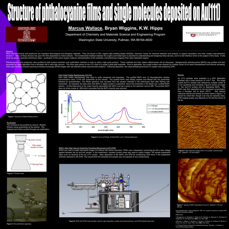
Marcus Wallace, Bryan Wiggins, K.W. Hipps Department of Chemistry and Materials Science and Engineering Program Washington State University, Pullman, WA 99164-4630 Abstract Metal phthalocyanines and porphyrins are important technological and biological materials. They are found in CDs, organic light emitting diodes (OLEDs), chemical “noses’ for chemical detection and analysis, in organic transistors, and other modern opto-electronic devices1,2. Many of these devices rely upon thin films deposited on metal contacts. This fundamental interface between the organic and metal layer is essential to understanding and designing these devices, and it is that interface which is the subject of this study. In this study we perform essential preliminary steps – purification of the active organic material, characterization of the substrate, and preliminary imaging of the vapor deposited organic. Phthalocyanines and porphyrins were purified by both solvent extraction and sublimation methods in order to yield a high purity product. These methods and their relative effectiveness will be discussed. Manganese(II) phthalocyanine [MnPc] was purified and then deposited by vapor deposition from a Knudsen cell in ultra high vacuum. Thin films were grown on the (111) face of a single crystal gold substrate. Prior to deposition, the crystal surface was cleaned by multiple cycles of ion beam bombardment and thermal annealing. Clean gold surfaces showed scanning tunneling microscopy (STM) images with well defined surface reconstruction patterns. Following deposition, the metal phthalocyanine/Au(111) system was studied by STM.. Ultra Violet Visible Spectroscopy (UV-Vis) Ultra violet visible spectroscopy was used to verify compound and impurities. The purified MnPc and 1,2 dicynobenzene solution concentrations were 1.0-3M with acetonitrile as the solvent. The purified MnPc was initially washed and filtered with hot acetonitrile followed by presublimation. The filtrate spectrum was recorded from the solution collected in the erlenmeyer flask after filtering with acetonitrile. As expected, the data show that MnPc is slighly souble in acetonitrile, hence the similarities in the filtrate and purified MnPc spectra. 1,2-Dicynobenzene is the major impurity associated with MnPc and has a peak on the spectrum around 288. The purified MnPc does not show a peak at ~288 which concludes that the MnPc impurity was removed. 2 absorbance 1.5 Figure 1: Structure of Metal Phthalocyanine Purification The compound was purified by vacuum filtration filtration using acetonitirile as the solvent. This material was further purified through pre-sublimation. Results Au (111) samples were prepared in a UHV deposition chamber with a base pressure of 1x10⁻¹⁰ Torr. The single crystal Au(111) sample was cleaned by multiple cycles of Ar ion sputtering and annealing. Figure 6 shows an image of the Au(111) surface prior to depositing MnPc. The MnPc was then deposited at sub-monolayer concentration from a Knudsen cell at about 375C. Well defined molecular islands are formed as shown below in figure 7. It should be noted that, though a tip may be optically sharp, STM imaging quality only depends on the last few atoms of the tip. Filtrate 1 1,2 Dicyanobenzene Purified MnPc 0.5 0 265 365 465 565 665 765 865 wavelength (nm) Figure 4: UV-vis of Filtrate, Purified MnPc, and 1,2-Dicyanobenzene RHK’s Ultra High Vacuum Scanning Tunneling Microscope (UHV-STM) STM’s are used to create real-space images of surfaces with atomic resolution. STM’s use a sharpened, conducting tip with a bias voltage applied between the tip and the sample. In this experiment, constant current mode was used to collect images.3 All sample preparation steps such as cleaning of the Au (111) crystal, vapor deposition of the MnPc, and STM tip conditioning were done in the preparation chamber attached to the STM. This insured that the substrate and sample was not exposed to any containments. Figure 6: High resolution image of Au (111) crystal: reconstruction line. Setpoint: 1.0V and 200pA Figure 2: Filtration setup Figure 7: Islands of MnPc deposited on Au(111). Setpoint: 1.0V and 200 pA Acknowledgements: I acknowledge the NSF for support in the form of grants CHE1058435 and DMR-1062898. References: 1.Macagnano, A.; Zampetti, E.; Pistillo, B. R.; Pantalei, S.; Sgreccia, E.; Paolesse, R.; d'Agostino, R. Thin Solid Films 2008, 516, 7857-7865. Figure 5: RHK UHV-STM: prep chamber used for vapor deposition, sputter and anneal (red box) and STM chamber (blue box) Figure 3: Pre-sublimation apparatus 2.Brunink, J.; Natale, C.; Bungaro, F.; Davide, F.; D’Amico, A.; Paoless, R.; Boshci, T.; Faccio, M.; Ferri, G. Anal. Chim. Acta 1996, 325, 53-64. 3. “Practical Guide to Scanning Tunneling Microscopy,” Benatar, J.; Howland, A., Thermal Microscopes (2000).


