SPECTRAL PROPERTIES OF LIGHT CONDUCTED IN STEMS OF
advertisement
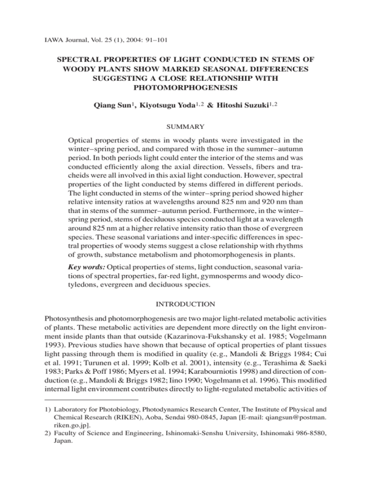
IAWA Journal, Vol. 25 (1), 2004: 91–101 SPECTRAL PROPERTIES OF LIGHT CONDUCTED IN STEMS OF WOODY PLANTS SHOW MARKED SEASONAL DIFFERENCES SUGGESTING A CLOSE RELATIONSHIP WITH PHOTOMORPHOGENESIS Qiang Sun1, Kiyotsugu Yoda1,2 & Hitoshi Suzuki1,2 SUMMARY Optical properties of stems in woody plants were investigated in the winter–spring period, and compared with those in the summer–autumn period. In both periods light could enter the interior of the stems and was conducted efficiently along the axial direction. Vessels, fibers and tracheids were all involved in this axial light conduction. However, spectral properties of the light conducted by stems differed in different periods. The light conducted in stems of the winter–spring period showed higher relative intensity ratios at wavelengths around 825 nm and 920 nm than that in stems of the summer–autumn period. Furthermore, in the winter– spring period, stems of deciduous species conducted light at a wavelength around 825 nm at a higher relative intensity ratio than those of evergreen species. These seasonal variations and inter-specific differences in spectral properties of woody stems suggest a close relationship with rhythms of growth, substance metabolism and photomorphogenesis in plants. Key words: Optical properties of stems, light conduction, seasonal variations of spectral properties, far-red light, gymnosperms and woody dicotyledons, evergreen and deciduous species. INTRODUCTION Photosynthesis and photomorphogenesis are two major light-related metabolic activities of plants. These metabolic activities are dependent more directly on the light environment inside plants than that outside (Kazarinova-Fukshansky et al. 1985; Vogelmann 1993). Previous studies have shown that because of optical properties of plant tissues light passing through them is modified in quality (e.g., Mandoli & Briggs 1984; Cui et al. 1991; Turunen et al. 1999; Kolb et al. 2001), intensity (e.g., Terashima & Saeki 1983; Parks & Poff 1986; Myers et al. 1994; Karabourniotis 1998) and direction of conduction (e.g., Mandoli & Briggs 1982; Iino 1990; Vogelmann et al. 1996). This modified internal light environment contributes directly to light-regulated metabolic activities of 1) Laboratory for Photobiology, Photodynamics Research Center, The Institute of Physical and Chemical Research (RIKEN), Aoba, Sendai 980-0845, Japan [E-mail: qiangsun@postman. riken.go.jp]. 2) Faculty of Science and Engineering, Ishinomaki-Senshu University, Ishinomaki 986-8580, Japan. 92 IAWA Journal, Vol. 25 (1), 2004 plants. Thus, understanding the optical properties of plant tissues and the characteristics of the light within plants is necessary for a full understanding of their light-regulated metabolic activities. However, prior to the present study, research has been based mostly on leaf and seedling tissues, and restricted to studies of optical properties generally at one specific growth stage. In an earlier report we characterized the optical properties of the stems and roots of woody plants from May to November (summer–autumn period), when plants are generally in an active state of growth (Sun et al. 2003). We demonstrated that light can enter the interior of the stems and roots and be efficiently conducted in the axial direction via certain elements of their vascular tissue (vessels, fibers and tracheids). Investigation of the spectral properties of stems and roots showed that far-red and near infra-red light, including a composition at a wavelength around 730 nm, are the mostly efficiently conducted. The attenuation patterns of light intensity in axial conduction were also revealed and indicated that the conducted light can leak to the surrounding living tissues, which thus is probably of photomorphogenetic significance. Plants undergo continuous developmental processes in which various changes involve not only the number, size and shape of cells but also the composition and amount of various metabolites. These changes in plant tissues should, on the one hand, influence their optical properties to lead to a change in internal light environment conditions. On the other hand the varied internal light environment may reversely regulate certain developmental processes of plant tissues themselves too. Thus, a grasp of the dynamic changes of the optical properties of plant tissues and the internal light environment is essential for a better understanding of the light-mediating metabolic activities of plants. However, little information is available at present. Having revealed the optical properties of the stems of woody plants in the summer–autumn period, our present study provides comparisons with stems in the winter–spring period, which is generally a period of dormancy or reduced activity and growth. The aim was to clarify any features in the winter–spring period in the light conduction properties of stem tissues and in the spectral properties of their internal light environment. MATERIALS AND METHODS Twenty-two species of woody plants (six gymnosperms and sixteen dicotyledons) including both evergreen and deciduous species were used in the present investigation (Table 1). Samples are obtained from the national forest near the Photodynamics Research Center, RIKEN (38° 14' 46" N, 140° 49' 34" E) or the Botanical Garden, Tohoku University (38° 15' 23" N, 140° 51' 00" E), Sendai, Japan. Sample collection was carried out from the same trees generally one to three times per month from mid-December 2001 to late May 2002 for all species. Special attention was paid to the period of leaf emergence of deciduous species, and additional sample collection for each tree was made on the evergreen species once per week, and on the deciduous species one to three times before leaf emergence and after leaf-size stability, respectively. Two or three stem lengths were obtained freshly from each sampled tree at every collection. Each sample was 20 cm long and 0.5–3 cm in diameter. A smooth transverse Sun, Yoda & Suzuki — Light conduction in woody stems 93 Table 1. List of species investigated. Evergreen gymnosperms: Abies firma Siebold et Zucc. Cryptomeria japonica (L.f.) D.Don Chamaecyparis obtusa (Siebold et Zucc.) Endl. Pinus densiflora Siebold et Zucc. Deciduous gymnosperms: Ginkgo biloba L. Metasequoia glyptostroboides Hu et W.C. Cheng Evergreen dicotyledons: Camellia japonica L. var. hortensis Photinia glabra (Thunb.) Maxim. × P. serratifolia (Desf.) Kalkman Quercus myrsinaefolia Blume Aucuba japonica Thunb. Deciduous dicotyledons: Rhododendron obtusum (Lindl.) Planch. Quercus crispula Blume Zelkova serrata (Thunb.) Makino Acer sieboldianum Miq. Salix triandra L. subsp. nipponica (Franch. et Sav.) A.K. Skvortsov Prunus yedoensis Matsum. Akebia trifoliata (Thunb.) Koidz. Wisteria floribunda (Willd.) DC. Pourthiaea villosa (Thunb.) Decne. Populus tremula L. Fagus japonica Maxim. Aesculus turbinata Blume surface was made at the lower cut end with a sharp razor blade. The stem length was inserted, with the lower cut end downwards, into a light-tight box through a hole at the top and sealed with black gum to maintain dark conditions inside the box (refer to Sun et al. 2003 for details of the experimental setup). Directly under the lower cut end a microscope with an attached far red-sensitive CCD camera (Wat-902H, Watec Corp., Japan) had previously been positioned. Outside the box light from a microscope halogen lamp, passed through a light guide, was used to illuminate the protruding part of the length unilaterally. The site of illumination was usually 2–4 cm from the lower cut end. The image of conducted light at the surface of the lower cut end was observed with the microscope, and the video signals were fed into an image processing system (Argus-20 with Aquacosmos version 1.10; Hamamatsu Photonics K.K., Japan) to clarify differences in the efficiency of light conduction in different cell types of the stem. Spectral properties of the conducted light were investigated in the range 190–950 nm by replacing the microscope with a photonic multi-channel analyzer (PMA-11, Hamamatsu). Transmission spectra of stems were used to analyze differences in relative ratio of transmission intensity at different wavelengths. The above trimming and measurements of the materials were performed rapidly to obviate any effects of desiccation. Routine tissue sections of the materials investigated were also made after measurements to identify the cell types involved in light conduction. 94 IAWA Journal, Vol. 25 (1), 2004 Sun, Yoda & Suzuki — Light conduction in woody stems 95 Table 2. Comparisons of spectral properties of transmitted light by stems of deciduous and evergreen plants in the winter-spring period. Species WTP1 WTP a (nm) WTP2 RLI b WTP3 RLI1 RLI 2 Deciduous species Ginkgo biloba Metasequoia glyptostroboides Rhododendron obtusum Quercus crispula Zelkova serrata Salix triandra subsp. nipponica Prunus yedoensis Pourthiaea villosa Acer sieboldianum Akebia trifoliata Wisteria floribunda Populus tremula 733.3 ± 1.5c 740.7 ± 2.3 740.0 ± 1.7 733.5 ± 2.1 737.0 ± 1.7 730.0 ± 1.4 745.3 ± 3.2 736.0 ± 1.0 734.3 ± 4.6 730.5 ± 0.7 730.0 ± 5.2 732.7 ± 1.5 825.7 ± 2.9 821.7 ± 6.0 823.3 ± 4.0 828.5 ± 0.7 827.3 ± 5.7 821.0 ± 5.7 821.3 ± 5.5 825.0 ± 0 829.3 ± 2.5 831.0 ± 5.7 826.7 ± 3.2 825.7 ± 3.1 919.7 ± 2.5 922.0 ± 0.0 922.0 ± 12.5 920.0 ± 11.3 922.0 ± 0.0 —d 915.0 ± 9.9 919.0 ± 2.7 920.3 ± 2.9 909.5 ± 3.5 — 917.3 ± 10.5 1.08 ± 0.05 0.88 ± 0.08 0.83 ± 0.05 0.67 ± 0.09 0.76 ± 0.03 0.53 ± 0.01 0.94 ± 0.08 0.72 ± 0.04 1.28 ± 0.01 1.20 ± 0.00 1.26 ± 0.09 0.67 ± 0.34 0.42 ± 0.01 0.32 ± 0.04 0.27 ± 0.07 0.26 ± 0.06 0.33 ± 0.06 732.6 ± 0.5 730.5 ± 1.7 736.3 ± 2.1 734.5 ± 3.1 733.3 ± 3.2 733.4 ± 3.5 733.0 ± 3.6 734.0 ± 1.4 826.6 ± 3.1 823.7 ± 2.6 830.0 ± 1.7 826.2 ± 2.3 824.3 ± 2.5 826.9 ± 2.6 818.3 ± 2.3 822.0 ± 1.4 923.2 ± 2.7 921.0 ± 3.6 924.0 ± 8.2 918.5 ± 5.8 920.7 ± 6.2 922.1 ± 5.5 916.7 ± 8.4 927.5 ± 9.2 0.66 ± 0.06 0.54 ± 0.04 0.59 ± 0.08 0.61 ± 0.06 0.54 ± 0.07 0.66 ± 0.10 0.58 ± 0.04 0.53 ± 0.03 0.29 ± 0.08 0.23 ± 0.05 0.22 ± 0.05 0.20 ± 0.04 0.20 ± 0.03 0.28 ± 0.06 0.19 ± 0.03 0.19 ± 0.04 0.30 ± 0.01 0.26 ± 0.01 0.61 ± 0.03 0.51 ± 0.05 0.32 ± 0.04 Evergreen species Abies firma Cryptomeria japonica Chamaecyparis obtusa Pinus densiflora Camellia japonica var. hortensis Photinia glabra × P. serratifolia Quercus myrsinaefolia Aucuba japonica a) WTP-wavelength of transmission peak, WTP 1-wavelength of the first transmission peak, WTP2-wavelength of the second transmission peak, WTP3-wavelength of the third transmission peak. b) RLI-ratio of light intensity among different transmission peaks, RLI -ratio of light intensity at a trans1 mission peak around 825 nm to that around 735 nm, RLI2-ratio of light intensity at a transmission peak around 920 nm to that around 735 nm. c) each datum represents mean ± SD of 5–12 samples. d) no obvious transmission peak. ← Fig. 1. Images of transmitted light from the transversely cut stem end of selected species in the winter–spring period. A & B: Zelkova serrata. C & D: Photinia glabra × P. serratifolia. – A: Light conduction in secondary xylem; brightness differences according to cell types show their differences in light conduction efficiency. – B: Efficient light conduction in vessels (ve) and fibers (xf) via their lumens and walls, respectively, and inefficient light conduction in rays (ra) and axial parenchyma (ap). – C: A one-year-old stem, showing that the phloem fiber ring (pf) is the most efficient in light conduction, followed by secondary xylem (sx), and that primary xylem bundles (px) in stem corners are the poorest light conductors. – D: Enlarged image of a vascular bundle (xy-xylem and ph-phloem) in C, showing much more efficient light conduction in the walls of phloem fibers (pf). – E: Light conduction in secondary xylem of a gymnosperm species, Metasequoia glyptostroboides. – F: Pinus densiflora, showing more efficient light conduction via the walls of tracheids in latewood (lw) than in earlywood (ew). — Scale bars in C & E = 1 mm, in A = 300 μm, in F = 115 μm and in B & D = 100 μm. 96 IAWA Journal, Vol. 25 (1), 2004 RESULTS In all species investigated, whether evergreen or deciduous, axial light conduction was observed in stems of the winter–spring period, and the tissues or cell types involved in light conduction were characteristic of their phylogenic groups (Fig. 1). All the dicotyledons showed light conduction in both xylem and phloem. In xylem, different cell and tissue types showed differences in the efficiency of light conduction (Fig 1A). Vessels and xylem fibers were the most efficient conductors of light axially along the stems. In vessels, light conduction was mainly via the large lumens but in xylem fibers it was via the thick walls; while vessel walls and fiber lumens were not efficient for axial light conduction (Fig. 1B). The brightness in vessel lumens generally showed no obvious differences whatever their diameter and arrangement, but it differed in the walls of xylem fibers according to thickness, with greater brightness in fibers of latewood than in those of earlywood. Other cell types in xylem (i.e. ray parenchyma cells and axial parenchyma cells) were not involved in efficient axial light conduction (Fig. 1B). Phloem fibers in woody dicotyledons were observed to conduct light axially only via their thick walls, but other cell types in phloem (sieve tube elements, companion cells and parenchyma cells) were not involved in efficient axial light conduction (Fig. 1C & D). In the six gymnosperms, axial light conduction occurred mainly in the xylem of stems (Fig. 1E ). Efficient light conduction was observed in tracheids via their thick walls (Fig. 1F ). The efficiency of the light conduction in tracheids was dependent on the period of their formation: tracheids of latewood were thicker-walled and more efficient in light conduction than those of earlywood (Fig. 1F ). Other cell types in xylem (such as ray cells) were not efficient axial light conductors, compared with tracheids. In species with sclerenchyma cells in the phloem, axial light conduction in phloem occurred via the thick walls of the sclereids. Other phloem elements (sieve cells, parenchyma cells etc.) did not conduct light efficiently in stems. Concerning spectral properties of the light conducted by stems in the winter–spring period, transmission spectra of stems with halogen light source as incident light were used for comparisons of relative transmission intensity at different wavelengths. In all the species investigated (whether gymnosperms or dicotyledons), a very small part of the incident light below the wavelength 700 nm was conducted along the axial direction of the stem (Fig. 2). However, between 700 nm and 940 nm, in which light was conducted efficiently by the stem tissues, there were usually three transmission peaks around 735 nm, 825 nm and 920 nm (Table 2 and Fig. 2 D–I). Therefore the transmission spectra of stems in this period differed from those in the summer–autumn period, which showed only a sole transmission peak around 735 nm (Fig. 2 B & C; Sun et al. 2003). Compared to the other two, the transmission peak around 920 nm was relatively small, usually less than 0.4 times the intensity relative to that around 735 nm (Table 2), or even unobvious in a few species. Evergreen and deciduous species showed differences in the ratio of light intensity at the transmission peak around 825 nm to that around 735 nm. The ratio ranged between 0.5 and 1.3 (with a mean of 0.9) in the deciduous species (Table 2), demonstrating an obvious peak around 825 nm (Fig. 2 D–F); and between 0.5 and 0.7 (with a mean of 733 828 823 928 921 Aucuba japonica Metasequoia glyptostroboides 733 738 823 816 934 922 Photinia glabra × P. serratifolia Acer sieboldianum Acer firma 738 729 733 823 832 921 922 Wavelength (nm) 192 255 318 381 444 507 570 633 696 759 822 885 948 192 255 318 381 444 507 570 633 696 759 822 885 948 192 255 318 381 444 507 570 633 696 759 822 885 948 Acer firma Ginkgo biloba 738 Acer sieboldianum 730 Fig. 2. Spectra of light transmitted through stems of representative deciduous and evergreen species in the winter–spring period (D–I). Figures indicated are the wavelengths of transmission peaks. – A: Incident light. – B: Spectral properties of a stem in a deciduous species after stability of the leaf size in early May. – C: Spectral properties of a stem in an evergreen species in middle May. – D–F: Deciduous species. G–I: Evergreen species. Light intensity (arbitrary units) Incident light 657 Sun, Yoda & Suzuki — Light conduction in woody stems 97 98 IAWA Journal, Vol. 25 (1), 2004 Table 3. Duration of the formation of transmission spectra characteristic in stems of the summer–autumn period. Date* Species Evergreen species Abies firma Cryptomeria japonica Chamaecyparis obtusa Pinus densiflora Camellia japonica L. var. hortensis Photinia glabra × P. serratifolia Quercus myrsinaefolia Aucuba japonica 10–15 May 16–26 April 25 April– 6 May 24–26 April 12–18 April 9–16 April 15–23 April 4–12 May Deciduous species Ginkgo biloba Metasequoia glyptostroboides Rhododendron obtusum Quercus crispula Zelkova serrata Acer sieboldianum Salix triandra subsp. nipponica Prunus yedoensis Akebia trifoliata Wisteria floribunda Pourthiaea villosa Populus tremula Fagus japonica Aesculus turbinata 7–13 May 29 April– 4 May 6–11 May 3–9 May 18–24 April 4–9 May 14–18 April 20–27 April 22– 30 April 5–9 May 22 April– 4 May 19–25 May 2–8 May 2–13 May * Dates apply to spectral measurements and analyses on 3–6 individual trees for each species. 0.6) in the evergreen species (Table 2), rendering a much less obvious peak or shoulder at this wavelength (Fig. 2 G–I). The transmission peaks around 825 nm and 920 nm in the stems of the evergreen species investigated disappeared in the period from mid-April to mid-May, depending on the species, and led to formation of the spectral properties characteristic in the stems of the summer–autumn period (Table 3 and Fig. 2 C). Disappearance of these two peaks occurred in the stems of the deciduous species investigated after their leaves emerged or became size-stable (Table 3 and Fig. 2B). DISCUSSION The present investigations of the optical properties of stems in the winter–spring period indicated that stems can conduct light efficiently along its axial direction, and that vessels, fibers (of both xylem and phloem) and tracheids are involved in this light conduction via the lumens (in vessels) or via the walls (of fibers and tracheids). Stems in Sun, Yoda & Suzuki — Light conduction in woody stems 99 this period are therefore similar to those in the summer–autumn period, with regard to the cell or tissue types involved in light conduction (Sun et al. 2003). However, although the spectral range conducted efficiently in stems of the winter–spring period is between the wavelength 700 nm and 940 nm and similar to that in stems of the summer– autumn period, the stems in the winter–spring period conduct light around 825 nm and 920 nm at a higher relative intensity ratio compared to those in the summer– autumn period. In addition, stems of deciduous species usually showed a much higher relative transmission ratio to the light around 825 nm than those of evergreen species in the winter–spring period. The cause of this seasonal variation in spectral properties of stems is still unknown, but it seems that the answer lies in the annual rhythm of plant growth and development. This annual rhythm influenced by periodical changes in the external environment is manifested in both growth and substance metabolism. In temperate zones, the most obvious differences of substance metabolism for woody plants occur between the summer–autumn and winter–spring periods. Under favorable environmental conditions in the summer–autumn period, anabolic activities in stem tissues predominate, increasing the amount and variety of ergastic substances in stems as well as their growth. In the winter–spring period, total metabolic activities in the stem tissues are much reduced and catabolism predominates, especially in early spring when leaf emergence begins. Catabolism may lead to a general reduction in the amount and varieties of ergastic substances within the stem tissues. The seasonal differences in the spectral properties of stems may well be considered relevant to the periodical variations in the amount and variety of ergastic substances in the stem. In the summer–autumn period, the substance accumulation in the stem probably increases the amount and/or kinds of the substances which absorb the two spectral regions around 825 nm and 920 nm to a relative greater extent, and thus lead to lower transmission ratio at the two spectral regions in the stem of this period. In the winter–spring period, catabolism in the stem reduces the amount and varieties of ergastic substances, by which the reduction or dissolution of the substances absorbing the spectral regions around 825 nm and 920 nm may also make these regions to be conducted much more efficiently besides that around 735 nm. However, identification of substance(s) influencing the absorption of the spectral regions around 825 nm and 920 nm has not been done and will be an important topic for further investigation. Regulation by light of developmental processes of plants occurs via the perception of specific light signals by their intrinsic photoreceptors (Briggs & Olney 2001). Research on plant photomorphogenesis has revealed five red/far-red receptors (phytochromes A–E: Quail 1998; Fankhauser 2001) and three UV-A/blue receptors (one phototropin and two cryptochromes: Cashmore et al. 1999). Recently UV-B receptors have also been reported (Briggs et al. 2001). These photoreceptors react to specific wavelengths, among which phytochromes respond to longer wavelengths, up to 780 nm in some cases (Shinomura et al. 1996; Shinomura et al. 2000). As clarified in our previous research, far-red light including that around 730 nm is efficiently conducted by vessels, tracheids and fibers in the stems and roots in the summer–autumn period, and also leaked to their surrounding living tissues in the axial conduction (Sun et al. 2003). Thus, these 100 IAWA Journal, Vol. 25 (1), 2004 living tissues are bathed in an internal light environment, enriched far-red so that phytochromes-mediated photomorphogenetic responses probably occur in stems and roots. Far-red light around 735 nm was also present in the transmission spectra of stems during the winter–spring period, and the same far-red perception by phytochromes as that in the summer–autumn period should also occur. The other two spectral regions around 825 nm and 920 nm, however, also conducted much more efficiently by the stem of the winter–spring period, lie outside the identified action spectral range of phytochromes, and it remains difficult to understand their photomorphogenetic significance. However, photomorphogenetic research has so far been restricted to very few species, and investigations are still lacking concerning any photomorphogenetic effects of the light beyond 800 nm. Also, as pointed out by Fankhauser (2001), the list of photoreceptors identified is expected to be still incomplete. Thus, further investigation on the possible physiological roles of the two spectral peaks beyond 800 nm will be necessary to reveal any photomorphogenetic significance in stems of the winter–spring period. ACKNOWLEDGEMENTS We are grateful to Dr. Fumio Takahashi, Dr. Kumi Sato-Nara and Prof. Mitsuo Suzuki for useful discussions; to Prof. Ian G. Gleadall for critical reading and his comments on the manuscript; and to Prof. Pieter Baas and two anonymous referees for valuable suggestions. REFERENCES Briggs, W.R., C.F. Beck, A.R. Cashmore, J.M. Christie, J. Hughes, et al. 2001. The phototropin family of photoreceptors. Plant Cell 13: 993–997. Briggs, W.R. & M.A. Olney. 2001. Photoreceptors in plant photomorphogenesis to date. Five phytochromes, two cryptochromes, one phototropin, and one superchrome. Plant Physiol. 125: 85–88. Cashmore, A.R., J.A. Jarillo, Y. J. Wu & D. Liu. 1999. Cryptochromes: blue light receptors for plants and animals. Science 284: 760–765. Cui, M., T.C. Vogelmann & W.K. Smith. 1991. Chlorophyll and light gradients in sun and shade leaves of Spinacia oleracea. Plant Cell Environ. 14: 493–500. Fankhauser, C. 2001. The phytochromes, a family of red / far-red absorbing photoreceptors. J. Biol. Chem. 276: 11453–11456. Iino, M. 1990. Phototropism: mechanisms and ecological implications. Plant Cell Environ. 13: 633–650. Karabourniotis, G. 1998. Light-guiding function of foliar sclereids in the evergreen sclerophyll Phillyrea latifolia: a quantitative approach. J. Exp. Bot. 49: 739–746. Kazarinova-Fukshansky, N., M. Seyfried & E. Schäfer. 1985. Distortion of action spectra in photomorphogenesis by light gradients within the plant tissue. Photochem. Photobiol. 41: 689–702. Kolb, C.A., M.A. Käser, J. Kopecky, G. Zotz, M. Riederer & E.E. Pfündel. 2001. Effects of natural intensities of visible and ultraviolet radiation on epidermal ultraviolet screening and photosynthesis in grape leaves. Plant Physiol. 127: 863–875. Mandoli, D.F. & W.R. Briggs. 1982. Optical properties of etiolated plant tissues. Proc. Natl. Acad. Sci. USA 79: 2902–2906. Mandoli, D.F. & W.R. Briggs. 1984. Fiber-optic plant tissues: spectral dependence in dark-grown and green tissues. Photochem. Photobiol. 39: 419–424. Sun, Yoda & Suzuki — Light conduction in woody stems 101 Myers, D.A., T.C. Vogelmann & J.F. Bornman. 1994. Epidermal focussing and effects on light utilization in Oxalis acetosella. Physiol. Plant. 91: 651–656. Parks, B.M. & K.L. Poff. 1986. Altering the axial light gradient affects photomorphogenesis in emerging seedlings of Zea mays L. Plant Physiol. 81: 75–80. Quail, P.H. 1998. The phytochrome family: dissection of functional roles and signalling pathways among family members. Philos. Trans. Roy. Soc. Lond. B 353: 1399–1403. Shinomura, T., A. Nagatani, H. Hanzawa, M. Kubota, M. Watanabe & M. Furuya. 1996. Action spectra for phytochrome A- and B-specific photoinduction of seed germination in Arabidopsis thaliana. Proc. Natl. Acad. Sci. USA 93: 8129–8133. Shinomura, T., K. Uchida & M. Furuya. 2000. Elementary processes of photoperception by phytochrome A for high-irradiance response of hypocotyl elongation in Arabidopsis. Plant Physiol. 122: 147–156. Sun, Q., K. Yoda, M. Suzuki & H. Suzuki. 2003. Vascular tissue in the stem and roots of woody plants can conduct light. J. Exp. Bot. 54: 1627–1635. Terashima, I. & T. Saeki. 1983. Light environment within a leaf. I. Optical properties of paradermal sections of Camellia leaves with special reference to differences in the optical properties of palisade and spongy tissues. Plant Cell Physiol. 24: 1493–1501. Turunen, M.T., T.C. Vogelmann & W.K. Smith. 1999. UV screening in lodgepole pine (Pinus contorta ssp. latifolia) cotyledons and needles. Intl. J. Plant Sci. 160: 315–320. Vogelmann, T.C. 1993. Plant tissue optics. Annu. Rev. Plant Physiol. Plant Mol. Biol. 44: 231– 251. Vogelmann, T.C., J.F. Bornman & D.J. Yates. 1996. Focusing of light by leaf epidermal cells. Physiol. Plant. 98: 43–56.

