Chapter 3 Physiology of Adrenal Steroids Regulation of Aldosterone Secretion
advertisement

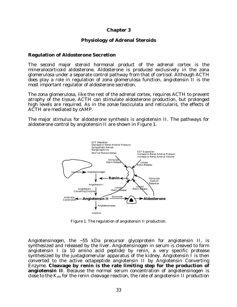
Chapter 3 Physiology of Adrenal Steroids Regulation of Aldosterone Secretion The second major steroid hormonal product of the adrenal cortex is the mineralocorticoid aldosterone. Aldosterone is produced exclusively in the zona glomerulosa under a separate control pathway from that of cortisol. Although ACTH does play a role in regulation of zona glomerulosa function, angiotensin II is the most important regulator of aldosterone secretion. The zona glomerulosa, like the rest of the adrenal cortex, requires ACTH to prevent atrophy of the tissue. ACTH can stimulate aldosterone production, but prolonged high levels are required. As in the zonæ fasciculata and reticularis, the effects of ACTH are mediated by cAMP. The major stimulus for aldosterone synthesis is angiotensin II. The pathways for aldosterone control by angiotensin II are shown in Figure 1. ECF Depletion Decrease in Renal Arterial Pressure Sympathetic Nerves Norepinephrine [Na+] at Macula Densa Stimulate Renin Release ECF Expansion Increase in Renal Arterial Pressure Increase in Renal Arterial Volume Inhibit Renin Release Angiotensinogen Renin Angiotensin I Inhibit Renin Release Angiotensin Converting Enzyme Arteriolar Constriction Angiotensin II Stimulate Stimulate Sodium Retention Inhibit Renin Release Aldosterone Angiotensinases Inactive Figure 1. The regulation of angiotensin II production. Angiotensinogen, the ~55 kDa precursor glycoprotein for angiotensin II, is synthesized and released by the liver. Angiotensinogen in serum is cleaved to form angiotensin I (a 10 amino acid peptide) by renin, a very specific protease synthesized by the juxtaglomerular apparatus of the kidney. Angiotensin I is then converted to the active octapeptide angiotensin II by Angiotensin Converting Enzyme. Cleavage by renin is the rate limiting step for the production of angiotensin II. Because the normal serum concentration of angiotensinogen is close to the Km for the renin cleavage reaction, the rate of angiotensin II production 33 Endocrine -- Dr. Brandt Chapter 3. The Adrenal Cortex -- Part II depends on both the concentration of renin and the concentration of angiotensinogen. The release of angiotensinogen from the liver is constitutive, but can be stimulated by cortisol, by estrogens, and by angiotensin II. Renin release from the kidney is stimulated by ECF depletion, by low serum sodium concentrations, and by decreases in blood pressure; renin release is inhibited by angiotensin II, aldosterone, and by elevated blood pressure. Note that angiotensin II also acts as a vasoconstrictor; thus both angiotensin II and aldosterone are directly involved in raising blood pressure. The zona glomerulosa responds to increases in serum potassium levels by increasing aldosterone synthesis. While decreases in serum sodium levels also stimulate aldosterone synthesis, the effect of sodium is almost certainly mediated by increased angiotensin II secondary to increased renin release from the kidney. In contrast, the regulation by increased potassium appears to be due to a direct effect of high serum potassium levels on the adrenal; recent evidence suggests that may be mediated by intra-adrenal production of angiotensin II. Angiotensin II action is probably mediated by the phosphatidyl inositol pathway. As with ACTH, the main action of angiotensin II is to increase P450scc activity; prolonged stimulation increases the levels of all of the glomerulosa enzymes, but particularly that of P450-18. A peptide hormone released by the heart, atrial natriuretic peptide (ANP), is the only known inhibitor of aldosterone production. ANP is released in response to elevated atrial pressure. The actions of appear ANP to be mediated by both inhibition of renin release by the kidney, and by direct effect on the adrenal to inhibit aldosterone synthesis. ANP is a vasodilator, and has additional effects related to reduction of blood pressure. At least three different natriuretic peptides exist in humans, produced not just in the heart but also in several other tissues. These peptides probably have endocrine, paracrine, and autocrine roles in regulating blood pressure, sodium levels, and fluid volume. ANP = atrial natriuretic peptide BNP = brain natriuretic peptide CNP = CNS natriuretic peptide Note that the localizations are not entirely consistent with the names of the peptides; the names (which could also be considered to correspond to the A, B, and C types of NP) are related to the tissues in which the peptides were discovered. The NPs are thought to bind at least one and probably more cell-surface receptors. These receptors appear to be transmembrane proteins, with the intracellular domains exhibiting guanylyl cyclase activity. However, it is still not established that elevated cGMP mediates all of the NP activities; some studies suggest that decreased cAMP levels either alone or in conjunction with elevated cGMP are required for full elaboration of NP activity. Receptors for the NPs are present in the adrenal, in kidney, and in a large variety of other tissues. During the majority of evolution, sodium was often difficult for land organisms to 34 Endocrine -- Dr. Brandt Chapter 3. The Adrenal Cortex -- Part II obtain. Ability to greatly suppress aldosterone secretion was therefore of relatively little benefit, and was in fact potentially deleterious. In the United States (and other developed countries), this is clearly no longer true; the usual health problem related to sodium levels is not insufficient retention, but rather excessive intake and therefore insufficient excretion. A genetically programmed inadequate suppression of aldosterone synthesis which was once survival-enhancing may be one reason for the relatively high incidence of hypertension in developed countries. Adrenal Androgens The adult adrenal cortex also produces DHEA and androstenedione in significant quantities. These steroids have very low affinity for the androgen receptor, but can be converted to testosterone in peripheral tissues, and are therefore called adrenal androgens. The production of adrenal androgens occurs primarily in the zonæ fasciculata and reticularis, and as you might predict, because P450scc is the rate limiting step for steroid biosynthesis, and because ACTH stimulates P450scc in the zonæ fasciculata and reticularis, ACTH is the primary control hormone for the adrenal androgens. However, there appear to be additional, poorly characterized mechanisms for control of adrenal androgen production, since the levels of cortisol and DHEA often vary divergently. The role of adrenal androgens is unclear. Adrenalectomized patients require glucocorticoid and mineralocorticoid replacement therapy for survival, but do not appear to suffer ill effects from loss of adrenal androgens. DHEA and DHEAS have little intrinsic androgenic activity, although they can be converted to physiological androgens (and estrogens) in peripheral tissues. No receptor has been found for either of these steroids. Although the PPAR (peroxisome proliferator-activated receptors, a sub-family of the steroid receptor superfamily) have been linked to DHEA action, neither DHEA nor DHEAS is thought to bind these proteins directly. DHEA and DHEAS levels decrease with aging, resulting in suggestions that DHEA replacement might reverse some of the symptoms of aging. A few studies have been done to address this question, but the results have not yet led to any definitive conclusions. In rodent studies, DHEA supplementation was associated with decreased obesity, diabetes, cancer and heart disease; however, it is not clear whether these results are applicable to humans. In humans, some studies suggest that low level DHEA replacement therapy may reduce heart disease risk, reduce some deleterious alterations in immune system function, and improve perception of well-being, supporting the suggestion that DHEA (or DHEAS) may have a direct physiological role distinct from its ability to serve as an androgen precursor. Rumors about the possible beneficial effects of DHEA have reached the popular literature. At present, nothing is known about the appropriate level of DHEA replacement that is most useful, or even if DHEA replacement has any useful effect at all. Little is known about long term deleterious effects, although liver toxicity has been observed in some cases, elevated DHEA has been associated with increased risk of prostate disease, and high levels of DHEA cause virilization in women. DHEA is currently available over the counter in the United States; the fact that DHEA has no definite physiological function in non-pregnant individuals has not stopped people from trying to make money selling it or writing books about it. 35 Endocrine -- Dr. Brandt Chapter 3. The Adrenal Cortex -- Part II Ascorbic Acid The adrenal cortex stores large amounts of Vitamin C (ascorbic acid), which is concentrated in the adrenal cortex by an active transport mechanism to 100-1000 times the serum concentration. The adrenal contains about 1% of the total body stores of the vitamin. The reason for these stores in the adrenal cortex is not known; it is perhaps relevant that the one of the adrenal medulla enzymes (dopamine βhydroxylase) involved in catecholamine synthesis requires Vitamin C. Vitamin C is released from the cortex in response to ACTH. Transport of Adrenal Steroids All steroid hormones are synthesized and then immediately secreted. The steroids are thought to travel into and out of the tissues by passive diffusion. Therefore synthesis and release of steroid hormones are synonymous terms; the control of steroid hormone release occurs at the level of P450scc, the rate limiting step for synthesis. Table I. Normal Physiological Levels of the Adrenal Steroids Steroid Amount Secreted (mg/day) Classification Average Total Serum Concentration (nM) (µg/dL) Cortisol Major Glucocorticoid 20 400 14 Corticosterone Minor Glucocorticoid 3 11 0.4 Aldosterone Major Mineralocorticoid 0.15 0.2 0.007 DOC Weak Mineralocorticoid 0.20 0.2 0.006 DHEA Adrenal Androgen 16 27 0.8 DHEAS Adrenal Androgen 10 7000 250 Table I lists approximate physiological levels of steroids produced and released from the adrenal in normal individuals. Several comments arise from these numbers. First, recall that cortisol (and aldosterone) synthesis varies significantly during the day; the “average concentration” therefore may be quite different from the concentration that would be observed at a given time of day. Note that almost equal amounts of DOC and aldosterone are produced and are present at any given time; however, DOC is normally essentially irrelevant, because it has only ~3% of the affinity of aldosterone for the mineralocorticoid receptor. The total amount of DHEA 36 Endocrine -- Dr. Brandt Chapter 3. The Adrenal Cortex -- Part II and DHEAS produced each day is more than that of cortisol, and the circulating amount of DHEAS is far higher than that of cortisol; in spite of this, the function of DHEA(S) remains unclear. Finally, and most importantly, note that while aldosterone is produced in amounts of about 1% of those of cortisol, the average concentration of aldosterone is about 0.05% of that of cortisol. This factor of 20 discrepancy is due to differences in the rate of clearance of the two steroids, as a result of differences in their methods of transport in serum. About 40% of aldosterone in serum circulates free in solution. In contrast, only 5% of cortisol is free in solution, while the rest circulates bound to serum proteins. Cortisol has a specific binding protein called cortisol binding globulin (CBG), which binds about 80% of the cortisol in serum; the remaining 15% of bound cortisol is associated with serum albumin, a ubiquitous serum protein that binds many hydrophobic molecules. Aldosterone has a low affinity for CBG and therefore much less is bound in circulation. The free steroid is the only form that can enter cells, and therefore is the only form that affects cellular processes. In addition, only the free steroid can act as a substrate for the metabolizing enzymes. The differences in transport mechanisms between cortisol and aldosterone are reflected in the halflives for the two steroids: cortisol has a half-life of one to two hours, while aldosterone has a half-life of only about 10 to 20 minutes. Another result of the fact that the majority of cortisol is bound to CBG is that the free cortisol concentration will not change rapidly; CBG acts as a buffer. This is logical, because cortisol is a fairly long term regulator of energy metabolism; in contrast, aldosterone is used to more rapidly modulate sodium and potassium levels. The levels of CBG are regulated by several hormones, including glucocorticoids. After a prolonged period with low cortisol levels, the amount of CBG will be low as well, and as a result, the individual will be considerably more sensitive to a relatively small increase in cortisol. In contrast, prolonged high cortisol leads to high CBG levels, and therefore, a decreased sensitivity to an increase in cortisol secretion. It is, however, possible to saturate CBG at high physiological levels of cortisol, after which free cortisol concentration increases essentially linearly with rising total cortisol concentration. Some synthetic steroids have low affinity for CBG. Consequently, a larger proportion of a given dose of these drugs are available for receptor binding. This is an important consideration in pharmacological treatment with glucocorticoid analogs. Metabolism of the Adrenal Steroids Most metabolism of cortisol and aldosterone occurs in the liver. The following discussion only illustrates the general types of reactions. There are a large number of other reactions as well. Cortisol is used as an example. The metabolic reactions all convert the hormonal steroid to an inactive form. Many of the reactions also make the steroids more soluble in aqueous media. Figure 2 shows two pathways of metabolism of cortisol. One, catalyzed by 11β-HSD, converts cortisol to the inactive cortisone (see Chapter 1). This reaction is reversible by some 37 Endocrine -- Dr. Brandt Chapter 3. The Adrenal Cortex -- Part II forms of 11β-HSD or by another enzyme called 11-oxidoreductase (or 11-ketosteroid reductase). Cortisone can be used as a drug because target cells can convert it to the active form, cortisol. An alternative pathway involves various modifications of other functions in the steroid: reduction of the 4-double bond, reduction of the 3-ketone to a 3-hydroxyl (by one of several 3-hydroxysteroid dehydrogenase isozymes, which are different from the 3β-HSD/∆5-∆4-isomerase mentioned in the discussion of steroid hormone synthesis). The final reaction in that pathway is the conjugation to glucuronic acid. The product of each of these steps is inactive. The tetrahydrocortisol and its glucuronide are major excreted metabolites of cortisol. Cortisone is also metabolized in this pathway. OH OH O HO O OH HO 5-reductase O Cortisol 11 -HSD OH O O OH dihydrocortisol 3-HSD HO HO tetrahydrocortisol 11-oxidoreductase transferase OH OH O O O OH O HO OH glucuronic–O acid Cortisone OH tetrahydrocortisol glucuronide Figure 2. Examples of cortisol metabolism. There is one especially important aspect of the metabolites in Figure 2 of which you should be aware: the steroid ring is not degraded. As a result, the urine and feces can be analyzed for the steroid metabolites, which can be very useful for diagnostic purposes. Physiological Actions of the Adrenal Steroid Hormones Glucocorticoids and mineralocorticoids are absolutely required to sustain life. Cortisol is necessary to survive stress and to mobilize energy stores. Aldosterone is necessary to maintain sodium and potassium balance and blood pressure. As discussed in Chapter 1, most of the effects of these hormones are mediated by alterations in the rate of gene transcription. As a result, the effects are relatively slow: full elaboration of the effect requires from 15 minutes to several hours. In addition, the effects also tend to be fairly long lasting, usually several hours to several days. There are two general classes of effects: direct and indirect. 38 Endocrine -- Dr. Brandt Chapter 3. The Adrenal Cortex -- Part II For direct effects, gene products produced in response to the steroid, or gene products produced in response to the steroid-regulated proteins elicit the observed effect. The chain of events may involve several steps, but the steroid hormone is both necessary and sufficient as a causative agent. Direct effects can be divided into two types: physiological and pharmacological. Physiological effects are mediated by “normal” concentrations of the hormone; pharmacological effects require very high concentrations of hormone, concentrations that are rarely if ever achieved without exogenous administration of the hormone or of synthetic agonists. For indirect (or permissive) effects the steroid is required to allow the action of another hormone. The steroid alone has no effect unless the other hormone is present. For example, cortisol induces the synthesis of a receptor for another hormone. Without the other hormone nothing happens (except production of the receptor). Without cortisol, the other hormone has little effect due to low levels of the receptor. Together the two hormones elicit a response from the cell. Effects of Cortisol on Intermediary Metabolism As the name “glucocorticoid” implies, one major function of cortisol is to raise serum glucose concentration. It does this through a number of direct and indirect effects in the liver, in muscle, and in adipose tissue. In liver, cortisol directly stimulates gluconeogenesis, glycogenesis, and amino acid uptake, thereby increasing both glucose serum levels and glucose storage. Cortisol is also required for glucagon-stimulated gluconeogenesis during fasting; this is an example of an indirect effect. In muscle, cortisol decreases glucose and amino acid uptake, and protein synthesis, and increases protein catabolism. The net result of these effects is to increase free amino acids for use by the liver, and to decrease muscle glucose utilization. In adipose tissue, cortisol directly decreases glucose uptake. An additional, indirect effect is the potentiation of the epinephrine effect on the hormone-sensitive lipase; the result (especially in the presence of epinephrine) is an increase in triacylglycerol hydrolysis to free fatty acids and glycerol. Note, however, that different fat tissues are affected differently, a fact which is manifested during cortisol hypersecretion disorders as a redistribution of fatty tissue. What is the reason for these effects of cortisol? Cortisol is a major hormone for ensuring that the heart and brain have sufficient substrate to survive. It thus has catabolic effects in various tissues (connective tissue, lymphoid tissue, adipose tissue, and muscle). Cortisol is anabolic in the liver, but only in the sense that it increases the capacity of the liver for glucose production and storage. The result is a breakdown of peripheral tissues to support the more important organs. Cortisol Effects on Liver Glucose Metabolism The role of cortisol in the liver is two-fold: 1) to increase serum glucose levels for 39 Endocrine -- Dr. Brandt Chapter 3. The Adrenal Cortex -- Part II immediate use, and 2) to increase the amount of stored glycogen. This is because cortisol is a long term effector and is concerned with more than the immediate effects. Cortisol induces an increase in the rate of transcription of several enzymes in the liver gluconeogenic and glycogenic pathways. The enzymes involved are underlined in Figure 3. Note that all of these enzymes are unidirectional: they do not catalyze the reverse reaction. These enzymes are therefore only involved in the synthesis pathways, and have no role in glycogen breakdown or glycolysis. For example, one of the enzymes, fructose-1,6-bis-phosphatase, catalyzes the reverse reaction to that of phosphofructokinase, which is the major control step for glycolysis. Some of these enzymes are also affected by other hormones; e.g., PEP carboxykinase is stimulated by glucagon, and inhibited by insulin. Hormonal regulation of glucose metabolism will be discussed further in Chapters 4, 5, and 6. glucokinase hexokinase Glucose Glycogen Synthase PGM Glucose-6-P Glucose-1-P PGI Glu-6-Pase Glycogen Phosphorylase Fructose-6-P FBPase PFK Fructose-1,6-P2 aldolase Triose PEP carboxykinase Phosphoenolpyruvate Oxaloacetate pyruvate kinase Pyruvate TCA intermediates Figure 3. Cortisol effects on glucose metabolism. The underlined enzymes are stimulated by cortisol. Additional Effects of Cortisol Although the effects of cortisol on glucose homeostasis and energy mobilization are the most well characterized, glucocorticoids also have a number of other functions. Glucocorticoids reduce the number and activity of lymphocytes, and decrease capillary permeability; this results in an increased susceptibility to infection, and in a marked decrease in inflammatory responses. The mechanisms for these responses are poorly understood, although glucocorticoids have been shown to stimulate apoptosis (a programmed cell death pathway) in lymphocytes. These effects require high levels of glucocorticoids, which might be reached during chronic stress. However, these effects are very useful pharmacologically; glucocorticoids are widely 40 Endocrine -- Dr. Brandt Chapter 3. The Adrenal Cortex -- Part II used clinically for their immunosuppressant and anti-inflammatory properties. Recent evidence suggests that at least some of the anti-inflammatory and immunosuppressant actions of glucocorticoids are mediated by inhibition of the action of two transcription factors: AP-1 and NF-κB. This inhibition appears to occur by both direct interaction between activated glucocorticoid receptor and AP-1 or NF-κB, and by induction of an inhibitory protein called IκBα. Assuming that these mechanisms are correct, it may be possible to design drugs that inhibit NF-κB and/or AP-1 activity directly and which lack other, more deleterious, glucocorticoid side effects. High levels of cortisol are required by the adrenal medulla for the induction of phenylethanolamine-N-methyl transferase, an enzyme in the epinephrine synthesis pathway (see Chapter 4). Cortisol also decreases activity of catechol-O-methyl transferase, one of the enzymes responsible for inactivating epinephrine. Cortisol is also involved in a number of general permissive effects: it is required for excretion of a water overload, and for general kidney function. It is also required for maintenance of blood pressure; smooth muscle response to catecholamines requires the presence of glucocorticoids, and glucocorticoids have both direct and indirect actions necessary for maintaining cardiac function. Cortisol is required by the fetal lung for surfactant formation. In premature neonates, treatment with glucocorticoids is often used to induce proper lung development. Finally, and perhaps most importantly, an appropriate level of cortisol is required for survival of stress, although the mechanism by which this occurs is not understood. Adrenalectomized rats kept in a carefully controlled environment (including proper diet to maintain salt balance) will survive for some time. However, if subjected to essentially any type of stress (e.g., cold, forced swimming, or sham surgery), they die very easily, while control animals survive unharmed. On the other hand, chronically high levels (or long term pharmacological administration) of glucocorticoids have severely detrimental effects. Elevated glucocorticoid levels cause net loss of calcium from bones, and long term elevation of glucocorticoid levels is a severe risk factor for development of osteoporosis. At high levels, glucocorticoids also result in neuronal death, especially in the hippocampus (with consequent deleterious effects on memory), in infertility, in redistribution of fat and altered fat metabolism, in insulin resistance and diabetes, in cataracts, and in impaired survival of trauma and other severe stresses. Recently, there was a report (Nature 373:427-432, 1995) of a CRH-deletion mutation in mice. Mice heterozygous for the CRH knockout mutation were apparently normal. First generation homozygotes had apparently normal pituitary corticotroph and adrenal zona glomerulosa morphology, but the zona fasciculata was hypoplastic. Homozygotes produced some corticosterone (the primary rodent glucocorticoid), both under basal conditions and under stress, with females producing considerably more than males. Second generation homozygote knockout mice died about 12 hours postnatally due to incomplete lung development unless treated with glucocorticoids during gestation. (The difference in survival between first and second generation homozygotes suggests that maternal hormones are capable of substituting for fetal hormones during development; offspring of heterozygotes are 41 Endocrine -- Dr. Brandt Chapter 3. The Adrenal Cortex -- Part II exposed to maternal glucocorticoids (and possibly other hormones) that offspring of homozygotes are not.) Quoting from the Nature paper: “The apparent well being of the CRH-deficient mice, especially the males, contradicts the dogma of glucocorticoids being essential for adult survival.” However, a few caveats remain: 1) it is possible that the residual levels of glucocorticoids are sufficient to fulfill the glucocorticoid requirement (the dogma is based on experiments with adrenalectomized animals which completely lack glucocorticoids), 2) even adrenalectomized animals survive under controlled conditions, dying only when stressed; the stresses used in this study were fairly minimal, and again the residual glucocorticoid production may have been sufficient to confer survival ability, and 3) the homozygotes became hypoglycemic after 24 hours of fasting, which would probably be fatal fairly shortly thereafter. On the other hand, as mentioned in the paper, it is possible that the deleterious effects of adenalectomy may at least in part be a result of the high levels of CRH and ACTH which are not present in the knockout mutant mice. Full interpretation of the role of CRH and of glucocorticoids in adult animals will require further study. Mineralocorticoid Action Aldosterone is the other major essential product of the adrenal. Its physiology is much simpler: aldosterone increases sodium reabsorption and potassium excretion in the kidney. The primary action of aldosterone is to increase the activity of the basal membrane sodium/potassium ATPase pump and the apical membrane sodium channel in the kidney collecting ducts. The mechanisms by which this is accomplished are incompletely understood, since some effects occur within 1-2 minutes of increased aldosterone levels (too fast to be mediated by altered gene transcription), although aldosterone clearly exerts many of its actions at the genome level. The effects of glucocorticoids are mediated by both Type I (high affinity) and Type II (~10-fold lower affinity) receptors, but some Type I receptor containing cells also express 11β-HSD, and therefore are essentially unresponsive to glucocorticoids. Type II receptors have a much (~10-20-fold) stronger effect on gene transcription than Type I. Mineralocorticoid action is probably also mediated by both Type I and II receptors, but in cells lacking 11β-HSD, aldosterone levels are almost invariably too low to have a significant effect due to the much higher levels of cortisol. In some tissues, such as sweat glands, there is evidence (especially in adults) that cortisol mediates the sodium retention response. It appears, in fact, that the separate actions of mineralocorticoids and glucocorticoids are largely confined to a few selected tissues, and that, with those (important) exceptions, the primary adrenal hormone for physiological purposes is cortisol. Clinical Manifestations of the Adrenal Cortex Symptoms of Adrenal Insufficiency Adrenal insufficiency can result from either Addison’s disease and similar conditions (i.e. a general lack of adrenal function, regardless of cause), or from a congenital defect in the ability to synthesize one or more of the adrenal steroid 42 Endocrine -- Dr. Brandt Chapter 3. The Adrenal Cortex -- Part II hormones. In neonates, adrenal insufficiency results in failure to thrive (i.e. little weight gain), vomiting, diarrhea, dehydration, hyponatremia, hyperkalemia, hypotension, acidosis, hypoglycemia, and in some cases, hyperpigmentation. These symptoms are primarily (except for the last two) due directly or indirectly to low aldosterone levels. Note that the primary method for differentiating Addison’s disease from Congenital Adrenal Hyperplasia (CAH) is in the level of adrenal androgen production; adrenal androgens are also low in the case of Addison’s disease, while these steroids are elevated in nearly all forms of CAH (the exceptions are deficiencies in P450-17α, in P450scc, and in cholesterol transport). Achieving proper dosage during replacement therapy with glucocorticoids is somewhat difficult, since there is a wide “normal” range, and symptoms of mild cortisol excess or deficiency are not always readily apparent. In some cases it is necessary to revert to older tests for hypocortisol status (such as inability to secrete a water overload, hypoglycemia following mild insulin challenge, and muscle weakness). Congenital Adrenal Hyperplasia Congenital Adrenal Hyperplasia (CAH) results from an inherited partial or total deficiency in one of 5 enzymes (P450scc, P450-21, P450-11β, P450-17α, or 3β-HSD). These cause a decreased ability to synthesize cortisol, and lead (as a result of continual stimulation by ACTH) to hyperplasia of the adrenal and (usually) to elevated levels of other adrenal steroid products. (One additional (rare) cause of CAH, usually termed Congenital Adrenal Lipoid Hyperplasia, is due to a defect in the mitochondrial cholesterol transport pathway.) The most common form of CAH is due to defects in the gene for P450-21 (>90% of cases). P450-21 has a functional gene and a 98% identical pseudogene, and both are located in the HLA locus on chromosome 6. Splicing errors, gene deletions, and gene conversions have all been observed. Inactive P450-21 results in salt-wasting as well as cortisol deficiency; partial loss of activity (about 100-times more common) usually yields low to normal cortisol but elevated aldosterone levels (as little as 1% of normal levels of P450-21 is enough to produce sufficient aldosterone); both forms present with elevated androgen levels. Due to the fact that many of the possible mutations result in reduced rather than abolished 21-hydroxylase activity, and to the fact that there is significant variation between individuals in the ability to 21-hydroxylate adrenal steroids in peripheral tissues, the severity of 21-hydroxylase deficiency is quite variable. Patients may present with mineralocorticoid levels ranging from low to high, and cortisol levels ranging from zero to within the normal range. The most common form of the disorder is “non-classical” 21-hydroxylase deficiency, in which symptoms may not be apparent until early puberty or even adulthood. In general, these patients have sufficient 21-hydroxylase activity to produce both cortisol and aldosterone early in life; however, alterations in steroid synthesis patterns during early puberty often result in insufficient capacity to produce cortisol, and therefore in excessive androgen production. Individuals with non-classical 21-hydroxylase deficiency may have transient (life-threatening) salt-wasting episodes. Non-classical deficiency in 21-hydroxylase is one of the most common genetic disorders, with an incidence of 43 Endocrine -- Dr. Brandt Chapter 3. The Adrenal Cortex -- Part II ~1% in the general population. P450-11β deficiency (~4% of total cases of CAH) generally presents with hypertension (either due to elevated DOC levels or to elevated levels of other steroids with mineralocorticoid activity). Since a separate enzyme (P450-18) catalyzes the 11β-hydroxylation of DOC, most cases of P450-11β deficiency only require glucocorticoid supplementation. However, the zona glomerulosa may not recover immediately from suppression, and therefore patients must be monitored to prevent salt-wasting until the zona glomerulosa function is restored. In complete 3β-HSD deficiency, only some pathway intermediates and (large amounts of) DHEA are produced by the adrenal. For poorly understood reasons, only some CAH-causing mutations result in reduced aldosterone production. Most of these patients are able to make at least some sex steroids due to the activity of the Type I gene in peripheral tissues. While in males 3β-HBD deficiency often results in ambiguous genitalia at birth due to marked decrease in testicular androgen synthesis, in females the peripheral production of androgens may be sufficient to cause androgenital syndrome. P450-17α deficiency (a rare form of CAH) causes hypertension for reasons that are less well understood. In some cases, aldosterone synthesis is suppressed, and the hypertension is probably a result of zona fasciculata synthesis of DOC. In other cases, high levels of aldosterone are observed. In both types, renin and angiotensin II levels are suppressed by the high levels of mineralocorticoids. All forms of CAH are life-threatening disorders, and must be recognized and treated. Untreated complete 21-hydroxylase or 3β-HSD deficiencies are rapidly fatal within two weeks after birth due to lack of mineralocorticoids. While 21-hydroxylase deficiency in females is generally diagnosed (due to the associated androgenital syndrome), in males it may be missed or misdiagnosed (since the elevated androgens have little additional phenotypic effect in male infants and the hypoaldosteronism does not result in symptoms until several days postpartum), often with lethal consequences. P450-21 and P450-11β (and many cases of 3β-HSD) deficiencies usually lead to androgenital syndrome. Androgenital syndrome is a consequence of the extremely high levels of adrenal androgens produced in response to overstimulation of the adrenal by ACTH; in males it leads to early puberty, and in females to ambiguous genitalia at birth, and to virilization and hirsuitism later in life. There have been some (somewhat controversial) attempts to begin treatment in utero (beginning in first trimester) to prevent the formation of ambiguous genitalia in female infants, especially in cases of P450-21 deficiency. Addison’s Disease Adrenocortical hypofunction is a life-threatening condition caused by atrophy or dysgenesis of the adrenal. The most common form is the result of an autoimmune attack on the adrenal, but Addison’s disease can also arise from damage due to tuberculosis (a common cause until about 50 years ago, and currently rising in incidence) or other microorganisms, from a congenital abnormality in one of the 44 Endocrine -- Dr. Brandt Chapter 3. The Adrenal Cortex -- Part II transcription factors required for adrenal cortex development or the ACTH receptor, or from decreased ACTH secretion (e.g., as a result of loss of pituitary function). Like severe CAH, the congenital abnormality forms are usually rapidly fatal unless treated. On the other hand, the autoimmune, tubercular, and secondary forms of Addison’s disease tend to have slow onset, and may not be diagnosed until the individual is observed to be unable to properly respond to severe stresses such as disease or trauma. In most individuals >90% of the adrenal must be destroyed before symptoms are evident. The symptoms of Addison’s disease are decreased (or absent) aldosterone effect (and therefore hyponatremia, leading to loss of blood pressure due to decreased extra cellular fluid levels, and hyperkalemia, and as a result, cardiac arrhythmia), and decreased cortisol effects (inability to mobilize energy stores, rapid fatigue, and inability to survive stress). Addison’s disease differs from the common forms of CAH in that all adrenal cortex products are decreased. Glucocorticoid resistance Resistance to glucocorticoids is characterized by high cortisol and adrenal androgen production, with high ACTH levels, but absence of symptoms of Cushing’s syndrome. In severe cases, aldosterone levels are elevated as well (with corresponding hypertension and hypokalemia). The circadian pattern of cortisol release is maintained, and cortisol release is suppressible by dexamethasone, although abnormally high levels are required. In women the high androgen levels result in hirsuitism and menstrual irregularities. The disorder is caused by either point mutations in or decreased quantities of the glucocorticoid receptor (GR Type II). (Note: complete absence of the GR Type II has not been observed and may be incompatible with fetal survival.) Hypoaldosteronism Deficiencies in aldosterone synthesis or response without adrenal hyperplasia can result from several defects: 1) Pseudohypoaldosteronism (PHA) is due to a receptor defect, and can also be called mineralocorticoid resistance. In its recessive form, PHA is due to a complete lack of response to aldosterone in all cell types (Type I receptors are either absent or inactive due to mutations). In the dominant form, PHA is due to a lack of response in the kidney. In both cases developmental changes in kidney function decrease the severity of the disorder during entry into adulthood (the efficiency of the kidney increases, and other mechanisms compensate for inability to respond to mineralocorticoids). 2) Corticosterone methyl oxidase (= aldosterone synthetase = 18-methyl oxidase = 18-hydroxylase = P450-18) deficiency results in lowered or absent aldosterone synthesis. There are two forms, one of which is due to complete absence of activity (i.e. <1% of normal; mutants with activities >1% of normal are capable of producing normal amounts of aldosterone), while the other appears to result from a modified enzyme that lacks the dehydrogenase activity (resulting in elevated 18-OHcorticosterone levels). This disorder usually presents with failure to thrive and in 45 Endocrine -- Dr. Brandt Chapter 3. The Adrenal Cortex -- Part II some cases salt-wasting and dehydration. Deficiencies in P450-18 do not result in elevated cortisol or androgen secretion due to the relatively limited ability of the zona glomerulosa to produce steroids and to the ability of other steroids to compensate for the lack of aldosterone. The existence of other mineralocorticoids, in combination with some protective effects of cortisol, explains the lower fatality rate of untreated P450-18 deficiency relative to 21-hydroxylase deficiency. The initial symptoms of P450-18 deficiency are treated with oral sodium supplementation or intravenous fluids; thereafter, treatment involves synthetic mineralocorticoids. As with PHA, the severity of the disorder usually decreases during entry into adulthood, and mineralocorticoid therapy may not be necessary for adults. 3) Acquired hypoaldosteronism is a disorder that results from loss of zona glomerulosa response to ACTH and angiotensin II (hyperrenic hypoaldosteronism), usually associated with septic responses damaging the adrenal, or from kidney disorders (especially in older diabetics) that result in decreased renin secretion (hyporenic hypoaldosteronism). Cushing’s Syndrome Cushing’s syndrome results from chronically elevated glucocorticoid levels. It can be caused by an autonomous adrenal cortical tumor, by a CRH-secreting tumor, or, most commonly, by an ACTH-secreting tumor (either pituitary or ectopic). Disorders with symptoms similar to Cushing’s syndrome may be caused by long-term pharmacological administration of glucocorticoids or by alcohol abuse. The result of elevated glucocorticoid levels is an increase in protein catabolism (and therefore weakened muscles and skin), decreased growth rate in children, poor wound healing, increased susceptibility to infection, osteoporotic levels of bone loss, and elevated plasma glucose. The elevated plasma glucose can lead to diabetes due to cortisol inhibition of insulin release and antagonism of insulin action. Chronically high levels of cortisol cause the extremities to lose fat and muscle, while fat deposition is observed in the face, abdomen, and in a “buffalo hump” between the shoulder blades. Cushing’s syndrome is often associated with hypertension; this may be due to the prolonged high levels of cortisol overloading or actually inhibiting the 11β-HSD system and inducing hypertension similar to that observed in Syndrome of Apparent Mineralocorticoid Excess. Primary aldosteronism Elevated levels of aldosterone can result from a number of abnormalities. Primary aldosteronism is due to either an aldosterone secreting tumor or to bilateral adrenal hyperplasia, neither of which are usually suppressed by dexamethasone treatment. The symptoms are hypertension in spite of low levels of renin and angiotensin II (and therefore hypertension not treatable by captopril, an inhibitor of angiotensin converting enzyme), hypernatremia, and hypokalemia. Glucocorticoid-remediable hyperaldosteronism Dexamethasone-suppressible aldosteronism is a rare autosomal dominant disorder that results in production of the 18-hydroxylase in the zona fasciculata due to 46 Endocrine -- Dr. Brandt Chapter 3. The Adrenal Cortex -- Part II acquisition of the promoter from P450-11β. This is thought to be a result of unequal meiotic crossing-over between the highly related (~95% nucleic acid identity) P45018 and P450-11β genes which are located in close proximity on chromosome 8. The result is 3 copies of the gene on one chromosome. This disorder does not usually result in sexual developmental abnormalities, and therefore can be differentiated from partial P450-11β and P450-17α deficiencies. It is also characterized by >20-fold elevation in the levels of two steroids (18-hydroxycortisol and 18-oxocortisol) which are measurable in urine and provide a marker for the disorder. Aldosterone and the cortisol metabolites are both produced in the zona fasciculata, while the zona glomerulosa is suppressed; thus aldosterone levels are high in spite of very low renin and angiotensin II levels. The symptoms are hypertension, even in infants, although usually not hypokalemia. Treatment has involved use of glucocorticoid agonists, such as dexamethasone or the shorter-acting prednisone, or, especially in children, cortisol (due to the growth inhibitory effects of excess glucocorticoids), and/or the mineralocorticoid antagonist spironolactone; both have side effects (spironolactone is also antiandrogenic). Syndrome of Apparent Mineralocorticoid Excess The Syndrome of Apparent Mineralocorticoid Excess (AME) is a rare disorder characterized by normal cortisol levels, suppressed aldosterone levels, hypertension, and hypokalemia (see also Chapter 1). AME appears to result from impaired 11β-hydroxysteroid dehydrogenase (11β-HSD) function, but usually other enzymes (5β-reductase and 3α,20β-reductase) are observed to be inhibited also. The disorder may (at least in some cases) therefore be a result of elevated levels of endogenous inhibitors of these enzymes (some bile acid metabolites may fit this description). AME may be diagnosed by examining levels of urinary cortisol metabolites, since levels of tetrahydrocortisone are markedly reduced compared to tetrahydrocortisol and 5α-reduced tetrahydrocortisol. Periodically in the above discussion I have mentioned a drug called dexamethasone. This is 9αfluoro-16α-methyl-1,4-pregnadiene-11β,17α,21-triol-3,20-dione (below, left). The 9α-fluoro decreases the rate of metabolism (especially 11β-HSD action), and the ∆1 and 16α-methyl modifications decrease the GR Type I receptor affinity of the drug; as a result, dexamethasone is a long acting glucocorticoid with little mineralocorticoid activity, and is one of the major (although by no means the only) glucocorticoid drugs used. It is also a standard compound used in research concerning glucocorticoid action. The structure on the right: 11β-[4-(N,N-dimethylamino)-phenyl]-17α-(1-propynyl)-4,9-estradiene17β-ol-3-one was called RU 38486 and mifepristone by the company that makes it. It is in the news as the abortifacient drug RU 486 due to its action as an antiprogestin; however, in the literature on cortisol hypersecretion disorders, it is referred to as a glucocorticoid antagonist. RU-486 has a higher affinity for the progesterone receptor than for the glucocorticoid receptor, but it is an antagonist for both receptors. 47 Endocrine -- Dr. Brandt Chapter 3. The Adrenal Cortex -- Part II OH O HO N OH OH CH3 F O O Dexamethasone Mifepristone References Yanase, et al. (1991) “17α-hydroxylase/17-20-lyase deficiency: from clinical investigation to molecular definition.” Endocr. Rev. 12: 91-108. Deftos (1991) “Chromogranin A: Its role in endocrine function and as an endocrine and neuroendocrine tumor marker.” Endocr. Rev. 12: 181-187. Lamberts et al. (1992) “Long-term treatment with RU486 and glucocorticoid receptor resistance.” Trends Endocrinol. Metab. 3: 199-204. Miller & Crapo (1993) “The medical treatment of Cushing’s syndrome.” Endocr. Rev. 14: 443-458. Wick et al. (1993) “Immunoendocrine communication via hypothalamo-pituitaryadrenal axis in autoimmune diseases.” Endocr. Rev. 14: 539-563. Drewett & Garbers (1994) “The family of guanylyl cyclase receptors and their ligands.” Endocr. Rev. 15: 135-162. White et al. (1994) “Disorders of steroid 11β-hydroxylase isozymes.” Endocr. Rev. 15: 421-438. Miller (1994) “Clinical review: genetics, diagnosis, and management of 21hydroxylase deficiency.” J. Clin. Endocrinol. Metab. 78: 241-246. Marx (1995) “How the glucocorticoids suppress immunity.” Science 270: 232-233. 48


