R658
Dispatch
Cell division: Plant-like properties of animal cell cytokinesis
Bruce Bowerman and Aaron F. Severson
Recent evidence that a syntaxin is required for
cytokinesis in Caenorhabditis elegans embryos suggests
that the mechanism of cell division in plant and animal
cells may be more similar than previously imagined.
Address: Institute of Molecular Biology, 1370 Franklin Boulevard,
University of Oregon, Eugene, Oregon 97403, USA.
E-mail: bbowerman@molbio.uoregon.edu
Current Biology 1999, 9:R658–R660
http://biomednet.com/elecref/09609822009R0658
0960-9822/99/$ – see front matter © 1999 Elsevier Science Ltd.
All rights reserved.
Past studies of cytokinesis in plants and animals have
suggested that two key processes, contractile ring function and membrane addition at the cell surface, contribute to the partitioning of daughter cells at the end of
mitosis [1]. Genetic and molecular studies of cytokinesis
in vertebrate, insect, yeast and slime mold systems have
provided ample in vivo evidence that the actin cytoskeleton and a contractile ring are critical for cell division [2].
Genetic studies in plants have demonstrated a requirement for membrane addition, though not cortical contraction [3]. Now, a study of cytokinesis in the nematode
Caenorhabditis elegans, published recently in Current
Biology [4], has shown that a syntaxin — a type of protein
known in other contexts to play a part in specific membrane fusion — is necessary for cell division. This new
work provides genetic evidence that membrane addition
may be required during cytokinesis in an animal cell, and
shows that, in spite of their apparently very different
cytokinetic strategies, plants and animals may use conserved genetic programs to incorporate new membrane
during cytokinesis.
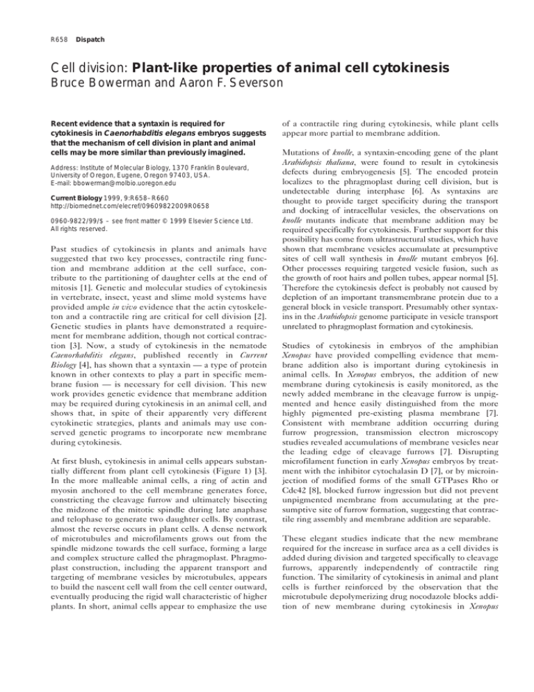
At first blush, cytokinesis in animal cells appears substantially different from plant cell cytokinesis (Figure 1) [3].
In the more malleable animal cells, a ring of actin and
myosin anchored to the cell membrane generates force,
constricting the cleavage furrow and ultimately bisecting
the midzone of the mitotic spindle during late anaphase
and telophase to generate two daughter cells. By contrast,
almost the reverse occurs in plant cells. A dense network
of microtubules and microfilaments grows out from the
spindle midzone towards the cell surface, forming a large
and complex structure called the phragmoplast. Phragmoplast construction, including the apparent transport and
targeting of membrane vesicles by microtubules, appears
to build the nascent cell wall from the cell center outward,
eventually producing the rigid wall characteristic of higher
plants. In short, animal cells appear to emphasize the use
of a contractile ring during cytokinesis, while plant cells
appear more partial to membrane addition.
Mutations of knolle, a syntaxin-encoding gene of the plant
Arabidopsis thaliana, were found to result in cytokinesis
defects during embryogenesis [5]. The encoded protein
localizes to the phragmoplast during cell division, but is
undetectable during interphase [6]. As syntaxins are
thought to provide target specificity during the transport
and docking of intracellular vesicles, the observations on
knolle mutants indicate that membrane addition may be
required specifically for cytokinesis. Further support for this
possibility has come from ultrastructural studies, which have
shown that membrane vesicles accumulate at presumptive
sites of cell wall synthesis in knolle mutant embryos [6].
Other processes requiring targeted vesicle fusion, such as
the growth of root hairs and pollen tubes, appear normal [5].
Therefore the cytokinesis defect is probably not caused by
depletion of an important transmembrane protein due to a
general block in vesicle transport. Presumably other syntaxins in the Arabidopsis genome participate in vesicle transport
unrelated to phragmoplast formation and cytokinesis.
Studies of cytokinesis in embryos of the amphibian
Xenopus have provided compelling evidence that membrane addition also is important during cytokinesis in
animal cells. In Xenopus embryos, the addition of new
membrane during cytokinesis is easily monitored, as the
newly added membrane in the cleavage furrow is unpigmented and hence easily distinguished from the more
highly pigmented pre-existing plasma membrane [7].
Consistent with membrane addition occurring during
furrow progression, transmission electron microscopy
studies revealed accumulations of membrane vesicles near
the leading edge of cleavage furrows [7]. Disrupting
microfilament function in early Xenopus embryos by treatment with the inhibitor cytochalasin D [7], or by microinjection of modified forms of the small GTPases Rho or
Cdc42 [8], blocked furrow ingression but did not prevent
unpigmented membrane from accumulating at the presumptive site of furrow formation, suggesting that contractile ring assembly and membrane addition are separable.
These elegant studies indicate that the new membrane
required for the increase in surface area as a cell divides is
added during division and targeted specifically to cleavage
furrows, apparently independently of contractile ring
function. The similarity of cytokinesis in animal and plant
cells is further reinforced by the observation that the
microtubule depolymerizing drug nocodazole blocks addition of new membrane during cytokinesis in Xenopus
Dispatch
R659
Figure 1
Higher plant cell cytokinesis
Landmark–preprophase band
Phragmoplast
Cell plate
Animal cell cytokinesis
Cleavage plane
specification
Contractile ring
assembly
Cleavage furrow
contraction
Cell separation
Current Biology
Cytokinesis in higher plants (top) requires vesicle fusion to generate the cell plate, while animal cell cytokinesis (bottom) requires contraction of an
acto-myosin ring.
embryos [9]. Thus, as for the plant phragmoplast, the targeting of membrane vesicles by microtubule-mediated
motor proteins may mediate the addition of new membrane during cytokinesis in animal cells.
Studies of cytokinesis in the early C. elegans embryo now
provide loss-of-function genetic evidence that membrane
addition is required for cytokinesis in an animal cell. This
advance comes from Jantsch-Plunger and Glotzer [4], who
have taken advantage of the nearly complete genome
sequence of C. elegans that is now available, and the powerful technique of RNA-mediated interference (RNAi), to
survey all known C. elegans syntaxin genes for requirements
during embryonic cytokinesis. They inactivated eight different syntaxin genes by RNAi and found one, called syn-4,
that appears to be required both for cytokinesis and for the
reassembly of nuclei after the completion of cell division.
C. elegans embryos lacking syn-4 function were found to
exhibit partially penetrant defects in cytokinesis: approximately two-thirds of the embryos analyzed failed to
complete the first two rounds of embryonic cytokinesis.
Some embryos failed to form ingressing furrows, whereas
in others the furrows ingressed substantially but ultimately regressed. Many of the syn-4-deficient embryos
also exhibited defects in the extrusion of polar bodies
during meiosis, apparently as a result of defects in meiotic
cytokinesis. Finally, mitotic spindles appeared to segregate chromosomes normally in the absence of syn-4 function, but supernumerary nuclei sometimes formed as
chromosomes decondensed and attempted re-assembly
into daughter nuclei.
Syn-4 protein, as detected by immunofluorescent labeling,
localizes prominently to cleavage furrows and to small
structures dispersed throughout the cytoplasm that
concentrate around re-forming nuclei. Syn-4 may therefore
be required for membrane addition at the cleavage furrow
during cytokinesis, and for subsequent membrane fusion
events that mediate nuclear re-assembly. Further supporting a role for membrane addition, the inactivation by RNAi
of rab-11 — which encodes a C. elegans member of the Rab
family of small GTPases that have also been implicated in
specific membrane fusion events — results in a cytokinesis
defect similar to that caused by inactivation of syn-4
(A. Skop and J. White, personal communication).
These results with C. elegans support the view that
membrane addition is required during cytokinesis in
animal cells, as in plant cells, but important issues remain
to be addressed. One is the question of whether new
membrane material is targeted specifically to the cleavage
furrow during mitosis in C. elegans embryos. Further
insight might come from membrane-labeling and
ultrastructural studies of vesicle fusion or accumulation at
the cleavage furrow during cytokinesis in wild-type and
syn-4-deficient embryos. Indeed, it remains possible that
Syn-4 and Rab-11 are required to transport transmembrane protein(s) essential for cytokinesis to the cell
surface, in which case the phenotypes caused by loss of
function of these proteins might not be due to defective
membrane addition. Such an indirect explanation is supported by the observation that syn-4 mutants fail to
secrete a normal chitinous eggshell following fertilization
[4], raising the possibility that Syn-4 might be required
R660
Current Biology, Vol 9 No 17
Figure 2
Nomarski micrographs of the first two cell
cycles in C. elegans embryos. (a) Pronuclear
migration stage embryo. The oocyte
pronucleus (left) migrates to meet the sperm
pronucleus (right). A transient furrow, the
pseudocleavage furrow, constricts the cell at
this stage. (b) The pronuclei meet in the
posterior of the embryo. The pseudocleavage
furrow has relaxed. (c) The first mitotic spindle
lies along the longitudinal axis of the embryo,
and is posteriorly displaced. (d) The first
cleavage furrow bisects the mitotic spindle,
separating the reforming nuclei. Note the
asymmetric shapes of the spindle poles.
(e) The two cell stage. (f) The nuclear
envelope has begun to break down in the
(a)
(b)
(c)
(d)
(e)
(f)
(g)
(h)
Current Biology
anterior blastomere (left), which divides before
the posterior daughter. (g,h) The second
more generally for the fusion of all secretory vesicles at
the cell surface.
The identification and analysis of temperature-sensitive
alleles of syn-4 may be necessary to dissect these different
requirements. Meanwhile, examining the localization of
transmembrane proteins expressed in the early embryo,
such as the Delta and Notch homologs Glp-1 and Apx-1,
might indicate whether syn-4 is required more generally
for vesicle transport. It will also be interesting to learn
whether microtubules are involved in targeting new
membrane addition in C. elegans embryos, and whether the
midzone microtubules that become constricted by the
contractile ring at the end of cytokinesis are in some ways
similar to the phragmoplast of dividing plant cells.
embryonic cytokinesis produces the four-cell
stage embryo.
insights. Using RNAi, one can go from gene to phenotype
in days, while reverse genetic screens for deletions allow
unambiguous determination of null-mutant phenotypes.
Moreover, classical genetic screens for non-conditional and
for temperature-sensitive mutations have begun to identify loci required for cell division processes, including
cytokinesis. Equally appealing, the relatively large size of
the C. elegans embryo makes it amenable to high-resolution
cytological observation and experimental manipulation
(Figure 2). While RNAi is largely responsible for the
increasing use of the early C. elegans embryo in cell biology,
a combination of powerful technologies should further
inform our understanding of the genetic programs that regulate and execute cytokinesis in a nematode embryo.
References
Intriguingly, a protein that from its sequence is likely to be a
member of the kinesin family of motor proteins is localized
to the spindle midzone in early C. elegans embryos [10,11].
This protein is called Zen-4 or CeMKLP1, and inactivation
of the zen-4 gene was found to result in a late defect in
cytokinesis, with cleavage furrows regressing only after substantial ingression. While the cytokinesis defect in zen-4
mutant embryos might be caused indirectly by spindle
defects, it also is possible that Zen-4/MKLP-1 is required to
target membrane vesicles to the cleavage furrow late in
cytokinesis, promoting the final separation of daughter cells
at the spindle midzone or stem body. Perhaps membrane
addition also occurs earlier, as syn-4 and rab-11 mutant
embryos exhibit a range of both early and late cytokinesis
defects, with variable degrees of furrow ingression.
While important questions remain, the genetic requirement for a syntaxin during cytokinesis in both C. elegans
and in Arabidopsis supports the view that plant and animal
cell cytokinesis may involve similar processes. The
emergence of the early C. elegans embryo as a powerful
genetic system for studying cell biology promises further
1. Glotzer M: The mechanism and control of cytokinesis. Curr Opin
Cell Biol 1997, 9:815-823.
2. Field C, Li R, Oegema K: Cytokinesis in eukaryotes: a mechanistic
comparison. Curr Opin Cell Biol 1999, 11:68-80.
3. Staehelin LA, Hepler PK: Cytokinesis in higher plants. Cell 1996,
84:821-824.
4. Jantsch-Plunger V, Glotzer M: Depletion of syntaxins in the early
C. elegans embryo reveals a role for membrane fusion events in
cytokinesis. Curr Biol 1999, 9:738-745.
5. Lukowitz W, Mayer U, Jurgens G: Cytokinesis in the Arabidopsis
embryo involves the syntaxin-related KNOLLE gene product. Cell
1996, 84:61-71.
6. Lauber MH, Waizenegger I, Steinmann T, Schwarz H, Mayer U, Hwang
I, Lukowitz W, Jurgens G: The Arabidopsis KNOLLE protein is a
cytokinesis-specific syntaxin. J Cell Biol 1997, 139:1485-1493.
7. Bluemink JG, Laat SD: New membrane formation during cytokinesis
in normal and cytochalasin B-treated eggs of Xenopus laevis. I.
Electron microscope observations. J Cell Biol 1973, 59:89-108.
8. Drechsel DN, Hyman AA, Hall A, Glotzer M: A requirement for Rho
and Cdc42 during cytokinesis in Xenopus embryos. Curr Biol
1997, 7:12-23.
9. Danilchik MV, Funk WC, Brown EE, Larkin K: Requirement for
microtubules in new membrane formation during cytokinesis of
Xenopus embryos. Dev Biol 1998, 194:47-60.
10. Raich WB, Moran AN, Rothman JH, Hardin J: Cytokinesis and midzone
microtubule organization in Caenorhabditis elegans require the
kinesin-like protein ZEN-4. Mol Biol Cell 1998, 9:2037-2049.
11. Powers J, Bossinger O, Rose D, Strome S, Saxton W: A nematode
kinesin required for cleavage furrow advancement. Curr Biol
1998, 8:1133-1136.

