Temperature dependent responses of live mammalian cells
advertisement

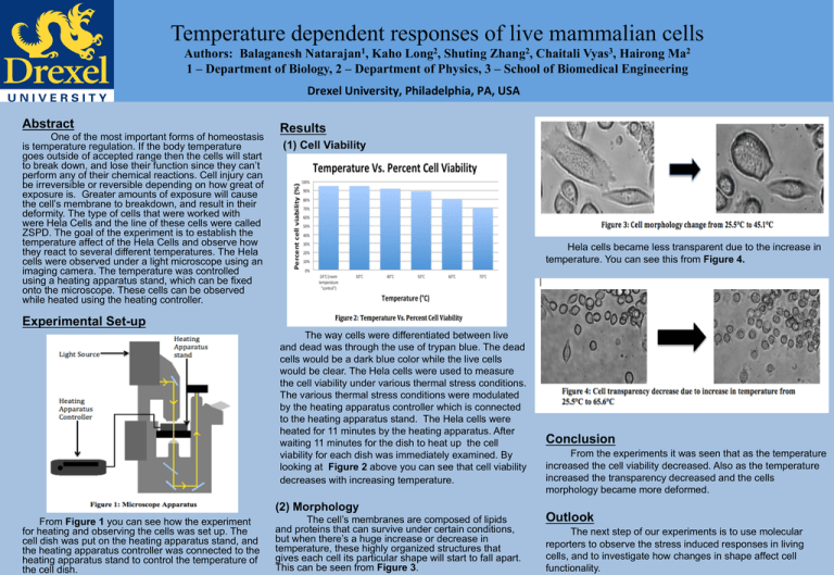
Temperature dependent responses of live mammalian cells Authors: Balaganesh Natarajan1, Kaho Long2, Shuting Zhang2, Chaitali Vyas3, Hairong Ma2 1 – Department of Biology, 2 – Department of Physics, 3 – School of Biomedical Engineering Drexel University, Philadelphia, PA, USA Abstract One of the most important forms of homeostasis is temperature regulation. If the body temperature goes outside of accepted range then the cells will start to break down, and lose their function since they can’t perform any of their chemical reactions. Cell injury can be irreversible or reversible depending on how great of exposure is. Greater amounts of exposure will cause the cell’s membrane to breakdown, and result in their deformity. The type of cells that were worked with were Hela Cells and the line of these cells were called ZSPD. The goal of the experiment is to establish the temperature affect of the Hela Cells and observe how they react to several different temperatures. The Hela cells were observed under a light microscope using an imaging camera. The temperature was controlled using a heating apparatus stand, which can be fixed onto the microscope. These cells can be observed while heated using the heating controller. Results (1) Cell Viability ! Hela cells became less transparent due to the increase in temperature. You can see this from Figure 4. ! Experimental Set-up The way cells were differentiated between live and dead was through the use of trypan blue. The dead cells would be a dark blue color while the live cells would be clear. The Hela cells were used to measure the cell viability under various thermal stress conditions. The various thermal stress conditions were modulated by the heating apparatus controller which is connected to the heating apparatus stand. The Hela cells were heated for 11 minutes by the heating apparatus. After waiting 11 minutes for the dish to heat up the cell viability for each dish was immediately examined. By looking at Figure 2 above you can see that cell viability decreases with increasing temperature. (2) Morphology From Figure 1 you can see how the experiment for heating and observing the cells was set up. The cell dish was put on the heating apparatus stand, and the heating apparatus controller was connected to the heating apparatus stand to control the temperature of the cell dish. The cell’s membranes are composed of lipids and proteins that can survive under certain conditions, but when there’s a huge increase or decrease in temperature, these highly organized structures that gives each cell its particular shape will start to fall apart. This can be seen from Figure 3. Conclusion From the experiments it was seen that as the temperature increased the cell viability decreased. Also as the temperature increased the transparency decreased and the cells morphology became more deformed. Outlook The next step of our experiments is to use molecular reporters to observe the stress induced responses in living cells, and to investigate how changes in shape affect cell functionality.


