9/18/2015
advertisement
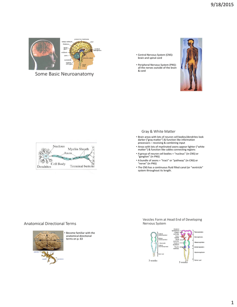
9/18/2015 • Central Nervous System (CNS): brain and spinal cord Some Basic Neuroanatomy • Peripheral Nervous System (PNS): all the nerves outside of the brain & cord Gray & White Matter • Brain areas with lots of neuron cell bodies/dendrites look darker (“gray matter”) & function like information processors – receiving & combining input • Areas with lots of myelinated axons appear lighter (“white matter”) & function like cables connecting regions • A group of neuron cell bodies = “nucleus” (in CNS) or “ganglion” (in PNS) • A bundle of axons = “tract” or “pathway” (in CNS) or “nerve” (in PNS) • The CNS has a continuous fluid filled canal (or “ventricle” system throughout its length. Anatomical Directional Terms Vesicles Form at Head End of Developing Nervous System • Become familiar with the anatomical directional terms on p. 63 3 weeks 5 weeks 1 9/18/2015 Telencephalon – outer part of forebrain Cerebral hemispheres Afferent Diencephalon – inner part of forebrain Thalamus & Hypothalamus 5 Chunks of of Brain Mesencephalon - midbrain Efferent Metencephalon – upper part of hindbrain Pons & Cerebellum Myelencephalon – lower part of hindbrain Medulla oblongata Book 4.6 Divisions of the Nervous System Structures Controlled by the ANS The Brainstem Reticular Activating System Thalamus Hypothalamus • The cells of the “reticular formation” have many other functions as well. Midbrain Cerebellum Pons Medulla 2 9/18/2015 2 and 3 on the “dorsal” surface of midbrain are the primitive visual (2) and auditory (3) processing centers, the superior (2) and inferior (3) colliculus. 3 2 1 Substantia Nigra Dopamine Neurons Red Nucleus is another motor region in the midbrain http://mindsci-clinic.com/rostral_midbrain.htm Middle layer added emotion & memory capabilities. Book 3.22 Substantia Nigra Dopamine Neurons Newest outer layer added judgment, reasoning, planning and self-control. Red Nucleus is another motor region in the midbrain http://mindsci-clinic.com/rostral_midbrain.htm Book 3.22 3 9/18/2015 The Reptilian Brain Periaqueductal Gray (PAG) The start of a descending pain suppression system • The brainstem, especially its core, is the most primitive portion of our brain, relatively unchanged from the time that dinosaurs roamed the earth. Most reptile behavior is reflexive response to stimuli. Mike, the Headless Chicken • Survived 18 months • Could still stand, sit on a perch, walk clumsily, and attempt to crow and preen. • These basic behaviors are like reflexes – built into the brainstem. A Sadder Example • Anencephaly – forebrain fails to develop. Baby has a flattened, open skull. Baby shows basic reflexive behaviors (can nurse, grasp, etc.) but with only hindbrain & midbrain structures intact, survival is brief (hours-days). http://en.wikipedia.org/wiki/Mike_the_Headless_Chicken Hypothalamus • Plays a role in lots of different basic behaviors/motivations necessary for survival of individual & survival of the species • The “four F’s” • • • • Means “beneath the thalamus” The Hypothalamus Feeding Fighting (aggression & rage) Fleeing (fear behaviors) Mating : ) • But also primitive parenting behaviors, temperature regulation, hormone regulation, biorhythms & sleep, mood/emotions 4 9/18/2015 The Brainstem Areas Again The Thalamus works closely with regions of cortex • Partially processing incoming sensations before passing input on to cortex • Part of the motor system • Works with higher cortical regions related to cognition, memory, personality etc. The Brain is Like a Tootsie Pop The Basal Ganglia Motor System 5 9/18/2015 In Front of Hypothalamus is the “Basal Forebrain” Another component is the nucleus basalis which sends arousing ACh messages to all of cortex for memory/cognition. This nucleus dies off in Alzheimer’s disease. One component of the basal forebrain is the nucleus accumbens, a hub of our pleasure/reward pathway Side View of Cortex & Cerebellum Corpus Callosum 6 9/18/2015 Motor cortex Central Sulcus Fig27 Association Somatosensory cortex cortex Association cortex FRONTAL LOBE PARIETAL LOBE Broca's area Lateral fissure TEMPORAL LOBE OCCIPITAL LOBE Visual cortex Auditory cortex Wernicke's area 2 More Regions in Neocortex Let’s add some common anatomical terminology Neocortex Has 6 Layers With Regional Variations in Thickness 7 9/18/2015 A “Processing Unit” Within the Cortex is a Column of Cells The Meninges completely enclose the CNS and help protect it. “-itis” = inflammation Meningitis= inflammation of meninges 8 9/18/2015 Filled with cerebrospinal fluid (CSF) which is replaced every few hours Enlarged Ventricles Due to Hydrocephalus A Shunt Tube Drains Away Excess CSF https://www.youtube.com/watch?v=WU HdkP278q0&list=PLJG4HdSoAx23j8Ev hgzuJ3sEtCiMsqNJg&index=6 Go to 20 Glia or Glial Cells (“supporting cells” of the nervous system) • 10X more numerous than neurons but one-tenth the size • make up about half of brain weight • several distinct types • assist neurons in multiple ways 9 9/18/2015 Form Myelin Sheath • Oligodendrocyte forms CNS myelin • Schwann cell forms PNS myelin • Multiple sclerosis – patchy loss of myelin sheaths that can interfere with any CNS function (depending on which neurons lose their insulation) or not at all http://www.youtube.com/watch?v=qgySDmRRzxY&feature=PlayList&p=ED18B251293C8C20&playnex t=1&index=2 Loss of White Matter in Multiple Sclerosis • Astrocytes exchange materials with neurons • Microglia remove debris and multiply to form scar tissue Blood-Brain Barrier Radial Glial Cells 10

