Photon Energy 8.1Photons Photon energies - Interactions of light with matter.
advertisement
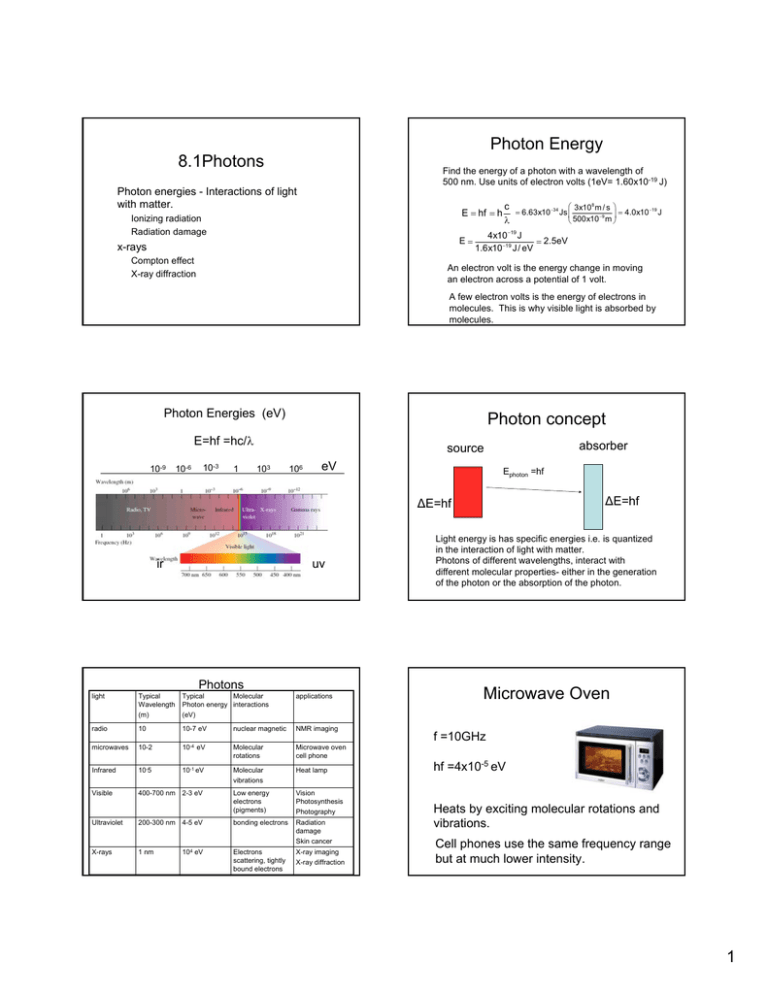
Photon Energy 8.1Photons Find the energy of a photon with a wavelength of 500 nm. Use units of electron volts (1eV= 1.60x10⁻¹⁹ J) Photon energies - Interactions of light with matter. E = hf = h Ionizing radiation Radiation damage E= x-rays Compton effect X-ray diffraction ⎛ 3x10⁸ m / s ⎞ c −19 = 6.63x10 −34 Js ⎜ ⎟ = 4.0x10 J −9 ⎝ 500x10 m ⎠ λ 4x10 −19 J = 2.5eV 1.6x10 −19 J / eV An electron volt is the energy change in moving an electron across a potential of 1 volt. A few electron volts is the energy of electrons in molecules. This is why visible light is absorbed by molecules. Photon Energies (eV) Photon concept E=hf =hc/λ 10-9 10-3 10-6 1 absorber source 103 106 eV Ephoton =hf ∆E=hf ∆E=hf ir uv Photons light Typical Wavelength (m) Typical Molecular Photon energy interactions (eV) applications radio 10 10-7 eV nuclear magnetic NMR imaging microwaves 10-2 10-4 eV Molecular rotations Microwave oven cell phone Infrared 10-5 10-1 eV Molecular vibrations Heat lamp Visible 400-700 nm 2-3 eV Low energy electrons (pigments) Vision Photosynthesis Photography Ultraviolet 200-300 nm 4-5 eV bonding electrons Radiation damage Skin cancer X-rays 1 nm 104 eV Electrons scattering, tightly bound electrons X-ray imaging X-ray diffraction Light energy is has specific energies i.e. is quantized in the interaction of light with matter. Photons of different wavelengths, interact with different molecular properties- either in the generation of the photon or the absorption of the photon. Microwave Oven f =10GHz hf =4x10⁻⁵ eV Heats by exciting molecular rotations and vibrations. Cell phones use the same frequency range but at much lower intensity. 1 Radiation damage to DNA UV light Altered DNA • UV radiation – 4-5 eV photons cause radiation damage by altering DNA • Ozone in the atmosphere protects by absorbing UV radiation. Sources of visible light tungsten lamp hf black body radiation of a heated metal filament hf e- The tungsten lamp is inefficient as a light source. Most of light is in the infrared region vis ir fluorescent bulb emission of excited atoms Hg hf semiconductor Long exposure to low intensity uv can be harmful. light emitting diode emission by electron across an energy gap in a semiconductor Fluorescent bulbs and LEDs are more efficient sources of light than incandescent lamps. X-rays Generation of x-rays by electrons X-rays are produced by electrons accelerated through high voltages V~ 104 V Photon energies ~ 104 eV 2 X-rays X-rays Rotating anode e- A Quantum Effect. The maximum photon energy is ~ equal to the kinetic energy of the electron. e∆V = hfmax = hc λmin X-ray source X-ray spectrum produced by 35 keV electrons hitting a molybdenum target. x-ray imaging Question Find the minimum x-ray wavelength for a 35 kev electron. e- KE hf=KE (neglect work functionit is small compared to x-ray energies) E=hf photon KE= 35x103 eV x (1.6x10-19 J/eV) = 5.6x10⁻¹⁵J E = hf = λ= λmin hc = KE λ −34 x-ray photograph of Wolfgang Roentgen’s wife’s hand. x-rays penetrate soft tissue (light atoms) but are absorbed by heavy metal atoms. eg.Calcium, Gold hc 6.63x10 Js(3.0x10⁸ m / s) = = 3.6x10 −11m 5.6x10⁻¹⁵ J KE 0.036 nm Compton scattering of x-rays. High energy photons knock electrons out light atoms such as carbon. The wavelength of a photon scattered from an electron is shifted due to loss of photon energy. Incident photon electron ∆λ = λ − λ o = h (1 − cos θ) me c X-ray diffraction • X-rays have wavelengths close to atomic dimensions • Crystalline solids have an ordered array of atoms that scatter x-rays much like a three-dimensional diffraction grating Scattered electron • The x-ray diffraction pattern from crystals of molecules can be used to determine the density of scattering electrons (i.e. the electron density) and thus the molecular structure. ө Scattered photon The Compton effect shows the particle nature of light. 3 NaCl Crystal – an ordered array of atoms 0.56 n Diffraction of x-rays from a crystal. Each atom acts as a wave source. Fig. 27-11, p.883 X-ray diffraction pattern of NaCl Condition for diffraction of x-rays from planes of atoms in a crystal Bragg’s Law 2dsinө = mλ m=1, 2, 3 ........ DNA structure determined by x-ray diffraction X-ray diffraction pattern from a crystalline fiber of DNA. Watson And Crick used this data to deduce the structure of the DNA molecule 4
