A. Cleavage B. Midblastula transition C. Cell movements/ gastrulation
advertisement
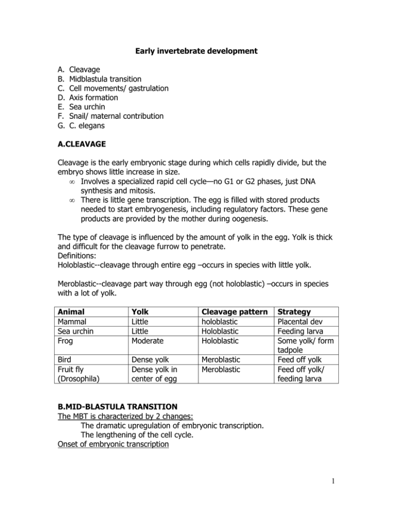
Early invertebrate development A. B. C. D. E. F. G. Cleavage Midblastula transition Cell movements/ gastrulation Axis formation Sea urchin Snail/ maternal contribution C. elegans A.CLEAVAGE Cleavage is the early embryonic stage during which cells rapidly divide, but the embryo shows little increase in size. • Involves a specialized rapid cell cycle—no G1 or G2 phases, just DNA synthesis and mitosis. • There is little gene transcription. The egg is filled with stored products needed to start embryogenesis, including regulatory factors. These gene products are provided by the mother during oogenesis. The type of cleavage is influenced by the amount of yolk in the egg. Yolk is thick and difficult for the cleavage furrow to penetrate. Definitions: Holoblastic--cleavage through entire egg –occurs in species with little yolk. Meroblastic--cleavage part way through egg (not holoblastic) –occurs in species with a lot of yolk. Animal Mammal Sea urchin Frog Yolk Little Little Moderate Cleavage pattern holoblastic Holoblastic Holoblastic Bird Fruit fly (Drosophila) Dense yolk Dense yolk in center of egg Meroblastic Meroblastic Strategy Placental dev Feeding larva Some yolk/ form tadpole Feed off yolk Feed off yolk/ feeding larva B.MID-BLASTULA TRANSITION The MBT is characterized by 2 changes: The dramatic upregulation of embryonic transcription. The lengthening of the cell cycle. Onset of embryonic transcription 1 At the MBT, there is a transition from maternal control of development to embryonic control. The time at which embryonic transcription begins is different in different species. Very often, a few genes are transcribed early, and then at a later time (the midblastula transition) embryonic transcription is dramatically increased. Lengthening of the cell cycle The cell cycle begins to include growth phases (G1 and G2). At this point, there is significant cell growth between divisions. As such, the embryo begins to increase in size. C. GASTRULATION The first morphogenetic movement—large scale, orchestrated cellular movements and rearrangements. Generates the three primary germ layers, ectoderm, mesoderm and endoderm. The prospective endoderm and mesoderm are internalized. ! Ectoderm will give rise to epidermis and nervous system ! Mesoderm gives rise to skeleton, muscles and blood ! Endoderm gives rise to the gut The pre-gastrulation embryo is called a blastula. It is basically a hollow sphere of cells. The hollow cavity is called the blastocoel. It functions to provide room for internalization of endodermal and mesodermal cells during gastrulation and to prevent premature cell-cell contacts. A dimple forms and protrudes into the hollow interior space. This forms a tube that extends through the blastocoel and attaches to the roof on the opposite side. The pore formed by the inward protrusion of the dimple is called the blastopore. The tube is called the archenteron, and represents the primitive gut. In protostomes (mollusks and worms) the blastopore forms the mouth. In deuterostomes (chordates and echinoderms) the blastopore becomes the anus. Several types of active cell movements are involved. (Fig 8.5) Invagination: cells change shape at apical and basal surfaces Involution: expanding epithelium folds and forms new layers Ingression: epithelial to mesenchyme transition Delamination: splitting of one sheet into two sheets Epiboly: epithelial sheets flatten; lateral expansion of the entire sheet Convergent extension:convergence of cell layers leads to extension (Fig 8.23) Cell movements involve cytoskeletal activity within the cells. 2 D. Axis formation During early embryogenesis, need to set up the major axes, anterior-posterior (A-P), dorsal-ventral (D-V), and sometimes left right. Variable timing and mechanisms. In some phyla, axes are established during oogenesis, prior to fertilization, in others they are established during gastrulation. E. Early SEA URCHIN Development Cleavage results in the formation of macromeres, mesomeres, micromeres. (Fig 8.7) Axis formation: The anterior-posterior axis of the sea urchin embryo is determined prior to fertilization. The vegetal pole (where the micromeres will form) will form posterior structures. Cytoplasmic determinants deposited in the oocyte by the mother are localized at the vegetal pole and will specify micromere identity. See Fig 8.11. Fate map Induction: • Micromeres induce posterior axis • Micromeres induce endodermal cell fates Potency Gastrulation sea urchin blastula is about 1000 cells—in an epithelial sheet 1.Ingression of primary mesenchyme (Fig 8.18) transition of epithelial cells to mesenchyme ! They lose affinity for the hyaline layer surrounding the embryo and for their neighbors. ! Gain affinity for ECM of the blastocoel ! Cells at the vegetal pole extend filopodia & interact with extracellular matrix in blastocoel. ! Attraction to ECM cues guides their migration. 2.Archenteron invagination(Fig. 8.20) Movement of a sheet of cells, driven by cell-shape changes and by modifications in the ECM/ hyaline layer. Invagination allows the vegetal plate to reach ~1/3 of the way into the blastocoel. 3.Archenteron convergent extension.(Fig. 8.22) 4.Filopodia reach for the roof of the animal hemisphere and complete formation of the intestinal tube (fig 8.24). 3 F. SNAILS/ MATERNAL CONTRIBUTIONS Snail shells contain spiral patterns. The “handedness” of the spiral is determined by the cleavage pattern. The spirals can be left-handed or right-handed. Controlled by a “maternal” gene. D confers right handedness d confers left handedness. DD or Dd females have right handed offspring, and dd females have left handed offspring, regardless of the genotype of the male parent. So consider the mating of a Dd female with a dd male: all the offspring will have right handed spiral shells, even though half will have the dd genotype. Similarly, a dd female mated with a DD male will produce all left handed offspring even though all the offspring carry a Dd genotype. The phenotype of the offspring is determined by the genotype of the mother ≡ ”maternal gene”. G. ELEGANS Specifying the A/P axis: The egg is oval. The A/P axis will be the long axis of the oval. The sperm entry point specifies the posterior end. The sperm nucleus centriole nucleates microtubule reorganizations. Blastomeres show both autonomous and conditional specification. If AB and P1 are separated, P develops autonomously to produce a posterior half animal. However AB only produces a subset of the cell types it normally would. P cell derivatives are required to induce certain cell fates. Autonomous cell specification—asymmetric segregation of cytoplasmic determinants E.g. P-granules: specify germ cell fate. Are asymmetrically segregated at each mitotic division of early embryogenesis. Conditional (non-cell-autonomous) specification of certain cell fates in C. elegans. P2 induces EMS to form E. If separate, EMS cell generates 2 MS daughter cells. P2 necessary for E cell fate. Not sufficient for E fate because contact with ABp cell does not induce E fate. 4
