Mike Russell, Ph.D.
advertisement

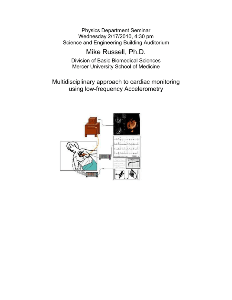
Physics Department Seminar Wednesday 2/17/2010, 4:30 pm Science and Engineering Building Auditorium Mike Russell, Ph.D. Division of Basic Biomedical Sciences Mercer University School of Medicine Multidisciplinary approach to cardiac monitoring using low-frequency Accelerometry Summary Abstract Heart disease is the leading cause of death in Georgia, and diabetics, the elderly, and African American women are most at risk. The cardiac stress test is the standard screening tool for assessing cardiac function, and electrocardiography (ECG), ultrasound (echocardiography, ECHO) and nuclear myocardial perfusion imaging (MPI) are the modalities commonly used for detecting ischemia and cardiac wall motion abnormalities. ECG is affected by metabolic state, and ECHO is not reliably predictive of outcome, as reliability is operator-dependent and it detects only high-grade ischemia. MPI shows only regional differences in myocardial perfusion, rather than overall perfusion efficiency, and it contributes the highest single radiation exposure of commonly used nuclear diagnostics. A method for detecting cardiac dysfunction is needed that 1) is inexpensive enough to provide widespread patient access, 2) is safe and non-invasive, 3) is at least as sensitive as ECHO, without the inherent dependence on operator proficiency, and 4) is independent of ionizing radiation. We hypothesized that measurement of cardiac accelerations within a defined frequency range, coupled to recently developed time- and frequency-domain analytical techniques, could provide the safety, sensitivity, reliability, and low cost currently lacking in a cardiac function diagnostic. Our goal is to clinically assess the diagnostic value of an accelerometer-based, low-cost, non-invasive, ischemia detection and functional assessment system for cardiac monitoring during physical or chemical stress testing procedures. The proposed system utilizes a new class of commercially available, non-invasive, low-frequency triaxial accelerometer coupled with novel data acquisition and time- and frequency-domain analyses to quantify the mechanical activity of the heart. The utility of single-axis cardiac accelerometry (seismocardiography, SCG) as a reliable method for monitoring cardiac ischemia during MRI and for assessing cardiac contractility in weightless environments is well established. In preliminary studies time- and frequency-domain data analysis of the novel low-frequency, triaxial SCG (3D-SCG) data revealed cardiac accelerations in three dimensions that were consistently indicative of specific experimental conditions, yet were independent of cardiac loading, body type, or incidental body movement. Novel frequency domain analysis showed that changes in wall motion and defects in valve function could both be identified using low-frequency 3D-SCG. We intend to evaluate the novel 3D-SCG system as a stress test diagnostic in comparison with ECG, ECHO, and MPI modalities. For the secondary provider, 3D-SCG could 1) improve reliability as a low-cost addition or alternative to current stress test diagnostics, 2) provide improved ischemia detection independent of operator expertise, and 3) allow real-time constant cardiac monitoring of post-operative or chronically ill patients. Because of the potential low cost and widespread accessibility, 3D-SCG could ultimately be adapted for primary care to 1) provide long-term monitoring of cardiac mechanical ability in at-risk patients, or 2) provide pointof-care assessment of cardiac mechanical function in student athletes . Please join us for light refreshments at 4:15pm outside SEB 203


