Eggshell Matrix Protein Mimetics: Elucidation of Molecular Mechanism of Goose Eggshell
advertisement

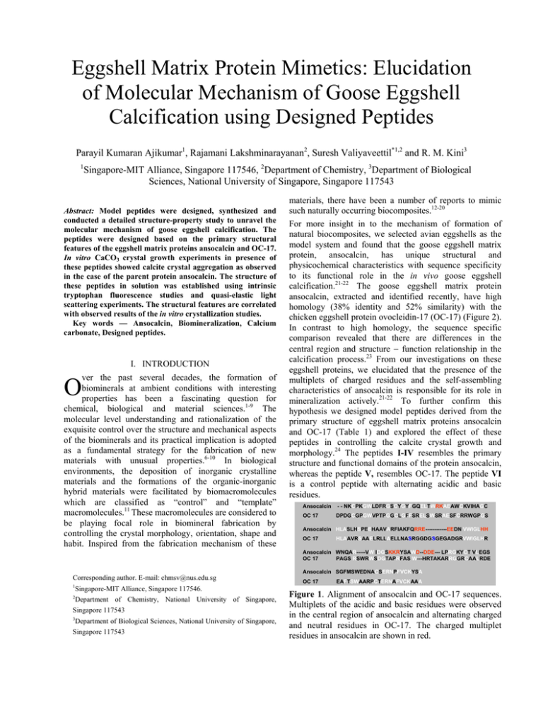
Eggshell Matrix Protein Mimetics: Elucidation of Molecular Mechanism of Goose Eggshell Calcification using Designed Peptides Parayil Kumaran Ajikumar1, Rajamani Lakshminarayanan2, Suresh Valiyaveettil*1,2 and R. M. Kini3 1 Singapore-MIT Alliance, Singapore 117546, 2Department of Chemistry, 3Department of Biological Sciences, National University of Singapore, Singapore 117543 Abstract: Model peptides were designed, synthesized and conducted a detailed structure-property study to unravel the molecular mechanism of goose eggshell calcification. The peptides were designed based on the primary structural features of the eggshell matrix proteins ansocalcin and OC-17. In vitro CaCO3 crystal growth experiments in presence of these peptides showed calcite crystal aggregation as observed in the case of the parent protein ansocalcin. The structure of these peptides in solution was established using intrinsic tryptophan fluorescence studies and quasi-elastic light scattering experiments. The structural features are correlated with observed results of the in vitro crystallization studies. Key words — Ansocalcin, Biomineralization, Calcium carbonate, Designed peptides. I. INTRODUCTION ver the past several decades, the formation of biominerals at ambient conditions with interesting properties has been a fascinating question for chemical, biological and material sciences.1-9 The molecular level understanding and rationalization of the exquisite control over the structure and mechanical aspects of the biominerals and its practical implication is adopted as a fundamental strategy for the fabrication of new materials with unusual properties.6-10 In biological environments, the deposition of inorganic crystalline materials and the formations of the organic-inorganic hybrid materials were facilitated by biomacromolecules which are classified as “control” and “template” macromolecules.11 These macromolecules are considered to be playing focal role in biomineral fabrication by controlling the crystal morphology, orientation, shape and habit. Inspired from the fabrication mechanism of these O materials, there have been a number of reports to mimic such naturally occurring biocomposites.12-20 For more insight in to the mechanism of formation of natural biocomposites, we selected avian eggshells as the model system and found that the goose eggshell matrix protein, ansocalcin, has unique structural and physicochemical characteristics with sequence specificity to its functional role in the in vivo goose eggshell calcification.21-22 The goose eggshell matrix protein ansocalcin, extracted and identified recently, have high homology (38% identity and 52% similarity) with the chicken eggshell protein ovocleidin-17 (OC-17) (Figure 2). In contrast to high homology, the sequence specific comparison revealed that there are differences in the central region and structure − function relationship in the calcification process.23 From our investigations on these eggshell proteins, we elucidated that the presence of the multiplets of charged residues and the self-assembling characteristics of ansocalcin is responsible for its role in mineralization actively.21-22 To further confirm this hypothesis we designed model peptides derived from the primary structure of eggshell matrix proteins ansocalcin and OC-17 (Table 1) and explored the effect of these peptides in controlling the calcite crystal growth and morphology.24 The peptides I-IV resembles the primary structure and functional domains of the protein ansocalcin, whereas the peptide V, resembles OC-17. The peptide VI is a control peptide with alternating acidic and basic residues. Ansocalcin - - NKCPKGWLDFRGSCYGYFGQELTWRKAEAWCKVIHAGC OC 17 DPDGCGPGWVPTPGGCLGFFSRELSWSRAESFCRRWGPGS Ansocalcin HLASLHSPEEHAAVARFIAKFQRRE------------EEDN VWIGLHH OC 17 HLAAVRSAAELRLLAELLNASRGGDGSGEGADGRVWIGLHR Ansocalcin WNQAR-----VW IDGSKKRYSAWD--DDE--- LPRGKYCT VLEGS OC 17 PAGS RSWRWSDGTAPRFAS W---HRTAKARRGGRCAALRDE Corresponding author. E-mail: chmsv@nus.edu.sg 1 Singapore-MIT Alliance, Singapore 117546. 2 Department of Chemistry, National University of Singapore, Singapore 117543 3 Department of Biological Sciences, National University of Singapore, Singapore 117543 Ansocalcin SGFMSWEDNACSERNPFVCKYSA OC 17 EAFTSWAARPCTERNAFVCKAAA Figure 1. Alignment of ansocalcin and OC-17 sequences. Multiplets of the acidic and basic residues were observed in the central region of ansocalcin and alternating charged and neutral residues in OC-17. The charged multiplet residues in ansocalcin are shown in red. Table 1. Amino acid sequence of the model peptides I II III IV V VI Peptide R2E2W2D2-16 R2E2W2D2P-17 K2E2W2D2-16 K2E2W2D2P-17 (RA)2(DA)2-16 (KE)2(WD)2-16 Amino acid sequence RREEWWDDRREEWWDD RREEWWDDPRREEWWDD KKEEWWDDKKEEWWDD KKEEWWDDPKKEEWWDD RADARADARADARADA KEKEWDWDKEKEWDWD II. RESULTS AND DISCUSSION Interaction of the peptides with CaCO3: In vitro CaCO3 crystallizations in presence of these peptides were performed to investigate the role of these peptides in calcite crystal growth and morphology. The crystallization experiments showed that there is a concentration dependent change in growth pattern of calcite crystals in presence of the peptides I-IV.25 Figure 2 shows the influence of these peptides on the morphology of the calcite crystals. The peptides I-IV showed the nucleation of a polycrystalline aggregate of calcite crystals. However, the peptides V and VI did not influence the crystal morphology. In our control experiments without peptide, exclusive nucleation of rhombohedral calcite crystals was observed. In addition to this, the replacement of the arginine residue of the peptide I and II with lysine in the peptide III and IV has also influenced the crystal size and morphology. Peptides I-IV showed similar influence on the crystal morphology in in vitro calcium carbonate crystallization as observed in the case of the ansocalcin.22 The peptide V showed crystal morphology similar to the calcite crystals nucleated by OC17.23 The aforementioned results showed that the observed multiplets of charged residues in protein ansocalcin has significant role in goose eggshell calcification. Investigation of the solution structure of the peptides Quasi-elastic light scattering studies (QELS): We performed QELS experiments to study the behavior of these peptides in solution. Table 2 summarizes the main parameters measured by these experiments. The mean diffusion coefficient (<D>) of the peptides I-V which relates to the motion of the molecules in solution, indicated that the peptides exist as large aggregates in solution. In addition to this the observed hydrodynamic radius (<RH>) confirmed the formation of these large aggregates of the peptides in solution. The observed very low <RH> and very high diffusion coefficient in the case of the control peptide (VI) indicates that it does not aggregate in solution. Intrinsic tryptophan Fluorescence: To understand the microenvironment around the tryptophan residues in peptides I to V, we investigated the change in fluorescence emission characteristics of these peptides in 7.5 mM CaCl2 solution. The concentration dependent aggregation of the peptides was monitored by the significant change in tryptophan fluorescence emission intensity (Figure 3). A B C D E F Figure 2. Representative SEM images of the calcite crystals in presence of the peptides (2 mg/mL). (A) R2E2W2D2-16 (I), (B) R2E2W2D2P-17 (II), (C) K2E2W2D216 (III), (D) K2E2W2D2P-17 (IV), (E) (RA)2(DA)2-16 (V), and (F) (KE)2(WD)2-16 (VI). Table 2. Average diffusion coefficient (<D>) and mean hydrodynamic radius (<RH>) of peptides I-VI Peptides I R2E2W2D2-16 II R2E2W2D2P-17 III K2E2W2D2-16 IV K2E2W2D2P-17 V (RA)2(DA)2-16 VI (KE)2(WD)2-16 <D> cm2/sec 8.80 × 10⁻⁷ 4.62 × 10⁻⁷ 13.5 × 10⁻⁷ 6.86 × 10⁻⁷ 5.17 × 10⁻⁷ 29.2 × 10⁻⁷ <RH> nm 3.3 6.4 4.1 8.1 5.7 1.8 In the case of the peptides I-IV there is a blue shift in the emission maxima as the concentration increased from 0.05 mg/mL to 2 mg/mL. In addition to this the observed florescence quenching, a large decrease in emission intensity (~85 %) at high concentration (2 mg/mL) may be due to the formation of large aggregate of the peptides in solution. However, in the case of the peptide VI the emission intensity linearly increased up to 1 mg/mL followed by a small decrease in intensity at 2 mg/mL. Also there is no considerable differences in the shift in emission maxima as the concentration of the peptide was increased. This shows the tryptophan residues in this peptide exist in exposed state compared to the peptide aggregates in I-IV. The lack of tryptophan residues in V limited the possibility of investigating the self-assembly in solution using fluorescence emission studies. F max 900 0 0 1 2 Peptide concentration (mg/mL) Figure 3. Intensity versus peptide concentration profile of fluorescence emission of synthetic peptides I (♦), II (■), III ( ), IV (•), VI (¥) in 7.5 mM CaCl2 solution. Emission intensity (Fmax) is expressed in arbitrary units. The present study was mainly focused on the understanding of the diverse characteristics of ansocalcin using model peptides with multiplets of charged residues, which believes to play a crucial role in goose eggshell mineralization. The structure-property relationship of two eggshell proteins ansocalcin and OC-17 indicated that ansocalcin exists in various non-covalent aggregated forms in CaCl2 solution, whereas OC-17 transforms into polymeric state at very high concentration.22-23 Ansocalcin induced aggregation of polycrystalline calcite crystals whereas OC-17 did not alter the size and shape of calcite crystals.23 The aggregate size is also reduced in presence of ansocalcin. Similar to these observations, the designed peptides (I-IV) exists in aggregated state at higher concentrations. The tryptophan fluorescence analysis showed a concentration dependent aggregation of these peptides in CaCl2 solution. The QELS analysis also showed the presence of large aggregates in CaCl2 solution. The self assembly and the charged amino acid multiplets may cooperatively accelerate the crystal nucleation and crystal aggregate formation. The observed results correlate the unique structural and physicochemical characteristics of the parent protein ansocalcin and the critical role of its self assembly nature and sequence specificity to function in in vivo eggshell mineralization when compared the chicken eggshell matrix protein OC-17. III. EXPERIMENTAL PROCEDURES Synthesis and Characterization of the Peptides: FmocPAL-PEG-PS and all the Nα-Fmoc-L-amino acid pentafluorophenyl esters were purchased from Novabiochem, San Diego, CA, USA. The peptides were synthesized using the Pioneer peptide synthesizer (Applied acid Biosystems) using the Nα-Fmoc-L-amino pentafluorophenyl ester/HOBt coupling method. The peptides I-VI were assembled on an Fmoc-PAL-PEG-PS (0.18 mmol/g) resin. All peptides were synthesized on a 0.1 mmol scale using an extended cycle protocol. The peptides were deprotected and cleaved using a cleavage cocktail consisting of 90% trifluoroacetic acid (TFA), 2.5% phenol, 2.5% water, 2.5% thioanizole, 2.5% ethanedithiol, for 3 hours at room temperature. The mixture was filtered and concentrated under reduced pressure. The peptide was precipitated using cold ether and centrifuged on an ultracentrifuge with repeated washing by cold ether to remove all reagents and impurities. Finally the peptides were lyophilized with 10% acetic acid solution and obtained as a white powder. All peptides were purified using the Phenominex (250 ×10 nm, 10µ) C18 reversephase column using a BioCAD workstation. The solvent systems used for the purification are as follows: solvent A, 0.1 % TFA and solvent B, 0.1% TFA in 80% acetonitrile. A linear gradient (flow rate 2 mL/min) 25% - 50% B over 50 min was used. The peptide elution was monitored at 215 nm. The purity of the peptides was confirmed by analytical HPLC and ESI-MS measurements. Crystal Growth Experiments: CaCO3 crystals were grown on glass cover slips placed inside the CaCl2 solution kept in a Nunc dish with 6 × 4 wells. Typically, 1 mL of 7.5 mM CaCl2 solution was introduced into the wells containing the cover slips and the whole set up was covered with aluminum foil with a few pinholes on the top. To study the role of synthetic peptides in the CaCO3 crystallization, aliquots of peptides were dissolved in 7.5 mM CaCl2 solution to prepare a series of solutions in the concentration range of 50 µg/mL to 2 mg/mL and introduced into each well with a glass cover slip. Crystals were grown inside a closed desiccator for 2 days by slow diffusion of gases released by the decomposition of ammonium carbonate crystals placed at the bottom of the desiccator. After 2 days, the slides were carefully lifted from the crystallization wells, rinsed gently with Millipore deionized water, air dried at room temperature and used for further characterization. Quasi-elastic light scattering studies (QELS): 120µL of the peptide solution (concentration of 5 mg/mL) was used to perform dynamic light scattering experiments using PDDLS/Batch (Precision Detectors, Franklin, MA) instrument. Intensity data from each sample were collected in five replicates and analyzed by using the PRECISION DECONVOLVE program and yielded size-versus-fraction distribution plots. Fluorescence Spectroscopy: The fluorescence emission spectra were collected on a Shimadzu RF-5301PC spectrofluorometer, with the emission and excitation band passes set at 3 nm. The excitation wavelength was set at 285 nm and the spectra were recorded from 300 to 400 nm at a scan rate of 50 nm/min. Emission spectra of the peptides were recorded in both 7.5 mM CaCl2 solution and in water. Scanning Electron Microscopy (SEM): SEM studies on the CaCO3 crystals were carried out using Philips XL-30 scanning electron microscope at 15/20 kV after sputter coating with gold to increase the conductivity. ACKNOWLEDGEMENT P. K.A. thanks SMA for the research fellowship. We also acknowledge the financial support from SMA. REFERENCES [1] H. A Lowenstam and S. Weiner, On Biomineralization, 1989, Oxford University Press, New York. [2] S. Mann, J. Web and R. J. P. Williams, Biomineralization: Chemical and Biochemical Perspectives, 1989, VCH publishers, Weinheim. [3] S Mann, Biomineralization: principles and concepts in bioinorganic materials chemistry, 2001, Oxford University Press, New York. [4] L. Addadi, S. Raz and S. Weiner, “Taking advantage of disorder: Amorphous calcium carbonate and its roles in biomineralization,” Adv. Mater., 15, pp. 959970, 2003. [5] B A.Gotliv, L. Addadi and S. Weiner, “Mollusk shell acidic proteins: In search of individual functions,” Chembiochem., 4, pp. 522-529, 2003. [6] J. S. Evans, “'Apples' and 'oranges': comparing the structural aspects of biomineral-and ice-interaction proteins,” Curr. Opin. Colloid In., 8, pp. 48-54, 2003. [7] A. H. Heuer, D. J. Fink, V. J. Laraia, J. L. Arias, P. D. Calvert, K. Kendall, G. L. Messing, J. Blackwell, P. C. Rieke, D. H. Thompson, A. P. Wheeler, A. Veis and A. I. Caplan, “Innovative material processing strategies-a biomimetic approach,” Science, 255, pp. 1098-1105, 1992. [8] S. Weiner and L. Addadi, “Design strategies in mineralized biological materials,” J. Mater. Chem., 7, pp. 689-702, 1997. [9] S.Weiner, L. Addadi and H. D. Wagner, “Materials design in biology,” Mat. Sci Eng., 11, pp. 1-8, 2000. [10] E. Baeuerlein, Biomineralization: from biology to biotechnology and medical application, 2000, WileyVCH publishers, New York. [11] L. Addadi and S. Weiner, “Control and design principles in biological mineralization,” Angew. Chem. Int., 31, pp. 153-169, 1992. [12] H. Colfen and S. Mann, “Higher-order organization by mesoscale self-assembly and transformation of hybrid nanostructures,” Angew. Chem. Int., 42, pp. 23502365, 2003. [13] Z. Y. Tang, N. A. Kotov, S. Magonov and B. Ozturk, “Nanostructured artificial nacre,” Nature Mater., 2, pp. 413-418, 2003 [14] P. J. Li, “Biomimetic nano-apatite coating capable of promoting bone ingrowth,” J. Biomed. Mater. Res., 66, pp. 79-85, 2003. [15] K. Ichikawa, N. Shimomura, M. Yamada and N. Ohkubo, “Control of calcium carbonate polymorphism and morphology through biomimetic mineralization by means of nanotechnology,” Chem-Eur. J., 9 pp. 32353241, 2003. [16] Y. J. Han and J. Aizenberg, “Face-selective nucleation of calcite on self-assembled monolayers of alkanethiols: Effect of the parity of the alkyl chain” Angew. Chem. Int. Ed., 42, pp. 3668-3670, 2003. [17] G. He, T. Dahl, A. Veis and A. George, “Nucleation of apatite crystals in vitro by self-assembled dentin matrix protein” Nature Mater., 2, pp. 552-558, 2003. [18] N. Hosoda, A. Sugawara and T. Kato, “Template effect of crystalline poly(vinyl alcohol) for selective formation of aragonite and vaterite CaCO3 thin films,” Macromolecules, 36, pp. 6449-6452, 2003. [19] J. M. Slocik, and D. W. Wright, “Biomimetic mineralization of noble metal nanoclusters,” Biomacromolecules, 4, pp. 1135-1141 2003. [20] H. Song, E. Saiz, and C. R. Bertozzi, “Preparation of pHEMA-CP composites with high interfacial adhesion via template-driven mineralization,” J. Eur. Ceram. Soc., 23, pp. 2905-2919, 2003. [21] R. Lakshminarayanan, S. Valiyaveettil and R. M. Kini, “Investigation of the role of ansocalcin in the biomineralization in goose eggshell matrix”, Proc. Natl. Acad. Sci. USA, 99, pp. 5155-5159, 2002. [22] R. Lakshminarayanan, S. Valiyaveettil, V. S. Rao and R. M. Kini, “Purification, characterization, and in vitro mineralization studies of a novel goose eggshell matrix protein, ansocalcin,” J. Biol. Chem., 278, pp. 29282936, 2003. [23] R. Lakshminarayanan, S. Valiyaveettil, V. S. Rao and R. M. Kini, “Comparative study of structure-property of eggshell proteins”, Proc. Second Int. Conf. on Str. Biol. and Funct. Geno., pp. 71, 2002. [24] P. K. Ajikumar, R. Lakshminarayanan, B. T. Ong, S. Valiyaveettil and R. M. Kini, “Eggshell matrix protein mimics: Designer peptides to induce the nucleation of calcite crystal aggregates in solution” Biomacromolecules, 4, pp. 1321-1326, 2003. [25] P. K. Ajikumar, R. Lakshminarayanan, S. Valiyaveettil and R. M. Kini, “Structure function relationship in eggshell matrix proteins: The role of multiplets of charged residues and self-assembly of protein ansocalcin in calcification of goose eggshell” (unpublished results).


