441
Cell–cell signaling in the shoot meristem
Achim Haecker and Thomas Laux*
The shoot meristem is a proliferating, changing cell population
yet displays a stable organization. Recent studies have
addressed how signaling processes coordinate the behaviour
of shoot meristem cells.
Addresses
Institute of Biology III, Schänzlestr.1, 79104 Freiburg, Germany
*e-mail: laux@biologie.uni-freiburg.de
Corresponding author: Thomas Laux
Current Opinion in Plant Biology 2001, 4:441–446
1369-5266/01/$ — see front matter
© 2001 Elsevier Science Ltd. All rights reserved.
Abbreviations
AG
AGAMOUS
ANT
AINTEGUMENTA
CLV
CLAVATA
CZ
central zone
LFY
LEAFY
OC
organizing center
NPA
N-(1-naphthyl)phtalamic acid
PID
PINOID
pin1
pin-formed1
pol
poltergeist
PZ
peripheral zone
RZ
rib zone
SAM
shoot apical meristem
STM
SHOOTMERISTEMLESS
WUS
WUSCHEL
that direct the two meristem functions: stem cell homeostasis
and organ initiation.
Regulation of stem cells
Clonal analyses have demonstrated that all of the
postembryonic structures of the plant shoot are derived
from about three stem cells in every layer of the meristem
[3]. We use the term stem cells in the operational sense
that they give rise to both daughters that renew the stem
cell pool and that differentiate into a variety of cell types.
The stem cells cannot be distinguished histologically but,
for geometric reasons, are thought to be located at the very
tip of the CZ. This position roughly coincides with the
expression domain of the CLAVATA (CLV)3 gene, which has
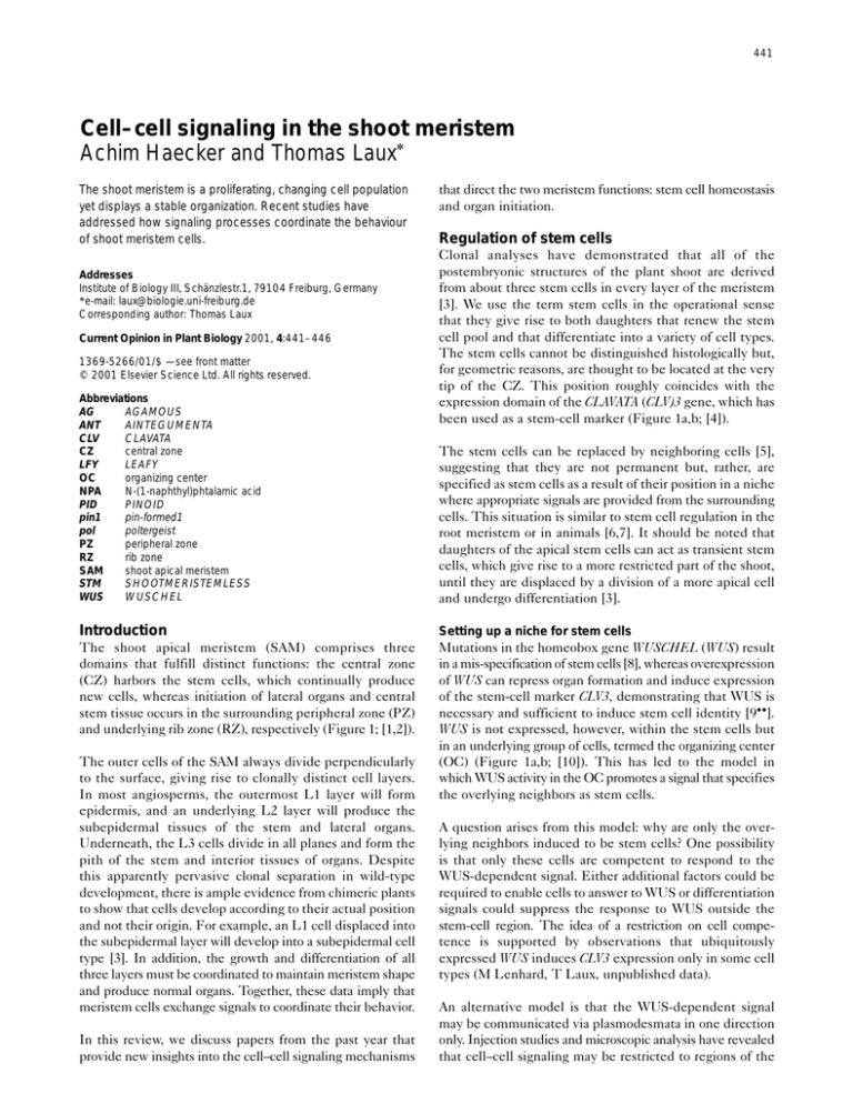
been used as a stem-cell marker (Figure 1a,b; [4]).
The stem cells can be replaced by neighboring cells [5],
suggesting that they are not permanent but, rather, are
specified as stem cells as a result of their position in a niche
where appropriate signals are provided from the surrounding
cells. This situation is similar to stem cell regulation in the
root meristem or in animals [6,7]. It should be noted that
daughters of the apical stem cells can act as transient stem
cells, which give rise to a more restricted part of the shoot,
until they are displaced by a division of a more apical cell
and undergo differentiation [3].
Introduction
Setting up a niche for stem cells
The shoot apical meristem (SAM) comprises three
domains that fulfill distinct functions: the central zone
(CZ) harbors the stem cells, which continually produce
new cells, whereas initiation of lateral organs and central
stem tissue occurs in the surrounding peripheral zone (PZ)
and underlying rib zone (RZ), respectively (Figure 1; [1,2]).
Mutations in the homeobox gene WUSCHEL (WUS) result
in a mis-specification of stem cells [8], whereas overexpression
of WUS can repress organ formation and induce expression
of the stem-cell marker CLV3, demonstrating that WUS is
necessary and sufficient to induce stem cell identity [9••].
WUS is not expressed, however, within the stem cells but
in an underlying group of cells, termed the organizing center
(OC) (Figure 1a,b; [10]). This has led to the model in
which WUS activity in the OC promotes a signal that specifies
the overlying neighbors as stem cells.
The outer cells of the SAM always divide perpendicularly
to the surface, giving rise to clonally distinct cell layers.
In most angiosperms, the outermost L1 layer will form
epidermis, and an underlying L2 layer will produce the
subepidermal tissues of the stem and lateral organs.
Underneath, the L3 cells divide in all planes and form the
pith of the stem and interior tissues of organs. Despite
this apparently pervasive clonal separation in wild-type
development, there is ample evidence from chimeric plants
to show that cells develop according to their actual position
and not their origin. For example, an L1 cell displaced into
the subepidermal layer will develop into a subepidermal cell
type [3]. In addition, the growth and differentiation of all
three layers must be coordinated to maintain meristem shape
and produce normal organs. Together, these data imply that
meristem cells exchange signals to coordinate their behavior.
In this review, we discuss papers from the past year that
provide new insights into the cell–cell signaling mechanisms
A question arises from this model: why are only the overlying neighbors induced to be stem cells? One possibility
is that only these cells are competent to respond to the
WUS-dependent signal. Either additional factors could be
required to enable cells to answer to WUS or differentiation
signals could suppress the response to WUS outside the
stem-cell region. The idea of a restriction on cell competence is supported by observations that ubiquitously
expressed WUS induces CLV3 expression only in some cell
types (M Lenhard, T Laux, unpublished data).
An alternative model is that the WUS-dependent signal
may be communicated via plasmodesmata in one direction
only. Injection studies and microscopic analysis have revealed
that cell–cell signaling may be restricted to regions of the
442
Cell signalling and gene regulation
Figure 1
(a)
(b)
L1
L2
L3
L1
CLV3
L2
L3
WUS
CLV1
Current Opinion in Plant Biology
Organization of the shoot apical meristem. (a) Schematic view of SAM
domains. The CZ (blue) contains slowly dividing cells that stain weakly
with cytoplasmic dyes. The apical stem cells (red) form part of the CZ.
Cells in the PZ (green), where initiation of organ primordia takes place,
divide more rapidly and stain more strongly. In the RZ (yellow),
differentiation of central pith tissue is initiated. Redrawn after [37].
(b) Outline of the central part of (a), showing the approximate mRNA
expression domains of CLV1, CLV3 and WUS.
shoot meristem by regulating the plasmodesmal coupling
of meristem cells [11,12]. At least for the epidermal
cells, facilitated transport was observed in the CZ domain
[11,12]. Thus, it is conceivable that the movement of the
WUS-dependent signal is directed by preferential coupling
between stem cells and the OC.
secretory signal peptide [4] that appears to be present in planta
as a small soluble complex [17••].
The reply from the stem cells
The function of WUS is counteracted by the CLV signaling
pathway [8,9••]. Mutations in any one of the three CLAVATA
genes (CLV1, CLV2 and CLV3) lead to a progressive
enlargement of the stem-cell population, and genetic data
suggest that all three genes act in the same pathway [13].
Several results suggest that one of the main targets of the
CLV pathway is WUS. The effects of wus mutations are
epistatic to clv1, clv2 and clv3 phenotypes; the WUS
expression domain is enlarged in clv mutants, indicating
that the CLV pathway suppresses WUS at the transcript
level; and enlarging the WUS expression domain in wild-type
plants can phenocopy the clv mutant defect [9••]. Genetic
analysis of the poltergeist (pol) mutant suggests that POL
acts downstream of CLV signaling and may have functions
that overlap with those of WUS [14••]. In double mutants,
pol mutations suppress the clv phenotype whereas pol alone
has no effect, and pol and wus show dominant interactions.
CLV1 encodes a putative receptor kinase with an extracellular leucine-rich repeat receptor domain and an
intracellular serine/threonine kinase region. It is
expressed mainly in the corpus of the SAM, but possibly
also in the L2 (Figure 1b; [15]). CLV2 encodes a protein
with an extracellular domain similar to that of CLV1 but
that lacks the kinase domain and probably forms a
membrane-associated complex with CLV1 [16]. CLV3,
which as mentioned above is expressed in the apical
stem cells, encodes a small polypeptide with a putative
Does CLV3 act as a signal in the shoot meristem? There is
convincing biochemical evidence that CLV3 represents a
ligand for the CLV1 receptor kinase: the formation of
the apparently active CLV1 complex of 450 kiloDaltons
is dependent on the presence of CLV3 [18]. CLV3 coimmunoprecipitates with CLV1 and this binding requires
an active intracellular kinase domain, suggesting that kinase
activity stabilizes the receptor–ligand interaction [17••].
In addition, CLV3 specifically binds to intact yeast cells
expressing CLV1 and CLV2, suggesting that CLV3 may
function as an extracellular signal that binds to
CLV1–CLV2 at the cell surface. It should be noted,
however, that other scenarios are also possible: as WUS is
repressed by the CLV pathway in the L3 stem cells, in
which CLV1 and CLV3 expression overlap (Figure 1b;
[4,9••,15]), the possibility that the CLV1–CLV3 interaction
is intracellular cannot be excluded.
The above results suggest the following model for the
regulation of stem-cell homeostasis (see also ‘Update’).
Stem cells are specified by, as yet unknown, WUS-dependent
signals from the underlying OC and in turn signal back via
the CLV3 molecule, delimiting the OC by repression of
WUS. This negative-feedback loop enables the stem cells
to autoregulate their number indirectly through size control
over the OC (Figure 2a).
Localized CLV3 signal
Transgenic plants that express CLV3 throughout the SAM
mimic the wus mutant phenotype [19••], indicating that
CLV3 activity is sufficient to repress WUS. This implies
that the availability of CLV3 function is the rate-limiting
Cell–cell signaling in the shoot meristem Haecker and Laux
factor in the repression of WUS; however, assuming that
CLV3 can travel within the meristem, why is WUS
repressed only in the cells above and possibly lateral to the
OC, but not in the OC itself? There must be a mechanism
that prevents CLV3 from being active in OC cells. One
conceivable model is that all CLV3 protein produced in the
apical stem cells is bound — for example in the third cell
layer — preventing it from entering the OC cells underneath (Figure 2b). Consistent with this idea, 75% of the total
amount of CLV3 protein in cauliflower meristem extracts
was found to be associated with CLV1 [17••]. Doubling the
number of CLV3 gene copies was not sufficient to overcome
this barrier and repress WUS in the OC [19••], suggesting
that, in accordance with the model, the OC is safely protected
from CLV3 signaling by a large excess of binding sites.
Anchoring the organizing center
Although the shoot meristem consists of a population of
dividing cells, expression domains remain stable at the same
position relative to the organization of the shoot apex. In the
case of the OC, periclinal divisions of the overlying L3 stem
cells result in a flow of cells through the OC: cells that enter
the OC from above activate WUS expression, whereas cells
that leave the OC towards the RZ switch it off.
This raises the question of how the organizing center is
maintained at a given distance from the shoot summit,
underneath the third cell layer. One possibility would be
that the stem cells not only send a repressive signal but also
a positive signal that induces WUS expression in the OC.
Although the strength of the repressive signal in this model
would drop sharply because of CLV3 sequestration at the
third cell layer, the activating signal would reach the OC.
An alternative, though not mutually exclusive, scenario is
that the position of the OC depends on signals from underlying or lateral cells. This view is consistent with findings
that meristem maintenance requires the presence of young
leaf primordia and is influenced by genes expressed in the
leaves and the vasculature [20–22].
Organ initiation
Cells that leave the CZ lose stem cell identity and initiate
differentiation and organ formation. How are the cells
instructed to do so? Consistent with the niche concept, loss
of stem cell identity could simply result from the cell leaving
the range of WUS signaling; however, absence of WUS
signaling is not sufficient to initiate organ formation, as the
central cells in wus mutant apices do not do so. Given the
continuity of tissues, it is conceivable that differentiated
cells instruct the new cells. Such a mechanism is indicated by
experiments in the root; when undifferentiated root cells
were disconnected from older tissue by cell ablation, they lost
their ability to differentiate according to their position [23].
Several recent studies have addressed the role of signaling
in organ initiation in the SAM. Reinhardt and coworkers
[24••] exposed excised tomato apices to the auxin transport
443
Figure 2
(a)
(b)
L1
Stem cells
L2
CLV3
L3
WUS
WUS
Organizing
center
WUS
Current Opinion in Plant Biology
Signaling between stem cells and the organizing center (OC).
(a) WUS expression in the OC promotes a yet unidentified signal that
specifies the overlying cells as stem cells. The stem cells signal back
via CLV3 and restrict the size of the OC. (b) Model for the protection
of the OC from CLV3 signaling. Binding of CLV3 (red circles) to the
CLV1 complex (blue crescents) results in repression of WUS in L3. An
excess of CLV1 receptor complex prevents CLV3 from entering the
underlying OC cells, allowing WUS to be expressed there.
inhibitor NPA (N-[1-naphthyl]phtalamic acid). Although
the histological organization of the SAM remained
unaffected, organ formation was blocked, indicating a
requirement for auxin transport. This phenotype is reminiscent of the pin-formed1 (pin1) mutant, in which polar
auxin transport is reduced by about 90% as a result of a
defect in the putative auxin efflux carrier PIN1 [25,26].
As the site of auxin production in the apex is unknown, the
phenotype of NPA-treated or pin1 shoots could be caused
by an increased or decreased concentration of auxin. To
address this question, auxin was locally applied at the
periphery of naked NPA-treated and pin1 mutant apices,
and in both cases induced organ formation at the corresponding sites. The number of cells that were incorporated
into an organ primordium increased with the amount of auxin
applied; however, only the PZ was competent to respond
to auxin, whereas the CZ of the meristem was not. Together,
these experiments indicate that local maxima of auxin are
sufficient to induce organ initiation in the PZ and that
polar auxin transport is required to establish such maxima.
This model is supported by an analysis of marker gene
expression in the pin1 mutant [27••]. In wild-type plants,
SHOOTMERISTEMLESS (STM) is expressed throughout
the PZ but is absent from sites of incipient organ primordia
[28]. In contrast, AINTEGUMENTA (ANT) and LEAFY
(LFY) are expressed in incipient and outgrowing organ primordia in a pattern that is complementary to that of STM
expression [29,30]. In the pin1 mutant, STM expression was
completely absent in the PZ, whereas ANT and LFY were
expressed throughout the PZ. PZ cells showed a mixed
identity, however: the same PZ cells that expressed LFY
and ANT also expressed CUP-SHAPED COTYLEDON2,
444
Cell signalling and gene regulation
which in wild-type plants marks the boundary of organs
[31]. This indicates that PIN1 is required to separate these
expression domains — that is, to delimit organ anlagen.
Target genes of LFY were not expressed in pin1 mutants,
suggesting that the cells in the periphery had initiated
organs but were blocked soon thereafter. Whether this
block is a consequence of the mixed cell identity or
whether PIN1 is directly required for progression of organ
development remains to be determined.
Mutations in PINOID (PID), which encodes a serine/
threonine kinase, also result in naked shoot apices [32••];
however, overexpression analysis suggests that PID
antagonizes auxin signaling. As PID is expressed in young
organ anlagen on the flanks of the SAM, these results
suggest that dampening of auxin signaling is also required
for organ formation.
Together, these findings indicate that polar auxin transport
is required for the organization of the peripheral zone into
primordia and non-primordial cells and, furthermore, that
auxin is necessary for organ outgrowth. What is the source
of auxin in the shoot apex and by which method is it transported? If NPA-treated shoots are allowed to recover in the
absence of NPA and auxin, they first form leaves at random
positions but, later, phyllotaxis became normal, confirming
that wild-type phyllotaxis requires signals from existing
organs. It is questionable, however, whether this signal is auxin
as organ formation is repressed in the vicinity of existing
organs. This signal could, therefore, antagonize auxin activity.
Update
In contrast to the Arabidopsis shoot meristem, floral meristems are determinate and produce only a limited number
of organs. Recent work suggests that floral meristem determinacy is regulated by a negative feedback mechanism, in
which WUS activates expression of the AGAMOUS (AG)
gene, which in turn represses WUS and thus terminates
stem-cell maintenance [38••,39••].
In vitro WUS protein binds to consensus homeodomain
target sites within the regulatory region of the AG gene.
These sites are necessary for expression of an AG reporter
gene construct in planta [38••]. However, as the AG gene is
still active in wus mutants [8], other yet unknown proteins
could also bind to the target sites or other cis-regulatory
elements could play a role. The activation of AG expression by WUS, and thus stem-cell termination, is restricted
to determinate floral meristems, apparently because of the
requirement for additional flower-specific factors. As
endogenous WUS does not appear to activate AG in lfy
mutants, one of these factors could be the floral meristem
identity gene LFY [39••]. However, overexpressed WUS
can still activate the AG promoter in the absence of LFY
[38••], suggesting that this requirement is not absolute.
Acknowledgements
We gratefully acknowledge support of grants from the Deutsche
Forschungsgemeinschaft to Thomas Laux. We thank the members of the
Laux laboratory for helpful comments on the manuscript. We apologize to
those colleagues working in the field whose work was not mentioned
because of space constraints.
Conclusions
References and recommended reading
Genetic and molecular analyses have revealed a complex
network of signaling between cells within the SAM and
between the SAM and other parts of the plant.
Papers of particular interest, published within the annual period of review,
have been highlighted as:
What are the roads along which the signals travel? Smaller
secreted molecules, such as CLV3, could travel through
the extracellular space and bind to receptors at the cell
surface. Larger molecules, however, may have to use plasmodesmata in order to move from cell to cell [33]. For
example, the transcription factor LFY was shown to be
present in cells in which the LFY gene was not expressed,
indicating that LFY protein itself can travel [34••].
Recently, a transient increase in plasmodesmal coupling
between all meristem cells has been observed immediately
after floral induction [35••]. This increase was due to
establishment of secondary plasmodesmata and occurs
independently of cell division. What is the significance of
a varying density of cell–cell connections? A conceivable
model, taking into account the growing number of reports
of non-cell-autonomous functions of floral regulators, holds
that during floral induction the roads between meristem
cells are opened briefly to allow a rapid and co-ordinated
response of all cells to the floral stimulus [36••]. In this
view, the regulation of cell–cell coupling can provide a
means with which to spatially and temporally modulate
cell–cell communication.
• of special interest
•• of outstanding interest
1.
Lyndon RF: The Shoot Apical Meristem: its Growth and
Development. Cambridge: Cambridge University Press; 1998.
2.
Steeves TA, Sussex IM: Patterns in Plant Development. Cambridge:
Cambridge University Press; 1989.
3.
Stewart RN, Dermen H: Determination of number and mitotic
activity of shoot apical initial cells by analysis of mericlinal
chimeras. Am J Bot 1970, 57:816-826.
4.
Fletcher JC, Brand U, Running MP, Simon R, Meyerowitz EM:
Signaling of cell fate decisions by CLAVATA3 in Arabidopsis shoot
meristems. Science 1999, 283:1911-1914.
5.
Ruth J, Klekowski EJ, Stein OL: Impermanent initials of the shoot
apex and diplontic selection in a juniper chimera. Am J Bot 1985,
72:1127-1135.
6.
van den Berg C, Willemsen V, Hendriks G, Weisbeek P, Scheres B:
Short-range control of cell differentiation in the Arabidopsis root
meristem. Nature 1997, 390:287-289.
7.
Xie T, Spradling A: A niche maintaining germ line stem cells in the
Drosophila ovary. Science 2000, 290:328-330.
8.
Laux T, Mayer KFX, Berger J, Jürgens G: The WUSCHEL gene is
required for shoot and floral meristem integrity in Arabidopsis.
Development 1996, 122:87-96.
9.
••
Schoof H, Lenhard M, Haecker A, Mayer KFX, Jürgens G, Laux T:
The stem cell population of Arabidopsis shoot meristems is
maintained by a regulatory loop between the CLAVATA and
WUSCHEL genes. Cell 2000, 100:635-644.
The interactions between WUS and the CLV genes are studied genetically
and molecularly. WUS is necessary for stem cell specification, and is
Cell–cell signaling in the shoot meristem Haecker and Laux
sufficient to inhibit organ initiation and to induce the stem cell marker CLV3
in the meristem. On the other hand, the CLV pathway represses WUS. From
these data, a model is derived in which stem cell homeostasis is regulated
by a feedback loop between stem cells and the underlying OC.
10. Mayer KFX, Schoof H, Haecker A, Lenhard M, Jürgens G, Laux T:
Role of WUSCHEL in regulating stem cell fate in the Arabidopsis
shoot meristem. Cell 1998, 95:805-815.
11. Rinne PL, van der Schoot C: Symplasmic fields in the tunica of the
shoot apical meristem coordinate morphogenetic events.
Development 1998, 125:1477-1485.
12. Gisel A, Barella S, Hempel FD, Zambryski PC: Temporal and
spatial regulation of symplastic trafficking during development
in Arabidopsis thaliana apices. Development 1999,
126:1879-1889.
13. Clark SE: Organ formation at the vegetative shoot meristem. Plant
Cell 1997, 9:1067-1076.
14. Yu LP, Simon EJ, Trotochaud AE, Clark SE: POLTERGEIST functions
•• to regulate meristem development downstream of CLAVATA loci.
Development 2000, 127:1661-1670.
The authors provide a thorough genetic analysis of the poltergeist (pol)
mutation. The pol mutant was isolated as a suppressor of clv mutations. The
pol and wus mutations show dominant interactions, but pol has no phenotype
of its own. The data suggest that POL is a downstream target of CLV signaling,
and may act redundantly with WUS.
15. Clark SE, Williams RW, Meyerowitz EM: The CLAVATA1
gene encodes a putative receptor-kinase that controls
shoot and floral meristem size in Arabidopsis. Cell 1997,
89:575-585.
16. Jeong S, Trotochaud AE, Clark SE: The Arabidopsis CLAVATA2
gene encodes a receptor-like protein required for the stability of
the CLAVATA1 receptor-like kinase. Plant Cell 1999,
11:1925-1934.
17. Trotochaud AE, Jeong S, Clark SE: CLAVATA3, a multimeric ligand
•• for the CLAVATA1 receptor-kinase. Science 2000, 289:613-617.
An excellent biochemical analysis of CLV3–CLV1 protein interactions is
presented. The results of experiments in planta and in yeast strongly suggest
that a soluble CLV3 complex is a ligand that binds to the membrane-associated
CLV1 receptor kinase complex at the cell surface. These data support previous
genetic studies on the function of CLV genes.
18. Trotochaud AE, Hao T, Wu G, Yang Z, Clark SE: The CLAVATA1
receptor-like kinase requires CLAVATA3 for its assembly into a
signaling complex that includes KAPP and a Rho-related protein.
Plant Cell 1999, 11:393-406.
19. Brand U, Fletcher JC, Hobe M, Meyerowitz EM, Simon R:
•• Dependence of stem cell fate in Arabidopsis on a feedback loop
regulated by CLV3 activity. Science 2000, 289:617-619.
Ubiquitous expression of CLV3 resulted in a phenocopy of the wus
mutant, consistent with CLV3 being sufficient for repression of WUS.
Doubling the dose of CLV3 expressed in the stem cells had no effect,
suggesting that if the CLV3 signal is limited by sequestration, the receptor
sites are in large excess.
20. Moussian B, Schoof H, Haecker A, Jürgens G, Laux T: Role of the
ZWILLE gene in the regulation of central shoot meristem cell
fate during Arabidopsis embryogenesis. EMBO J 1998,
17:1799-1809.
21. Waites R, Hudson A: Phantastica: a gene required for dorsoventrality
of leaves in Antirrhinum majus. Development 1995, 121:2143-2154.
22. Shabde M, Murashige T: Hormonal requirements of excised
Dianthus caryophyllus L. shoot apical meristem in vitro. Am J Bot
1977, 64:443-448.
23. van den Berg C, Willemsen V, Hage W, Weisbeek P, Scheres B: Cell
fate in the Arabidopsis root meristem is determined by directional
signalling. Nature 1995, 378:62-65.
24. Reinhardt D, Mandel T, Kuhlemeier C: Auxin regulates the initiation
•• and radial position of plant lateral organs. Plant Cell 2000,
12:507-518.
This paper provides a straightforward analysis of the role of auxin in the
regulation of phyllotaxis. Exposing shoot apices to a combination of auxin
transport inhibitor and locally applied auxin, the authors demonstrate that
auxin transport is necessary for the radial patterning of organ initiation in the
meristem periphery. The meristem tip is not competent to respond to auxin
with organ initiation. The conclusion from this work is that the establishment
of auxin maxima determines the position of organ initiation.
445
25. Okada K, Ueda J, Komaki MK, Bell CJ, Shimura Y: Requirement of
the auxin polar transport system in early stages of Arabidopsis
floral bud formation. Plant Cell 1991, 3:677-684.
26. Gälweiler L, Guan C, Müller A, Wisman E, Mendgen K, Yephremov A,
Palme K: Regulation of polar auxin transport by AtPIN1 in
Arabidopsis vascular tissue. Science 1998, 282:2226-2230.
27.
••
Vernoux T, Kronenberger J, Grandjean O, Laufs P, Traas J:
PIN-FORMED1 regulates cell fate at the periphery of the shoot
apical meristem. Development 2000, 127:5157-5165.
Through a careful molecular analysis of the effects of the pin1 mutation,
this paper shows that in the pin1 mutant, which has reduced polar auxin
transport, establishment of organ boundaries is defective and lateral organ
initiation is blocked.
28. Long JA, Moan EI, Medford JI, Barton MK: A member of the
KNOTTED class of homeodomain proteins encoded by the STM
gene of Arabidopsis. Nature 1996, 379:66-69.
29. Weigel D, Alvarez J, Smyth DR, Yanofsky MF, Meyerowitz EM: LEAFY
controls floral meristem identity in Arabidopsis. Cell 1992,
69:843-859.
30. Elliott RC, Betzner AS, Huttner E, Oakes MP, Tucker WQJ,
Gerentes D, Perez P, Smyth DR: AINTEGUMENTA, an APETALA2like gene of Arabidopsis with pleiotropic roles in ovule
development and floral organ growth. Plant Cell 1996, 8:155-168.
31. Aida M, Ishida T, Tasaka M: Shoot apical meristem and cotyledon
formation during Arabidopsis embryogenesis: interaction among
the CUP-SHAPED COTYLEDON and SHOOT MERISTEMLESS
genes. Development 1999, 126:1563-1570.
32. Christensen S, Dagenais N, Chory J, Weigel D: Regulation of auxin
•• response by the protein kinase PINOID. Cell 2000, 100:469-478.
Mutations in the PINOID (PID) gene result in naked shoot apices, similar
to those seen in plants with reduced polar auxin transport. However, overexpression of PID, which encodes a serine/threonine kinase, suggests that
PID acts as a negative regulator of auxin signaling.
33. Lucas WJ, Bouché-Pillon S, Jackson DP, Nguyen L, Baker L, Ding B,
Hake S: Selective trafficking of KNOTTED1 homeodomain protein
and its mRNA through plasmodesmata. Science 1995,
270:1980-1983.
34. Sessions A, Yanofsky MF, Weigel D: Cell–cell signaling and
•• movement by the floral transcription factors LEAFY and
APETALA1. Science 2000, 289:779-782.
The authors of this paper convincingly demonstrate that the LFY transcription
factor can migrate into adjacent cells and activate downstream genes there.
As LFY mRNA is present in all cell layers, the authors suggest that the
mobility of LFY may be important to ensure that all of the cells within a group
have equal amounts of LFY protein. In contrast to LFY, the AP1 protein
appears to cell autonomously activate early downstream genes.
35. Ormenese S, Havelange A, Deltour R, Bernier G: The frequency of
•• plasmodesmata increases early in the whole shoot apical
meristem of Sinapsis alba during floral induction. Planta 2000,
211:370-375.
Analysing Sinapsis alba shoot meristem apices by microscopy, the authors
find that, soon after floral induction, there is an increase of plasmodesmal
coupling between cells throughout the meristem. The increase is due to the
formation of secondary plasmodesmata and is transient.
36. Schultz E, Carpenter R, Doyle S, Coen E: The gene fimbriata
•• interacts non-cell autonomously with floral regulatory genes.
Plant J 2001, 25:499-507.
The FIMBRIATA (FIM) gene, encoding an F-box protein that may be involved
in targeting proteins to ubiquitin-dependent degradation, is thought to mediate
between the floral meristem identity regulator FLORICAULA (FLO) and floral
organ identity genes. Using an unstable fim allele, the authors find that FIM,
like FLO itself, acts non-cell autonomously; however, the non-cell-autonomous
function of FLO is not solely mediated by FIM.
37.
Esau K: Anatomy of Seed Plants. New York: John Wiley and
Sons; 1977.
38. Lohmann JU, Hong RL, Hobe M, Busch MA, Parcy F, Simon R,
•• Weigel D: A molecular link between stem cell regulation and floral
patterning in Arabidopsis. Cell 2001, 105:793-803.
The authors show that ectopically expressed WUS can induce expression
from the AG regulatory region. They demonstrate that WUS protein binds
in vitro to consensus homeodomain target sites in the regulatory region of
the AG gene, and that these sites are necessary for expression of a
reporter gene construct in planta. Although activation of an AG::lacZ
reporter gene in yeast requires binding of both WUS and LFY proteins,
overexpression of WUS in floral meristems results in activation of an
446
Cell signalling and gene regulation
AG::GUS reporter gene even in the absence of LFY. The identification of
WUS as a region-specific activator of AG expression links general meristem
function to floral meristem patterning.
39. Lenhard M, Bohnert A, Jürgens G, Laux T: Termination of stem cell
•• maintenance in Arabidopsis floral meristems by interactions
between WUSCHEL and AGAMOUS. Cell 2001, 105:805-814.
This work shows that AG is required to terminate WUS expression in the
later stages of flower development and that this repression is necessary for
floral meristem determinacy. Ectopically expressed WUS induces expression
from the regulatory region of the AG gene. Together, these findings indicate
that floral meristem determinacy is regulated by a feedback mechanism, with
WUS contributing to the activation of AG expression in the center of the
flower, which in turn represses WUS and thus terminates stem-cell maintenance.
Endogenous WUS is not sufficient to activate the AG regulatory region to
detectable levels in the absence of LFY, suggesting that the requirement for
flower-specific factors such as LFY restricts stem cell’s termination and the
formation of determinate floral meristems.

