The Importance of Digital Holographic Microscopy for Automated, Real-Time
Monitoring of Human Adult Stem Cell Confluence in Large-Scale Cultures
F. Collignon, 1J. Goffinet, 1J.-C. Drugmand, 1Q. Vanwalleghem, 1M. Egloff (megloff@atmi.com), 2P. Mathuis, 2S. Jooris,
1
J.Castillo • 1ATMI LifeSciences, rue de Ransbeek 310, B- 1120 Brussels, Belgium,
2
Ovizio Imaging Systems, Engelandstraat 555, B-1180 Brussels, Belgium
1
Introduction
To guarantee robust cell expansion on a large-scale, an automated control method becomes essential for obtaining sustainable and useful stem-cell-based
products. Given that both stem cell behavior and the differentiation mechanisms are sensitive to cell density, monitoring of cell confluence is mandatory.
Actual observation protocols of traditional polystyrene T-flasks, or multitray stacks, are ineffective for large scale manufacturing.
Integrity™ Xpansion™ multiplate bioreactors have been developed to enable noninvasive, real-time observation of stem cell growth at large scales. The
specific design of the bioreactor, combined with the iLine™ – a differential digital holographic microscope (DDHM) of the newest generation – enables
automatic multiplate cell monitoring. The DDHM technology captures 3-D information, enabling label-free image processing and automatic cell
confluence counting.
iLine Microscope
Ovizio’s quantitative imaging technology is based on Differential Digital Holographic
Microscopy (DDHM) and can be used for label-free multilayer monitoring. In digital
holography, the scattered light beam from an illuminated object interferes with a
reference beam on a CCD camera. This interference pattern is used for a 3-D numerical
reconstruction of the object. Differential digital holography is where the scattered light
beam from an object, by means of a small lateral shift, interferes with itself on a CCD
camera. Advanced processing is used for the 3-D reconstruction of the object. In DDHM the light source can be distant from the objective and can
thus be used to monitor large objects. Holograms contain information of the volume observed and are used to construct intensity images, quantitative
phase images and 3-D images covering the shape and density of an object. The phase image is equivalent to the optical thickness of the sample. One
of the key features of holography is the capability to refocus images post acquisition.
The iLine device that is used to monitor Xpansion bioreactors consists of a microscope mounted on top of the bioreactor, a high power LED source
mounted underneath the reactor and a controller. The platform monitors cells on multiple layers in real time and measures cell count and confluency
per layer. The setup is completely automated and easy to operate. The iLine controller runs Ovizio’s OsOne software.
Xpansion Design for Microscopic Observation
Xpansion bioreactors are made up of stacked polystyrene plates within a closed
system. Their compact, efficient design enables an automatically-controlled
aeration system which provides advanced gas diffusion to control the environment
to user-defined optimum values of pH and dissolved oxygen. The Xpansion
Multiplate Bioreactor is designed to promote cell growth on the top of each plate.
Each plate of the bioreactor has been specially engineered to include specific
optical slots enabled by stacking the plates to form optical channels. Thanks to these
optical channels, light can cross the bioreactor from the bottom of the bioreactor to
the top plates. Such microscopic observation from the top plates is possible.
Results
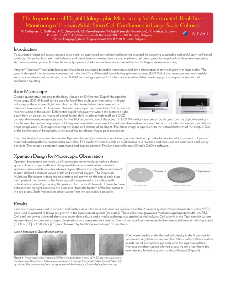
iLine microscope was used to monitor, and finally assess, Human Adult stem cell confluence in the Xpansion system. Mesenchymal stem cells (MSC)
were used as a model to follow cell growth in the Xpansion 50 system (50 plates). These cells were grown in a medium supplemented with 10% FBS.
Cell confluence was achieved after six to seven days culture and a media exchange was applied at mid-culture. Cell growth in the Xpansion 50 system
was monitored by iLine microscopic observations and compared to a control. Control was a cell culture leaded in the same conditions in multitray stacks
(T-Flask [TF] or Cell stack [CS]) and followed by traditional microscopic observations.
iLine Microscope: Growth Monitoring
a
b
c
d
Figure 1: Microscopic observations (DDHM; magnification x 100) of MSC growth evolution in
the Xpansion 50 system. Pictures were taken after 1 day (a), 3 days (b), 5 days (c) and 7 days (d)
of culture. The pictures are from the upper part of the bioreactor (second plate from the top.
MSC were seeded at the desired cell density in the Xpansion 50
system and regulations were switched 8 hours after cell inoculation
in order to let cells adhere properly onto the Xpansion plates.
Microscopic observations allowed assessing cell attachment the
next day and following growth until confluence (Figure 1).
DDHM Microscope: Confluence Assessment
3-D images from microscopic observation have been automatically analyzed via
OsOne software. Cell confluency have been assessed using OsOne software and is
similar to visual observation (Figure 2).
a
b
Xpansion Fixation: Homogeneity Study
A second part of the work focused on homogeneity and studied how cells colonized
the entire bioreactor. For this, MSC were grown in an Xpansion 50 system and
cultures were stopped at different confluence levels based on DDHM microscopic
observations. Cells were then fixed inside the system with formaldehyde (3 % v/v)
and stained with violet cristal (10 % w/v). The Xpansion system was finally dismantled
plate-by-plate in order to observe cell distribution at macroscopic and microscopic
levels.
c
Figure 2: OsOne software (a) and cell confluence calculated via OsOne
software at 15% (b) and 85% (c).
Macroscopic Level
Macroscopic analysis was realized by visually comparing cell repartition from plate to plate. Figure 3 shows MSC fixed and stained in the Xpansion 50
system at confluence. Pictures showed that cells colonized all plates of the bioreactor with the same distribution pattern on each plate.
Microscopic Level
Analysis at the microscopic level observed specific areas to
check cell layer homogeneity per plate. The most relevant and
critical points were selected and compared between all plates of
the Xpansion 50 bioreactor (Figure 4).
Plate 5
Plate 20
Plate 35
Plate 50
Figure 3: MSC distribution pattern on Xpansion plates. Cells were fixed and stained at confluence
inside the Xpansion 50 system. Pictures were taken from several levels of the bioreactor from the top
(Plate 1) to the bottom (Plate 50).
Figure 4: Plate segment of an Xpansion
bioreactor; schematic representation of
microscopic observation fields intended to
compare cell homogeneity inside Xpansion;
Point 1 & Point 2.
Plate 5
Plate 20
Plate 35
Plate 50
Figure 5: MSC fixed and stained in the Xpansion 50 system at confluence, illustrated through
microscopic pictures from the optical channel. Note the cell confluence comparison of several
plates of the bioreactor (plates #5, #20; #35; #50).
Results of microscopic analysis showed that, for each observed
area, cell distribution and cell density was equivalent on each
plate of the system, as illustrated on Figure 5. This demonstrates
that cells were able to grow homogeneously inside the
bioreactor. Finally, global observation of the Xpansion system
showed that the optical channel provided the view that best
illustrated the average confluence of the plate (Figure 6).
Point 1
Point 2
Figure 6: MSC fixed and stained in the Xpansion
50 system at confluence through microscopic
observations of the most relevant areas. The
pictures are from plate #30. Point 2 is the
observation at the optical channel position.
Conclusion
Fixing and staining cells in the Xpansion system proved that cells were able to adhere and grow homogeneously on each plate of the bioreactor
and confirmed efficiency of DDHM microscopy analysis. Moreover, the optical channel, which allows real-time observations and pictures of the
top 10 plates of the bioreactor, revealed to be best representative of the cell confluence. Additional QC analysis also proved that cells grown in the
Xpansion system remained undifferentiated, as in multitray stack controls, and so kept their functionality. Given that cell confluence levels are critical
for guaranteeing cell quality, it’s important to track and monitor cell growth. The DDHM microscope is a key element for defining cell harvest time,
depending on average cell confluence. The associated and automated confluence software is therefore a useful tool in assessing cell confluence in a
reproducible and consistent way for large-scale productions.
ATMI LifeSciences
The Source of Bioprocess Efficiency™
ATMI, Rue de Ransbeek 310, 1120 Brussels, Belgium
+32 2 264 18 80 • www.atmi-lifesciences.com
© 2012 ATMI, Inc. All rights reserved. ATMI, the ATMI logo, Integrity and Xpansion are
trademarks or registered trademarks of Advanced Technology Materials, Inc. in the U.S.,
other countries, or both. All other names are trademarks of their respective companies.


