Primer
advertisement
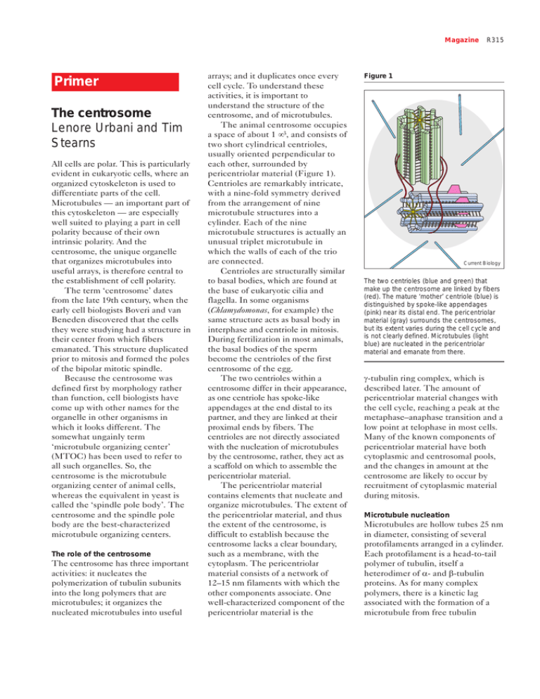
Magazine Primer The centrosome Lenore Urbani and Tim Stearns All cells are polar. This is particularly evident in eukaryotic cells, where an organized cytoskeleton is used to differentiate parts of the cell. Microtubules — an important part of this cytoskeleton — are especially well suited to playing a part in cell polarity because of their own intrinsic polarity. And the centrosome, the unique organelle that organizes microtubules into useful arrays, is therefore central to the establishment of cell polarity. The term ‘centrosome’ dates from the late 19th century, when the early cell biologists Boveri and van Beneden discovered that the cells they were studying had a structure in their center from which fibers emanated. This structure duplicated prior to mitosis and formed the poles of the bipolar mitotic spindle. Because the centrosome was defined first by morphology rather than function, cell biologists have come up with other names for the organelle in other organisms in which it looks different. The somewhat ungainly term ‘microtubule organizing center’ (MTOC) has been used to refer to all such organelles. So, the centrosome is the microtubule organizing center of animal cells, whereas the equivalent in yeast is called the ‘spindle pole body’. The centrosome and the spindle pole body are the best-characterized microtubule organizing centers. The role of the centrosome The centrosome has three important activities: it nucleates the polymerization of tubulin subunits into the long polymers that are microtubules; it organizes the nucleated microtubules into useful arrays; and it duplicates once every cell cycle. To understand these activities, it is important to understand the structure of the centrosome, and of microtubules. The animal centrosome occupies a space of about 1 µ3, and consists of two short cylindrical centrioles, usually oriented perpendicular to each other, surrounded by pericentriolar material (Figure 1). Centrioles are remarkably intricate, with a nine-fold symmetry derived from the arrangement of nine microtubule structures into a cylinder. Each of the nine microtubule structures is actually an unusual triplet microtubule in which the walls of each of the trio are connected. Centrioles are structurally similar to basal bodies, which are found at the base of eukaryotic cilia and flagella. In some organisms (Chlamydomonas, for example) the same structure acts as basal body in interphase and centriole in mitosis. During fertilization in most animals, the basal bodies of the sperm become the centrioles of the first centrosome of the egg. The two centrioles within a centrosome differ in their appearance, as one centriole has spoke-like appendages at the end distal to its partner, and they are linked at their proximal ends by fibers. The centrioles are not directly associated with the nucleation of microtubules by the centrosome, rather, they act as a scaffold on which to assemble the pericentriolar material. The pericentriolar material contains elements that nucleate and organize microtubules. The extent of the pericentriolar material, and thus the extent of the centrosome, is difficult to establish because the centrosome lacks a clear boundary, such as a membrane, with the cytoplasm. The pericentriolar material consists of a network of 12–15 nm filaments with which the other components associate. One well-characterized component of the pericentriolar material is the R315 Figure 1 Current Biology The two centrioles (blue and green) that make up the centrosome are linked by fibers (red). The mature ‘mother’ centriole (blue) is distinguished by spoke-like appendages (pink) near its distal end. The pericentriolar material (gray) surrounds the centrosomes, but its extent varies during the cell cycle and is not clearly defined. Microtubules (light blue) are nucleated in the pericentriolar material and emanate from there. γ-tubulin ring complex, which is described later. The amount of pericentriolar material changes with the cell cycle, reaching a peak at the metaphase–anaphase transition and a low point at telophase in most cells. Many of the known components of pericentriolar material have both cytoplasmic and centrosomal pools, and the changes in amount at the centrosome are likely to occur by recruitment of cytoplasmic material during mitosis. Microtubule nucleation Microtubules are hollow tubes 25 nm in diameter, consisting of several protofilaments arranged in a cylinder. Each protofilament is a head-to-tail polymer of tubulin, itself a heterodimer of α- and β-tubulin proteins. As for many complex polymers, there is a kinetic lag associated with the formation of a microtubule from free tubulin R316 Current Biology, Vol 9 No 9 subunits. This represents the time it takes for the appropriate number of tubulin subunits to be arranged into the microtubule structure. The centrosome provides a template for the assembly of the tubulin subunits, eliminating the kinetic lag. Microtubules nucleated from mammalian centrosomes typically have 13 protofilaments, whereas microtubules polymerized without the benefit of a centrosome have a range of protofilament numbers, suggesting that the centrosomal template directly determines the structure of the microtubule. What is the centrosomal template for microtubule polymerization? An answer comes from the study of γ-tubulin, a conserved component of all microtubule organizing centers. Though closely related to the α- and β-tubulins that make up the microtubule polymer, γ-tubulin is localized specifically to the pericentriolar material of the centrosome. Microtubule nucleation occurs in the pericentriolar material, and γ-tubulin is required for this nucleation activity. The γ-tubulin is part of a large protein complex that is organised into an open ring of roughly 25 nm diameter, the same as the diameter of a microtubule. Electron microscopic tomography of purified Drosophila centrosomes revealed many ring structures containing γ-tubulin in the pericentriolar material. When microtubules were grown from the centrosomes, these ring structures were found at the ends of the microtubules embedded in the centrosome, suggesting that the γ-tubulin rings provide a direct template for microtubule nucleation. An alternative hypothesis holds that the open ring of the γ-tubulin complex is actually a γ-tubulin protofilament, which acts to nucleate microtubules by interacting with the regular α/β protofilaments. Both of these models are based on the notion that γ-tubulin interacts directly with either α-tubulin or β-tubulin, or both. Although this seems likely, no such interaction has been proven as yet. The γ-tubulin complex consists of γ-tubulin and at least five other proteins. Two of these proteins, Spc97p and Spc98p, were first identified in yeast and have now been cloned from Drosophila, Xenopus and human. Interestingly, the γ-tubulin complex in Saccharomyces cerevisiae, which is much smaller than the animal cell γ-tubulin complex, consists of only γ-tubulin, Spc97p and Spc98p, and is unlikely to be able to form a ring. This suggests that the minimal functional unit for microtubule nucleation might be simpler than the large ring found in animal cells. What are centrosomes made of? The centrosome is a complex organelle that contains around 100 proteins in addition to the γ-tubulin complex. Proteins are usually defined as being centrosomal on the basis of immunofluorescence localization or, increasingly, localization of GFP fusion proteins. The resolution of such experiments is often only sufficient to reveal that a protein is in the general region of the centrosome. Some proteins localize to the centrosome independently of microtubules, whereas others require microtubules. It is useful to consider only those whose localization is microtubuleindependent as true components of the centrosome. Similarly, some proteins are present at the centrosome throughout the cell cycle, whereas others appear only at a particular time and then leave the centrosome or are degraded. There is increasing evidence that the centrosome is home to several proteins that have nothing to do with centrosomal function, and may use association with the centrosome as a means of ensuring segregation at mitosis, or as a way to increase local protein concentration. The centriole microtubules are assembled from α- and β-tubulin, and their special triplet structure also seems to require δ-tubulin, a recently discovered member of the tubulin family. The microtubules that form the centrioles are very stable, unlike most cytoplasmic microtubules, which exhibit dynamic instability, characterized by random transitions between lengthening and shortening. The stability of centriolar microtubules may derive from post-translational modifications, such as acetylation and glutamylation, of the tubulin in those microtubules. The pericentriolar material remains largely uncharacterized but some general themes are becoming apparent. For example, several proteins of the pericentriolar material have long stretches of predicted coiled-coil structure. Pericentrin is a large protein with extensive regions of predicted coiled-coil structure that has been reported to form a dynamic reticular lattice in the pericentriolar material. An example from the yeast spindle pole body is Spc110p, a large coiled-coil protein that links the γ-tubulin complex to the inner structure of the spindle pole body. These proteins are likely to be ‘struts’ upon which the functional elements of the centrosome are assembled. The core pericentriolar matrix has been studied in the large centrosomes found in Drosophila and in clam embryos, and seems to be of relatively simple constitution, although no components have been identified. Centrosome duplication The cell cycle is an ordered progression of events that leads to division of one cell into two, each with an identical copy of the genome. In normal divisions, both the chromosomal DNA and the centrosome must be duplicated only once per cycle. The structure of DNA, with its complementary strands, provides a simple mechanism for duplication. Although there have been several claims to the contrary, it is now believed that the centrosome does Magazine Figure 2 Duplication starts Elongation S G2 Cell division Separation G1 M Current Biology The centrosome duplication cycle. After cell division, each cell has one centrosome that contains two centrioles (green and blue). The centrioles separate during the G1 phase. During S phase, a ‘procentriole’ (light green and light blue) forms at each centriole. These small procentrioles grow longer during the G2 phase and migrate to form the poles of the mitotic spindle (bright blue) during mitosis. The cell divides to make two cells that each contain one centrosome. (The centrosomes are not drawn in proportion to the mitotic spindle.) not have specific nucleic acids associated with it, and therefore that it must employ some other mechanism of duplication. What happens during centrosome duplication? In the G1 phase of the cell cycle, there is one centrosome per cell, consisting of two centrioles and the associated pericentriolar material. The duplication process begins at the G1–S transition, at approximately the same time that DNA replication is initiated. The visible manifestation in the centrosome is that the centrioles move apart from each other (Figure 2). Once separated, new centrioles start to grow orthogonal to the parental centrioles. By G2, there are two centrosomes lying side by side, each with a pair of centrioles within. Centrosome duplication is semi-conservative, in that each centrosome of the duplicated pair has one old and one new centriole. Typical somatic cells must have an existing centriole to create a new centriole, although there are several well-characterized situations in both animal and plant cells in which basal body/centriole formation occurs de novo. This lack of a fundamental requirement for an existing centriole suggests that new centrioles are not strictly templated by old centrioles, and it is not known how the very regular structure of the centriole is propagated. At the G2–M transition the duplicated centrosomes move to opposite sides of the nuclear envelope. This migration is dependent on the action of kinesin microtubule motor proteins, in particular, those that work to slide apart anti-parallel microtubules. As the nuclear envelope breaks down, microtubules from the centrosomes interact with the chromosomes, and with overlapping microtubules from the opposite pole, creating the bipolar spindle. Segregation of chromosomes, followed by cytokinesis results in daughter cells with a single centrosome. How is centrosome duplication controlled so that it happens at the right time, and happens only once per cell cycle? Recent experiments have shown that cyclin E and its associated kinase Cdk2 are required for centrosome duplication. Cyclin E–Cdk2 reaches a peak of activity at the G1–S transition, and is also responsible for initiating DNA replication, consistent with the similar timing of these events. In vitro experiments have shown that separation of the centrioles is the first step in centrosome duplication requiring cyclin E–Cdk2 activity, although the relevant substrates of the kinase in this process are not yet known. An interesting difference between DNA replication and centrosome duplication is that DNA replication has a stringent once-andonly-once control that involves a mechanism termed ‘licensing’, which depends on selective access of replication factors to the DNA. Centrosome duplication seems to be less tightly controlled, as in both embryonic and somatic systems it is possible to have multiple rounds of centrosome duplication within one R317 cycle if the time spent in S phase is extended artificially. What are the unanswered questions in centrosome research? Among the most important are those concerning the molecular mechanisms behind the processes of microtubule nucleation and centrosome duplication. The molecules directly responsible for nucleation are in hand, and a detailed study of their function is now possible. For duplication, the link to the cell cycle has been established with the discovery of the the requirement for cyclin E–Cdk2 kinase activity, but little is known of what other components might be important, or of the events at the centrosome that lead to duplication of this complex structure. Perhaps the most interesting questions are the broader ones of how the cell makes use of this unique organelle, which lies at the center of the microtubule cytoskeleton and of the cell. Some of the more fanciful ideas proposed over the years — such as the potential for the centrioles of the centrosome to act as a cellular ‘gyroscope’ — are almost certainly wrong, but the steadily increasing number of proteins that localize to the centrosome suggests that considerably more than microtubule organization is going on. Key references Erickson HP, Stoffler D: Protofilaments and rings, two conformations of the tubulin family conserved from bacterial FtsZ to α/β β and γ tubulin. J Cell Biol 1996, 135:295-300. Kalt A, Schliwa M: Molecular components of the centrosome. Trends Cell Biol 1993, 3:118-128. Kellogg DR, Moritz M, Alberts BM: The centrosome and cellular organization. Ann Rev Biochem 1994, 63:639-674. Stearns T, Winey M: The cell center at 100: a meeting report. Cell 1997, 91:303-309. Zimmerman W, Sparks CA, Doxsey SJ: Amorphous no longer: the centrosome comes into focus. Curr Opin Cell Biol 1999, 11:122-128. Oakley BR: A nice ring to the centrosome. Nature 1995, 378:555-556. Address: Department of Biological Sciences, Stanford University, Stanford, California 94305-5020, USA.
