The Reproductive System
advertisement
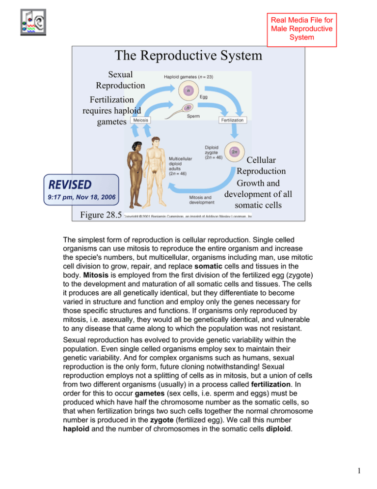
Real Media File for Male Reproductive System The Reproductive System Sexual Reproduction Fertilization requires haploid gametes 9:17 pm, Nov 18, 2006 Cellular Reproduction Growth and development of all somatic cells Figure 28.5 The simplest form of reproduction is cellular reproduction. Single celled organisms can use mitosis to reproduce the entire organism and increase the specie's numbers, but multicellular, organisms including man, use mitotic cell division to grow, repair, and replace somatic cells and tissues in the body. Mitosis is employed from the first division of the fertilized egg (zygote) to the development and maturation of all somatic cells and tissues. The cells it produces are all genetically identical, but they differentiate to become varied in structure and function and employ only the genes necessary for those specific structures and functions. If organisms only reproduced by mitosis, i.e. asexually, they would all be genetically identical, and vulnerable to any disease that came along to which the population was not resistant. Sexual reproduction has evolved to provide genetic variability within the population. Even single celled organisms employ sex to maintain their genetic variability. And for complex organisms such as humans, sexual reproduction is the only form, future cloning notwithstanding! Sexual reproduction employs not a splitting of cells as in mitosis, but a union of cells from two different organisms (usually) in a process called fertilization. In order for this to occur gametes (sex cells, i.e. sperm and eggs) must be produced which have half the chromosome number as the somatic cells, so that when fertilization brings two such cells together the normal chromosome number is produced in the zygote (fertilized egg). We call this number haploid and the number of chromosomes in the somatic cells diploid. 1 Cellular Reproduction Mitosis – exact duplication of genetic material to produce daughter cells which are genetically identical to parent cell. Diploid Cell Produces all somatic cells of the body; all cells except gametes (sperm and eggs). Mitosis produces all the somatic cells of the body (non-gametes), and they are all genetically identical. 2 Diploid Cell Meiosis Meiosis – division which reduces the number of chromosomes to produce haploid gametes. Homolgous pairs separate producing haploid cells. Meiosis I •Two division phases. •Homologous pairs separate. Meiosis II •Crossover magnifies the genetic variability. The division process which produces haploid cells is called meiosis. In fact, not only must the number be half in the gametes, it must be a specific half, namely one of each homologous pair of chromosomes. Each pair consists of chromosomes having the same genetic loci or genetic characteristics represented. If proper separation of these homologs fails to occur (called non-disjunction), the resulting zygote can have too many or too few chromosomes. For example, Down's Syndrome results from three of chromosome number 21. Meiosis occurs in two division phases. In the first division the homologs separate producing two haploid cells. Since there are 23 homologs with a choice of 2 for each, the number of different possible combinations of these in the haploid cells is 223 or 8 million. In practice there are many times this because the homologs cross over and exchange parts (synapsis) in prophase of meiosis I, changing the assortment of the genes. In meiosis II, the chromatids of each chromosome separate in a process reminiscent of mitosis. The potential number of gametes is four for each meiosis, but that varies in practice as seen below. 3 Spermatogenesis Spermatogonia Seminiferous tubules of testis 1o spermatocyte Meiosis I Meiosis II 2o spermatocyte Spermatids Sustentacular Sertoli cell (Sertoli) cells – nucleus stimulate spermatogenesis and manage the sperm’s environment. Maturation called spermiogenesis Mature sperm in epididymis The process of sperm formation occurs in the seminiferous tubules (See Figure 28.3). A man is born with stem cells or spermatogonia which have the potential to produce sperm. These cells divide continuously throughout the man's life producing more stem cells and, simultaneously, cells which undergo spermatogenesis. Spermatogenesis consists of two parts: meiosis which produces haploid pre-spermatozoa called spermatids, and spermiogenesis which is the maturation of these spermatids to produce mature sperm. Sustentacular cells manage the process in the seminiferous tubules, maintaining the environment of the spermatocytes and secreting ABP (Androgen Binding Protein) that, in combination with testosterone, stimulates the completion of spermatogenesis. Spermiogenesis begins in the seminiferous tubules, but is usually completed in the epididymis . 4 : : Oogonium : 1o oocyte Meiosis I Meiosis MeiosisIIIIonly only occurs if occurs if II Meiosis sperm sperm penetrates penetrates oocyte. Figure 28.19 oocyte. Primordial follicles Developing follicles 2o oocyte Ovulation 22oooocyte oocyte : Polar body = nonfunctional cell – all cytoplasm goes to oocyte. 1o oocyte arrested in prophase of I : At birth Oogenesis The oogonia have already matured before birth and women are born with a limited number of primary oocytes which have already begun, and are suspended in, prophase of the first meiotic division. Each month a small number of these primary oocytes continue meiosis I, usually from alternating ovaries, and usually only one becomes a secondary oocyte. (Fertility drugs are FSH derivatives and stimulate many follicles, which increases the probability that some will develop into secondary oocytes to be fertilized) It is the secondary oocyte which is ovulated. Surrounding each early primary oocyte is a primordial follicle. These follicles develop along with the oocytes, first becoming primary follicles and continuing as growing or secondary follicles, and ultimately becoming a mature (a.k.a. Graafian or vesicular) follicle which contains the secondary oocyte which is ovulated. 5 : Spermatogenesis vs. Oogenesis • Four gametes formed per meiotic division • Unlimited number of gametes may be formed throughout the man’s life • Only one functional gamete per meiotic division • Limited to those primary oocytes present at birth • Second division only occurs if fertilization occurs. 6 Brain-testicular Axis 1) Hypothalamus monitors L hormone levels. 2) FSH and ICSH are released. 3) FSH stimulates release of ABP (androgen binding protein). 4) ICSH causes release of testosterone -ABP binds testosterone to stimulate spermatogenesis. 5) Negative feedback by testosterone and inhibin suppresses gonadotropins. inhibin testosterone ICSH FSH Sustentacular cell Spermatogenesis is controlled by the gonadotropins of the anterior pituitary, which in turn are controlled by the hypothalamus. ICSH (Interstitial Cell Stimulating Hormone, a.k.a. LH) stimulates the interstitial cells to produce testosterone and other androgens. FSH stimulates the Sustentacular (Sertoli) Cells to produce a substance called Androgen Binding Protein (ABP) which, as its names suggests, binds to the androgen testosterone. The testosterone-ABP combination stimulates spermatogenesis. Feedback to the hypothalamus-pituitary controls the process. Testosterone in the blood feeds back to suppress ICSH release. This modulates testosterone levels, keeping them within the normal range. Testosterone is important for other processes such as the normal function of the seminal vesicles and prostate, as well as other masculinizing effects. A hormone product of the Sustentacular Cells called inhibin acts to suppress the secretion of FSH by the adenohypophysis. Testosterone and inhibin act independently in suppressing ICSH and FSH, but both suppress GnRH from the hypothalamus. In ways not completely understood FSH is also suppressed when sperm are not ejaculated and build up in the epididymis. Under these conditions spermatogenesis slows to a crawl. Conversely, if sperm are ejaculated often and therefore don't build up FSH is not suppressed and spermatogenesis is encouraged. 7 Control of Spermatogenesis Hypothalamus - f.b. - f.b. Adenohypophysis FSH ICSH Sustentacular cells Inhibin Regulates Regulates rate rateof ofsperm sperm formation. formation. ABP Interstitial cells Testosterone Spermatogenesis Regulates Regulates testosterone testosterone level. level. Under normal circumstances the level of testosterone feeds back to regulate ICSH release and therefore keep testoterone levels within the normal range. Likewise, inhibin regulates FSH release and spermatogenesis. But excessive levels of testosterone, e.g. when abused, will suppress GnRH and both FSH and ICSH release and therefore the body's own testosterone production and spermatogenesis fails. 8 Spermatic cord The Testes Testicular artery Head of Epididymis Testicular veins Rete testes Tunica vaginalis Tunica albuginea Seminiferous tubules Ductus (vas) deferens septum Cauda epididymis Testicles are suspended in a skin-and-muscular sac known as the scrotum into which the testes descend before birth. (See Figures 28.2 and 28.3) The scrotum is lined with a thick tunica vaginalis and each testis is covered by a whitish tunica albuginea which forms septa which divide the testis into lobes. The process of sperm formation occurs in the seminiferous tubules. From the seminiferous tubules the sperm migrate through the rete testes to the highly coiled epididymis (Figure 28.3). The epididymis actually stretches to 4 to 6 m. and consists of a head, a body, and a tail (the cauda epididymis), which wraps around the testis. The epididymis leads to the vas (ductus) deferens which carries sperm to the urethra. Sperm mature during their passage through the epididymis acquiring motility and the ability to fertilize an oocyte. 9 Cells of the Seminiferous Tubule Outer epithelium Spermatogonia Spermatocytes Spermatids Sustentacular cell 10 Sustentacular (Sertoli) Cells Sertoli (sustentacular) cell Spermatid Spermatocyte Sustentacular (Sertoli) cells surround the developing spermatocytes and manage their environment and protect them. Note the elongated spermatids entering the tubule’s lumen. 11 Interstitial Cells Spermatocytes Seminiferous epithelium Interstitial cells produce testosterone Interstitial cells are found in between the seminiferous tubules, and produce testosterone. 12 Epididymis Tubule h.p. Epididymis tubule (lined with pseudostratified columnar with stereocilia) Smooth muscle provides providesperistalsis peristalsisfor forsperm sperm movement toward the movement toward theductus ductus deferens. deferens. The lining of the epididymis is made of pseudostratified columnar epithelial cells, many of which possess microvilli (called stereocilia) which aid in secretion and absorption as the cells manage the sperm's environment. A thin layer (or two) of smooth muscle is also present. 13 Vas Deferens, lp. Three layers of smooth muscle: outer-longitudinal, middle-circular, and inner-longitudinal layers. The vas deferens has three layers of smooth muscle which propels the sperm during emission and ejaculation. Pseudostratified columnar with stereocilia (microvilli) are present, similar to the lining of the epididymis. 14 Male Reproductive Tract Seminal vesicle Produces 65% of semen, alkaline to neutralize acid Corpus cavernosum Ejaculatory duct Corpus spongiosum Prostate gland Bulbourethral gland Scrotum Scrotumkeeps keeps o o testis testisat at33oto to55o below belowbody bodytemp. temp. Vas deferens Epididymis Scrotum Testis The vas deferens is a continuation of the cauda epididymis and is histologically very similar, including the pseudostratified columnar epithelium with microvilli and three layers of smooth muscle. The vas deferens continues into the body cavity through the spermatic cord until it joins with the duct of the seminal vesicle to form the ejaculatory duct which runs through the prostate. [prostate in situ] (See Figure 28.1) The prostate is composed of secretory epithelial glands which secrete a sperm-activating semen during ejaculation. Acid phosphatase is among the substances secreted. In patients with prostatic cancer, blood levels of acid phosphatase are used to check for metastasis. Fibromuscular tissue of the prostate propels the semen into the urethra and wraps around the ejaculatory duct to function in ejaculation. The seminal vesicles secrete an alkaline fluid containing sugars and other substances which makes up 65% of the semen. The bulbourethral (Cowper's) glands secrete mucus into the urethra prior to ejaculation. 15 The Spermatic Cord Corpora cavernosa Corpus spongiosum Pampiniform plexus Cremaster muscle Venous Venousplexus plexus which acts which actsas asaa radiator radiatorto tocool cool incoming blood incoming blood to tothe thetestis. testis. Contracts Contracts during duringcold cold weather weatherto topull pull testes testescloser closer to tobody. body. Epididymis Testis The spermatic cord has a heat control system resulting from a network of veins called the pampiniform plexus. (See Figure 28.2) These veins absorb heat from the incoming testicular artery and radiate it away from the testicle, helping to maintain the optimum temperature for spermatogenesis of 5 to 7 degrees below body temperature. The dartos muscle of the scrotum along with the cremaster muscle of the spermatic cord help to pull the testes closer to the body during cold weather. 16 Spermiogenesis Head from nucleus Acrosome from lysosome Midpiece from mitochondria, etc. Flagellum from centrioles Spermiogenesis begins in the seminiferous tubules, but is usually completed in the epididymis . In spermiogenesis all non-essential components of the spermatids are lost in order that the sperm have only the chromosomes and the machinery required to propel them to the female oocyte. The nucleus of the cell becomes the head of the sperm, and the lysosomes become the acrosome. (See Figure 28.9) The acrosome contains digestive enzymes in order to digest the cumulous mass (derived from the corona radiata) around the oocyte. The midpiece of the sperm is derived from the mitochondria and other metabolic organelles of the cell, and the flagellum is derived from the centrioles. The flagellum returns to its role as centrioles after fertilization has occurred. 17 : Real Media file for Female Reproductive System Control of Oogenesis - f.b. + f.b. - f.b. high estrogen low estrogen FSH LH Follicle development Completion of meiosis I Ovulation Estrogen and progesterone Corpus luteum Estrogen and progesterone also cause endometrial buildup. Figure 28.21 1) Hypothalamus sends GnRH to ant. Pituitary causing the release of FSH. 2) FSH causes follicle development and secretion of estrogen. 3) Low levels of estrogen suppress FSH release thru – f.b. 4) High levels of estrogen stimulate release of LH in a positive feedback mechanism. (5) LH causes completion of Meiosis I, (6) ovulation, and (7) development of the corpus luteum. (8) The corpus luteum produces both estrogen and progesterone which exert – f.b. on GnRH secretion. 18 FSH Primordial follicles The Ovary Primary follicle 2o follicles estrogen Mature (Graafian or vesicular) follicle Corona radiata Corpus albicans 2o Estrogen progesterone Corpus luteum LH LH oocyte Figure 28.12 The oogonia have already matured before birth and women are born with a limited number of primary oocytes which have already begun, and are suspended in, prophase of the first meiotic division. Each month a small number of these primary oocytes continue meiosis I, usually from alternating ovaries, and usually only one becomes a secondary oocyte. (Fertility drugs are FSH derivatives and stimulate many follicles, which increases the probability that some will develop into secondary oocytes to be fertilized) It is the secondary oocyte which is ovulated. Surrounding each early primary oocyte is a primordial follicle. These follicles develop along with the oocytes, first becoming primary follicles and continuing as growing or secondary follicles, and ultimately becoming a mature (a.k.a. Graafian or vesicular) follicle which contains the secondary oocyte which is ovulated. After ovulation the follicle becomes a corpus luteum under the control of LH. If no fertilization occurs the corpus luteum will break down and produce a corpus albicans. 19 The Primordial Follicle The primordial follicle (red arrow). Atretic follicles (blue arrows). Atretic follicles are ones whose development has been curtailed. Usually only one follicle will complete the entire process to maturity and ovulation. A limited number of primordial follicles are present at birth. Each contains a primary oocyte arrested in prophase of Meiosis I. 20 Primary Follicle At least two layers of follicular cells identify the primary follicle (red arrow). A primordial follicle (blue arrow). Zona pellucida – a polysaccharide membrane surrounding the oocyte. Under the stimulation of FSH a few primordial follicles develop each month, first forming a primary follicle. 21 Secondary Follicle Antrum (A) – a fluid-filled space characterizes secondary follicles and beyond. Oocyte (arrow) Granulosa cells A secondary or growing follicle contains the beginnings of a fluid-filled antrum (space). 22 Mature (Graafian) Follicle Theca interna Theca externa Antrum Secondary oocyte Corona radiata Granulosa cells Zona pellucida Cumulous oophorus A mature (Graafian or vesicular) follicle contains a secondary oocyte which has resulted from Meiosis I. This occurs under stimulus of LH. 23 Test Diagram of Ovarian Structures Figure 28.20 Use this diagram to test yourself on the stages seen previously. 1. Primordial follicle 2. Primary follicle 3. through 5. Growing follicles 6. A mature or graafian follicle 7. A follicle at ovulation of the secondary oocyte 8. A corpus luteum 9. A corpus albicans 24 (1) Gonadotropin rise (FSH) starts cycle 1 Gonadotropins control oogenesis under the regulation of the hypothalamus. (See Figures 28. 21 and 28.22 modified) The cycle begins about day 4 with Follicle Stimulating Hormone (FSH) stimulating the development of some of the primordial follicles to become primary follicles with accompanying development of the oocyte. 26 (2) FSH causes development of follicles, which produce estrogen. Estrogen (and later, progesterone) causees endometrial buildup FSH continues to exert influence over these follicles causing them to secrete estrogen. Estrogen has two effects at this point. The low levels of estrogen which occur at first exert negative feedback through the hypothalamus to suppress FSH secretion. Estrogen also causes the proliferative phase of endometrial development. 27 Estrogen levels: (3) Estrogen rises from secretion by follicles. (4) Low estrogen exerts neg. feedback suppressing FSH 5 1 4 (5) High estrogen causes LH spike 3 Estrogen stimulates uterine proliferative phase. The low levels of estrogen which occur at first exert negative feedback through the hypothalamus to suppress FSH secretion. This prevents development of additional follicles. (Multiple births from fertility drugs usually result when they are not cut off as they should be when developing oocytes are discovered) The second effect of estrogen is to stimulate the buildup of the functional endometrium, the lining of the uterus which will accept a zygote. This first phase of endometrial buildup is called the proliferative phase of the uterus because it is characterized by the growth of large numbers of capillaries and secretory cells. Late in the follicular phase of the ovary estrogen levels peak. This estrogen surge actually exerts a positive feedback effect on the pituitary, causing the release of luteinizing hormone (LH). 28 LH Effects: (6) LH causes completion of meiosis I, ovulation, and development of corpus luteum. Estrogen Progesterone LH causes completion of Meiosis I and ovulation of the secondary oocyte produced. This occurs about day 14 of the cycle. LH also causes the follicle remaining in the ovary to become a glandular corpus luteum. The corpus luteum produces and secretes progesterone and estrogens. These hormones continue the buildup of the endometrium, now in its secretory phase. They also exert a feedback effect on the pituitary suppressing LH secretion. 29 (8) Gonadotropins suppressed The corpus luteum: 5 1 (7) Corpus luteum produces estrogen and progesterone. 8 4 3 7 Estrogen and progesterone secreted by the corpus luteum exerts negative feedback through the hypothalamus to suppress gonadotropin release. 30 X (9) Lack of LH causes degeneration of corpus luteum (10) Without corpus luteum estrogen and progesterone decline X Estrogen X X Progesterone X (11) Without hormones endometrium exfoliates This becomes self-defeating if no egg is fertilized because without LH secretion the corpus luteum will break down, at about day 25 of the cycle. And without the corpus luteum progesterone and estrogen secretion will cease and the endometrium will break down, an event called menstruation, which occurs by day 28. (The corpus luteum shrinks and shrivels into the white corpus albicans which remains in the ovary. Old ovaries show the presence of many corpi albicans. If an egg is fertilized resulting tissues (more below) will produce HCG, Human Chorionic Gonadotropin, which replaces LH to keep the corpus luteum intact and hormone levels high, continuing the maintenance of the endometrium. HCG together with other luteotropins, keeps the corpus luteum going for about 8 weeks into the pregnancy. It grows to 2 to 3 cm filling most of the ovary. After about 6 weeks of pregnancy the placenta will produce estrogen and progesterone to take over the function of the corpus luteum. HCG can be detected in the serum as early as 6 days after conception and in the urine as early as 10 to 14 days. An immunoglobulin response to HCG in urine is the basis for the modern pregnancy test. 31 Endometrium Uterine Structure Uterine gland Stratum functionalis Sinusoids Spiral artery Stratum basalis Myometrium – smooth muscle Figure 28.15 Uterine artery and vein Blood Supply - The uterine artery gives off 6 to 10 arcuate arteries which anastomose with one another in the myometrium. Branches from these called radial arteries enter the basal layer of the endometrium, giving off straight arteries which supply this region, and continuing upward into the functional layer as the highly-coiled spiral arteries. These spiral arteries lead to numerous arterioles which anastomose to supply a rich capillary bed that includes thin-walled dilated segments called lacunae. (See Figures 28.15 and 29.7) These lacunae play an important role in providing blood supply for the placenta. The uterus is composed of three layers: the endometrium or cervical mucosa made of columnar epithelium, the myometrium, a thick smoothmuscular layer, and the perimetrium, an external serosa or visceral peritoneum. Both the endometrium and myometrium exhibit substantial change during pregnancy. As the uterus enlarges, the myometrial cells may grow to 10X their original size and new muscle cells are produced as well. The endometrium has two layers: (See Figure 28.15) the stratum basale, a supporting layer, and the stratum functionalis which proliferates then degenerates during the menstrual cycle, varying from 1 to 6 mm in thickness. 32 Female Reproductive Organs Fertilization Fallopian tube Fundus of uterus Lumen of uterus Ampulla X Fimbriae Ovary Round ligament Ovarian ligament Cervix Broad ligament Vagina Figure 28.14 The vagina is a highly folded fibromuscular tube lined by non-keratinized stratified squamous epithelium. Vaginal mucosa contains no glands and is lubricated by mucus produced by the cervical glands. Vaginal mucosa undergoes changes during the menstrual cycle which somewhat mimics those of the endometrium. Uterus - Once the morula is received from the oviduct all embryonic and fetal development occurs in the uterus. It is a melon shaped organ which has a body, a fundus, and a cervix. The cervix is a barrel-shaped portion separated from the body by the isthmus. The lumen or cervical canal has a constricted opening or os at each end. The cervical mucosa is from 2-3 mm thick and undergoes little change in thickness, nor is it sloughed off during menstruation. At mid-cycle there is a ten-fold increase in the amount of mucus produced by the cervical glands, and this mucus is less viscous and provides a more favorable environment for sperm migration than at other times when it inhibits the passage of sperm into the uterus. The external os is the site of transition from vaginal stratified squamous to cervical simple columnar epithelium. Cervical epithelial cells are constantly exfoliated into the vagina, and stained preparations (called Papanicolaou cervical smears after the scientists who developed the technique) are used to screen for precancerous and cancerous lesions of the cervix. 33 Normal Uterus 1 4 6 7 5 8 2 3 Normal uterus with fundus (1), lower uterine segment (2), cervix, vaginal cuff (3), right fallopian tube (4), right ovary (5), fimbriae (6), ovarian ligament (7), broad ligament (8). 34 There is no audio file For this slide The Cervix Cervical os 35 There is no audio file For this slide Endometrial Proliferative Phase Red arrows = uterine glands, relatively straight during the proliferative phase, are cuoidal epithelial tubules which become more and more convoluted in the later secretory phase. 36 Fertilization Figure 29.2 Acrosome Granulosa cells Zona pellucida Cortical granules Enzymes from many sperm are necessary to digest cumulous mass surrounding oocyte, but only one can penetrate. The cortical reaction: enzymes released from cortical granules cause rearrangement of fibers in zona pellucida and prevent further sperm penetration. Sperm is engulfed in cytoplasm – nucleus becomes pronucleus for first division, midpiece breaks apart, flagellum returns to role as centrioles. 37 2 Days morula blastocyst 3+ days 4+ days Fertilization produces zygote Cell mass becomes embryo oocyte Implantation – 7 to 10 AAsplit splitin inmorula morula before days after ovulation. beforedifferentiation differentiation results in two embryos results in two embryos Cell Division ––identical identicaltwins. twins. Figure 29.4 The first mitotic division occurs within 24 hours after fertilization. Cell division continues producing a morula and about 3 days, and beginning differentiation as the blastocyst is produced at about 4 days. HCG must be produced by 6 or 7 days to prevent menstruation and permit implantation of the blastocyst at 7 to 10 days. 38 Chorion Chorion becomes becomesfetal fetal portion of portion of placenta. placenta. Early Development Amniotic cavity Figure 29.7 Intervillus spaces (lacunae) Chorionic villi Chorion embryo Yolk sac Tissue Tissuewhich which develops developsinto into chorion chorion secretes secretesHCG. HCG. The Placenta - (See Figure 29.7) The developing fetus is maintained by a placenta which develops from fetal and maternal tissues. The fetal portion, formed by the chorion, and the maternal portion, formed by the decidua basalis, are together involved in physiologic exchange of substances between fetal and maternal circulation. Blood begins to circulate through the primitive cardiovascular system and into chorionic villi at about 21 days. The fetal heart rate initially is about 65 bpm but increases to around 140 bpm before birth. Intervillus spaces or lacunae provide the site for exchange of nutrients, wastes, hormones, and respiratory gases between the fetal and maternal circulatory systems. pO2 is comparatively low in fetal blood, but fetal hemoglobin is more highly saturated than adult hemoglobin and its concentration is higher. CO2 diffusion from fetus to mother causes a double Bohr effect which works in both the maternal and fetal blood to enhance the release of oxygen from maternal hemoglobin and the increase in saturation of fetal hemoglobin. 39 Placental Development Decidua basalis – maternal portion of placenta Intervillus spaces (lacunae) containing maternal blood Chorionic villi containing fetal vessels. The placental barrier between maternal and fetal blood is similar in structure to the respiratory membrane in the lungs and does not exclude alcohol, nicotine, and toxins from crossing into the fetal circulation. Except for the rare rupturing of capillary walls, which may occur at delivery, fetal and maternal blood do not mix. The placenta also functions as an endocrine organ producing steroid and peptide hormones as well as prostaglandins that play an important role in the onset of labor. By the end of the eighth week the placenta has taken over the production of progesterone and estrogen from the corpus luteum. Other hormones include HCG, Insulin-like Growth Factors I and II, placental lactogen (prolactin), other growth factors and hormones (e.g. relaxin which prepares the cervix and pelvic ligaments for birth), and oxytocin which acts together with that from the mother in instigating labor and stimulating the ejection of milk. 40 There is no audio file For this slide Hormones Secreted During Pregnancy Placental Hormones: HCG – keeps corpus luteum intact through 6th week Estrogens and progesterone – stimulate breast and other tissue development Somatomammotropin – stimulates breast development Adenohypophysis – K 50% K corticosteroids, K T4 and T3 Ovaries – relaxin softens uterine ligaments for birth Estrogen and progesterone are important in the growth and development of the breasts, but their presence actually inhibits the secretion of much milk before birth of the infant. Prolactin is produced both by the adenohypophysis of the mother and by the placenta, along with somatomammotropin which stimulates the development of secretory cells in the breast. These cells secrete a small amount of fluid, called colostrum, before birth, but actual milk secretion cannot occur until the inhibition of estrogen and progesterone are eliminated after delivery. Growth hormone, glucocorticoids, and insulin are also increased during pregnancy, as are the thyroid hormones T4 and T3, and parathyroid hormone. 41 There is no audio file For this slide Changes During Pregnancy Cardiac output K 30% Respiration K 20% K Na+ and H2O reabsorption Fetal hemoglobin - carries 20% to 50% more oxygen Breast development – stimulated by estrogen and progesterone, prolactin, somatomammotropin, GH and others 42 There is no audio file For this slide Parturition Hormonal stimuli K estrogen/progesterone ratio oxytocin – from mother and fetus prostaglandins Mechanical stimuli Stretch of uterine muscle and irritation to the cervix Æ positive feedback Parturition, or delivery, occurs as a result of a combination of increased uterine muscle excitability and a positive feedback mechanism. Both hormonal and mechanical factors are involved. The hormone oxytocin is produced by both the placenta and the mothers neurohypophysis as a result of increasing estrogen:progesterone ratio. This hormone stimulates increasing contractions of the uterine fundus. These contractions cause more pressure to be exerted on the cervix by the head of the fetus. The cervix responds to this pressure through a neural and hormonal reflex to stimulate more forceful contractions of the uterine muscle. This positive feedback mechanism continues to result in ever more forceful contractions until birth occurs. Labor can often be induced by physically stretching the cervix. A hormone called relaxin causes the pelvic ligaments and cervix of "relax" and become more pliable for delivery and dilates the cervix to about 10 cm. 43 There is no audio file For this slide Disorders Related to Pregnancy Preeclampsia – high arterial pressure, protein deposited in glomerular basement membrane reduces GFR, produces edema. Gestational diabetes – hormones produced during pregnancy increase resistance of insulin receptors, especially in susceptible individuals. 44
