Session 4 – Thursday 15 May 2008
advertisement
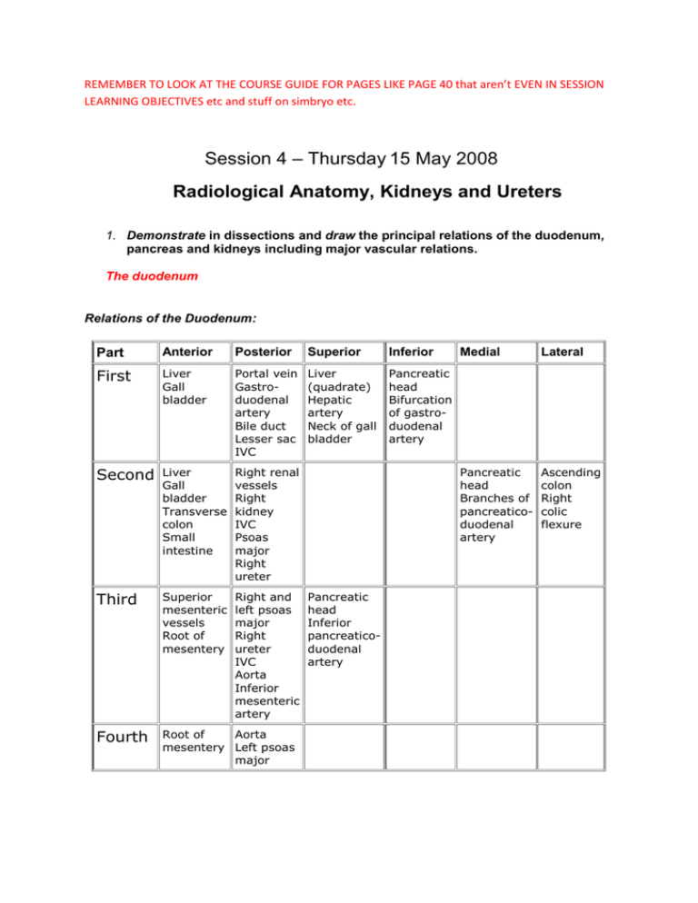
REMEMBER TO LOOK AT THE COURSE GUIDE FOR PAGES LIKE PAGE 40 that aren’t EVEN IN SESSION LEARNING OBJECTIVES etc and stuff on simbryo etc. Session 4 – Thursday 15 May 2008 Radiological Anatomy, Kidneys and Ureters 1. Demonstrate in dissections and draw the principal relations of the duodenum, pancreas and kidneys including major vascular relations. The duodenum Relations of the Duodenum: Part Anterior Posterior Superior Inferior First Liver Gall bladder Portal vein Gastroduodenal artery Bile duct Lesser sac IVC Liver (quadrate) Hepatic artery Neck of gall bladder Pancreatic head Bifurcation of gastroduodenal artery Second Liver Gall bladder Transverse colon Small intestine Right renal vessels Right kidney IVC Psoas major Right ureter Third Superior mesenteric vessels Root of mesentery Right and left psoas major Right ureter IVC Aorta Inferior mesenteric artery Fourth Root of Aorta mesentery Left psoas major Pancreatic head Inferior pancreaticoduodenal artery Medial Lateral Pancreatic head Branches of pancreaticoduodenal artery Ascending colon Right colic flexure • • • • First Part - partially in foregut mesenteries. Common Bile duct and Portal Vein pass posterior to it Second Part - Common Bile Duct and Pancreatic Ducts open into it, root of Transverse Mesocolon crosses it Third Part - Superior Mesenteric Vein lies anterior to it Fourth Part - leads into Jejunum 2. Name the main organs in contact with each of the left and right kidneys • • • • • • • • Adrenal glands on both sides Liver on right 2nd part of duodenum on right Ascending colon on right Descending colon and stomach on left Spleen on left Tail of pancreas on left Coils of small bowel especially on left 3. Describe the arterial supply of the kidneys and adrenal (suprarenal) glands 4. Name the principal macroscopic components of the kidney and relate these to microscopic organisation. For further information see kidney lectures. 5. Mark the likely positions of the kidneys and ureters in a living subject and demonstrate their positions in appropriate plain and contrast radiographs and in CT images. See Gray’s anatomy page 352 and 38-42 of course guide for details Courtesy of Ghannam et all the following website should be used for CT http://www.med.wayne.edu/diagradiology/Anatomy_Modules/Abdomen.html 6. Demonstrate how to palpate the kidneys in a living subject. An attempt should be made to palpate both the right and left kidneys. The ballottement method is normally used. Keep your anterior hand steady in the deep palpation position in the right upper quadrant lateral and parallel to rectus muscle. Attempt to ballot the kidney with the other hand in costophrenic angle. An enlarged kidney should be palpable by the anterior hand. Repeat the same maneuver for the left kidney. Alternate method for the right kidney: Place your left hand behind the patient between the rib cage and iliac crest and place your right hand below the right costal margin. While pressing your hands firmly together, ask the patient to take a deep breath. Attempt to feel the lower pole of the right kidney. Repeat the same maneuver for the left kidney. Normal: In an adult, the kidneys are not usually palpable, except occasionally for the inferior pole of the right kidney. The left kidney is rarely palpable. An easily palpable or tender kidney is abnormal. However, the right kidney is frequently palpable in very thin patients and children. 7. Demonstrate the main components of the posterior abdominal wall from diaphragm to pelvic inlet. Page 314 of Gray’s anatomy is a useful diagram. A summary will be given of this objective with further information below for understanding and further revision. The Posterior Abdominal Wall The posterior abdominal wall lies at the back of the abdomen, behind the posterior peritoneum. With the anterior structures removed (the stomach, jejunum and ileum) the retroperitoneal parts of the gut tube can be more easily identified. The duodenum The first part of the duodenum continues on from the pylorus of the stomach suspended in a mesentery. As the duodenum changes direction it becomes retroperitoneal in its descending second part and transverse third part. On the left the third part of the duodenum passes anteriorly and cranially to form the fourth part of the duodenum, continuous with the jejunum. The fourth part of the duodenum is suspended in a fold of pertitoneum, the ligament of Trietz. The first and second parts of the duodenum receive their blood supply from the anterior and posterior pancreaticoduodenal arteries which branch from the gastroduodenal artery. The third and fourth parts of the duodenum receive their arterial supply from the anterior and posterior inferior pancreaticoduodenal arteries. The pancreaticoduodenal arteries form an anastomoses between the celiac trunk and superior mesenteric artery. The portal vein is formed behind the neck of the pancreas and passes to the porta hepatis behind the first part of the duodenum. The superior mesenteric artery and vein lie anterior to the third part of the duodenum. The colon The ascending colon is retroperitoneal. The ileum enters the cecum at the ileocolic valve. The appendix is attached to the cecum and may lie in a variety of positions, including behind the cecum and within the pelvis. At the hepatic flexure the cecum is suspended in a mesentery as the transverse colon. The greater omentum is draped over the transverse colon. The descending colon begins at the splenic flexure, becoming retroperitoneal. As the descending colon reaches the iliac fossa it forms the sigmoid colon which has a short mesentery. The sigmoid colon moves towards the midline to form the rectum in the pelvis. The ileocolic artery, a terminal branch of the superior mesenteric artery, supplies the terminal ileum, caecum and appendix. A branch from the ileocolic or superior mesenteric, the right gastric, supplies the ascending colon. An early branch from the superior mesenteric, the middle colic, supplies the transverse colon. The inferior mesenteric artery supplies the descending and sigmoid colon and the rectum. The ascending left colic branch of the inferior mesenteric runs upwards to the splenic flexure and forms an anastomosis with the middle colic artery through the formation of the marginal artery. The abdominal aorta The aorta passes into the abdomen from the thorax in the midline lying on the vertebral bodies. The crura of the diaphragm form an opening so that the aorta passes behind the diaphragm under the median arcuate ligament. The aorta gives off four pairs of lumbar arteries that supply the abdominal wall (similar to the intercostal arteries of the thorax). Four other pairs are also given off: The inferior phrenic arteries supplying the diaphragm; the middle suprarenal arteries; the renal arteries; the gonadal arteries. There are three unpaired arteries which arise from the anterior aorta: the celiac trunk; the superior mesenteric artery; the inferior mesenteric artery. At the lower border of the L4 lumbar vertebra the aorta bifurcates into the common iliac arteries. The inferior vena cava The IVC lies on the right side of the aorta. The IVC is formed by the left and right common iliac veins which lie behind the right common iliac artery. The vein ascends the abdomen on the right side of the lumbar vertebrae and receives the right gonadal vein(s), the renal veins (the left gonadal vein drains into the left renal vein), the adrenal veins, the inferior phrenic veins. The IVC passes behind the liver being clasped in a groove at the back of the liver by the caudate lobe. Within the groove the IVC receives the hepatic veins. The lumbar veins drain into a pair of ascending lumbar veins which pass behind the diaphragm to become the azygos venous system in the thorax. There are connections between the ascending lumbar veins and the IVC so that blood from the azygos/ascending lumbar system can either drain through the azygos vein into the superior vena cava or through the connections to the inferior vena cava. (Note: all of the intra-abdominal gut drains through the hepatic portal vein.) The adrenal glands The adrenal glands lie on the superior pole of each kidney, with the left adrenal gland lying more on the medial aspect. The glands are separate from the capsule of the kidney. Each gland receives arterial supply from three sources: the renal artery; aorta; inferior mesenteric artery. As these branches reach the gland they break up into many small vessels. Venous drainage of the right adrenal is directly to the IVC. Venous drainage of the left adrenal is to the left renal vein. Pancreas The pancreas is for the most part retroperitoneal but becomes suspended in a mesentery (the lienorenal ligament) as the tail reaches the hilum of the spleen. The uncinate process, head and neck of the pancreas lie within the curvature of the duodenum. The pancreatic ducts drain into the duodenum. The main pancreatic duct drains the tail, body, uncinate process and part of the head. In the head the main pancreatic duct joins the bile duct to form the ampulla of Vater to drain into the second part of the duodenum. The sphincter of Oddi controls flow into the duodenum through the major duodenal papilla. The accessory pancreatic duct drains part of the head, either joining the main pancreatic duct or entering the duodenum separately as the minor duodenal papilla. The portal vein is formed behind the neck of the pancreas. The superior mesenteric artery and vein lie anterior to the uncinate process. The splenic artery supplies the body and tail of the pancreas. The neck and head of the pancreas are supplied by the anterior and posterior superior pancreaticoduodenal arteries which branch from the gastroduodenal artery. The uncinate process and part of the head are supplied by the anterior and posterior inferior pancreaticoduodenal arteries which arise from the superior mesenteric artery. The pancreatic veins drain into the portal vein. The sympathetic system The lumbar sympathetic trunk is a continuation of the sympathetic chain in the thorax. The splanchnic nerves arise from the thoracic sympathetic chain as preganglionic fibres. The greater splanchnic nerve arises from T5-T9 and passes into the abdomen through the crura of the diaphragm to synapse in the celiac ganglia. The celiac ganglia lie on either side of the celiac trunk and send postganglionic fibres with all of the branches of the celiac trunk. Similarly the lesser splanchnic (T10,11) and the least splanchnic nerves (T12) arise as preganglionic fibres in the thorax, pass to the abdomen where they synapse in preaortic ganglia (celiac, superior mesenteric, inferior mesenteric ganglia) before being distributed with the arteries to the tissues. A preaortic plexus of autonomic fibres is formed by sympathetic fibres from the preaortic ganglia and from the lumbar sympathetic chain, and by parasympathetic fibres from the vagus and S2,3,4. The pelvic organs receive their autonomic innervation from the superior and inferior hypogastric plexuses formed by extension of the preaortic plexus into the pelvis. The muscles The muscles of the posterior abdominal wall are the psoas, quadratus lumborum and the iliacus. The psoas muscle arises from the sides of the upper lumbar vertebrae and the intervertebral discs. The muscle runs downwards into the pelvis and out again under the inguinal ligament. It inserts into the lesser trochanter of the femur in common with the iliacus muscle. The psoas is innervated by the L2,3,4 lumbar nerves. The muscle is enclosed within the psoas fascia, a compartment which may limit the spread of a psoas abscess. The psoas muscle flexes the hip, or flexes the lumbar spine. Several structures such as the kidney and ureter, gonadal vessels, appendix and lumbar nerves have a close relationship to the muscle. Patients attempt to immobilize the psoas muscle when there is pain from any of these structures. This is accomplished by drawing the knees upwards passively. The quadratus lumborum muscle arises from the medial half of the twelfth rib and inserts into the iliac crest. It forms a bed for the kidney. It is innervated by the T12 and lumbar nerves. Its action is to fix the twelfth rib during inspiration. The iliacus muscle arises from the iliac fossa in the pelvis. It runs below the inguinal ligament to insert together with psoas into the lesser trochanter. It is innervated by the femoral nerve. The nerves The nerves of the posterior abdominal wall are the subcostal (T12), the ilioinguinal and iliohypogastric (L1), the lateral cutaneous nerve of the thigh (L2,3) and the femoral (L2,3,4) emerging from the lateral side of the psoas muscle; the obturator nerve (L2,3,4) emerging in the pelvis from the medial side of the psoas muscle; the genitofemoral nerve(l1,2) emerging through the psoas muscle onto its anterior surface.


