Clonorchis sinensis - Winona State University
advertisement
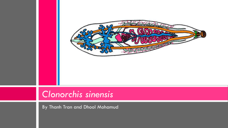
Clonorchis sinensis By Thanh Tran and Dhool Mohamud Taxonomy Kingdom: Animalia Phylum: Platyhelminthes Class: Trematoda Order: Opisthorchiida Family: Opisthorchiidae Genus: Clonorchis Species: Sinensis Clonorochis sinensis (Branched testes) Introduction Opisthorchis sinensis (lobed) Common Name: Oriental liver fluke, Chinese liver fluke Lives: in the liver of humans, and is found mainly in the common bile ducts and gall bladder. Disease: Clonorchiasis Clonorochis sinensis was first discovered by McConnell in 1875 in the bile passage of a Chinese carpenter in Calcutta. Clonorochis was formally known as Opisthorchis sinensis, however it 1907, Looss erected the genus Clonorochis because of the branched testes in contrast to the lobed testes of Opisthorchis. Hosts First intermediate host: Freshwater snail (Parafossarulus manchouricus) Occurrences Outside of Asia Second intermediate host: Fish (most commonly Grass carp) Definitive host: Humans, pigs, dogs, cats, rats, camels, Reservoir host: dogs, cats Geographic distribution: Japan, Korea, Taiwan, China, and Vietnam Currently infecting an estimated 30,000,000 humans. Believed to be the third most prevalent worm parasite in the world Clonorchiasis has been reported in non endemic areas (including the United States). In such cases, the infection was found in Asian immigrants, or individuals who have ingested pickled freshwater fish containing metacercariae imported from endemic areas. The freshwater snail vector is not found in the United States Morphology sinensis radiae - in the snail intestine Miracidium - This larval stage is ciliated and slightly oval in shape. It has 2 simple eyespots and lateral papillae which protrude outwards and serve as sensory organs. Sporocyst- The sporocyst resembles a hollow and simple sac. Rediae -It has a pharynx but no esophagus or intestine. sinensis eggs - released from adult worm Cercaria - resembles a small adult with a tail, which it loses upon penetration of the second intermediate host. The tail has dorsal and ventral fins on it to aid in locomotion. It is brownish in color Metacercaria - is encysted and does not look like a fluke. It has lost larval organs such as the eyespots, the stylet, and the tail. The cyst has thick walls and maturing fluke is visible sinensis cercariae - free-swimming sinensis metacercariae - encysted in fish Anatomy of Adult Fluke The adult worm measures to be 8-25 mm long and 1.5-5.0 mm wide. The oral sucker is slightly larger than the ventral oral sucker. The male reproductive organs consists of two large branched testes in the posterior end. The ovary consists of three lobes. The adult C. sinensis do not have a body cavity, blood circulatory system, scales or spine. The Fluke can be translucent gray or yellow due to the absorption of bile. Life Cycle Behavioral pattern: The cercaria emerges from the snail, and positions itself in an upside-down position, and sinks towards the bottom of the water. Though the exact way a C. sinensis cercaria sense stimuli is unknown, it is stimulated to swim by passing shadows and movement in the water. Any kind of stimulation will cause it to swim rapidly back up and then resumes its sinking,. This behavioral pattern favors its contact with another host. On touching the epithelium of the fish it attaches itself, castes its tail and bores through the skin. Life Cycle Embryonated eggs are discharged in the billiary ducts and in the stool Eggs are ingested by a suitable snail intermediate host . Each egg releases a miracidia , which go through several developmental stages (sporocysts , rediae , and cercariae ). The cercariae are released from the snail and after a short period of free-swimming time in water, they come in contact and penetrate the flesh of freshwater fish, where they encyst as metacercariae Infection of humans occurs by ingestion of undercooked, salted, pickled, or smoked freshwater fish After ingestion, the metacercariae excyst in the duodenum and ascend the biliary tract through the ampulla of Vater Maturation takes approximately 1 month. The adult flukes (measuring 10 to 25 mm by 3 to 5 mm) reside in small and medium sized biliary ducts. In addition to humans, carnivorous animals can serve as reservoir hosts. Symptoms In the second-order bile ducts, the flukes can cause erosion of epithelium lining. Excessive mucus production inflammation Epithelial cell proliferation Periductal fibrosis and necrosis Atrophy of surrounding liver cells In the acute phase, abdominal pain, nausea, diarrhea, and eosinophilia can occur Adult metacercaria may consume all the bile created in the liver (individual is unable to digest fats) causing obstruction of the biliary ducts. flukes (arrows) within the dilated bile ducts Clinical Diagnosis The most practical diagnostic method is microscopic observation of eggs in feces, bile, or duodenal aspirates. Computed Tomographic scan (CT) of the abdomen, and shows dilated common bile duct Treatment Triclabenzole, praziquantel, bithionol, albendazole, mebendazole The preferred treatment is Praziquantel. Praziquantel is an anthelmintic that alters ion flow across the worm membrane. This change in potential causes the worm to have muscle spasms and paralysis, helping a person's immune system attack and expel the worm. When administered at 25 mg/kg three times a day for one or two days, the cure rate is about 100% effective. Albendazole is also as effective when administered at 10 mg/kg twice a day for seven days. Public Health and Prevention The infection is not eradicable because of diversity and large numbers of nonhuman reservoirs. Many of the infected humans are asymptomatic and can shed eggs for decades. Prevalence can be reduced by promotion of sanitary disposal of human feces. Avoidance of raw fish would also reduce prevalence. Cantonese delicacy of raw carp, rice soup and soysauce Interesting Facts Adult liver flukes can produce up to 4000 eggs per day for at least six months. Worms can live up to 8 years in humans. An infected person may pass viable eggs up to 30 years. The redia is the first stage that actually feeds. It feeds actively on the tissues of the first intermediate host, normally the digestive and reproductive systems. A human host with an average infection will have two or three dozen worms; heavily infected individuals have been found with as many as 20,000 worms. Video References "Clonorchis Sinensis." Wikipedia, the Free Encyclopedia. Web. 20 Feb. 2011. <http://en.wikipedia.org/wiki/Clonorchis_sinensis>. "Clonorchiasis Site." Stanford University. Web. 20 Feb. 2011. <http://www.stanford.edu/group/parasites/ParaSites2001/clonorch/ClonorchiasisWebsite.html>. Eckroad, E. and H. Lee. 2001. "Clonorchis sinensis" (On-line), Animal Diversity Web. Accessed February 20, 2011 http://animaldiversity.ummz.umich.edu/site/accounts/information/Clonorchis_sinensis.html Roberts, Larry S., Gerald D. Schmidt, and John Janovy. Gerald D. Schmidt & Larry S. Roberts' Foundations of Parasitology. Boston: McGraw-Hill Higher Education, 2009.


