The Ankle
advertisement
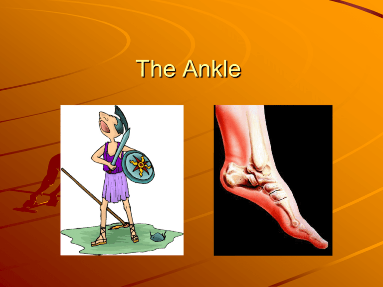
The Ankle 1) IN WHICH PLANE DOES INVERSION AND EVERSION OF THE ANKLE JOINT TAKE PLACE? Frontal or coronal plane 2) NAME THE LIGAMENTS THAT WOULD BE INJURED IN AN INVERSION INJURY OF THE ANKLE When an ankle is injured from twisting in towards the other foot, called an inversion injury, most commonly the Anterior Talofibular ligament is stretched. Calcaneofibular (CFL) is the second most common ligament injured after the anterior talofibular ligament (ATFL). 3) THE ARTICULATION OF WHICH BONES FORM THE ANKLE JOINT? The Ankle joint is made up of the Talus, the distal Tibia, and the distal Fibula The articulation between the Tibia and the Talus bears more weight. 4) ENUMERATE THE MOVEMENTS POSSIBLE IN THE ANKLE JOINT. DORSIFLEXION (Flexion): dorsal flexion; movement of the top of the ankle and foot toward the anterior tibia bone. PLANTAR FLEXION (extension): movement of the ankle and foot away from the tibia. EVERSION: turning the ankle and foot outward; abduction, away from the midline; weight is on the medial edge of the foot. INVERSION: turning the ankle and foot inward; adduction, toward the midline; weight is on the lateral edge of the foot TOE FLEXION: movement of the toes toward the plantar surface of the foot TOE EXTENSION: movement of the toes away from the plantar surface of the foot. PRONATION: a combination of ankle dorsiflexion, subtalar eversion, and forefoot abduction (toe-out) SUPINATION: a combination of ankle plantar flexion, subtalar inversion, and forefoot adduction (to-in). 5) NAME THE MUSCLE WHICH CONTRACTS CAUSING DORSIFLEXION OF ANKLE JOINT Tibialis anterior 6) WHAT IS THE DISTAL MEDIAL END OF TIBIA CALLED? The predominant structure at the distal end of the tibia is the medial malleolus 7) WHERE DOES THE TIBIA CONNECT TO THE FIBULA MEDIALLY? The fibula is placed on the lateral side of the tibia, with which it is connected superiorly and inferiorly by the Superior tibiofibular joint and the Inferior tibiofibular joint. 8) WHAT IS THE FUNCTION OF GASTROCNEMIUS MUSCLE? The function of the gastrocnemius muscle is ankle plantar flexion and knee flexion 9) WHERE IS THE INSERTION OF THE TIBIALIS POSTERIOR MUSCLE? The tibialis posterior has multiple insertions on the lower inner surfaces of the navicular, cuneiform, and second through fifth metatarsal bases. 10) Where can you palpate the tibialis anterior muscle? It is the first muscle to the lateral side of the anterior tibial border 11) Which nerve supplies the gastronemius muscle? Tibial nerve: S1, S2 12) The eversion of the ankle joint is governed by which muscles? peroneus longus, peroneus brevis 13. What is the function of flexor digiti minimi? Abduction of the proximal phalanx of the fifth phalange 14) Locate the following parts of the ankle and foot on a subject a- Lateral malleolus b- Medial malleolus c- Phalanges d- Metatarsal bone 15) Demonstrate the following movements. a- Plantar flexion b- Dorsal flexion c- Inversion d- Eversion e- Flexion of toes 16) List the plane in which the following movement takes place. a- Plantar flexion b- Flexion of toe Both take place in the sagittal plane. 17. If a person injures his lateral planter nerve then which movement is likely to get affected? Flexion of the 3rd, 4th, 5th phalanges 18) Where is the insertion of the Adductor hallucis muscle, and what is its function? Insertion is on the lateral aspect on the base of the 1st proximal phalanx. Function is adduction of the great toe and assists the flexor hallucis brevis in flexing the great toes at the metatarsophalangeal joint. 19) Which movement helps a person to walk in terrain plain? Per Dr. Ray’s instructions, omit this question. 20) What are the functions of the flexor hallucis longus muscle? Flexion of the great toe at the metatarsophalange al joint (MTP) and interphalangeal joint Inversion of the foot Plantar flexion of the ankle 21) Name the nerve supplying the following muscles a. Extensor Digitorum Longus deep peroneal nerve b. Peroneus brevis superficial peroneal nerve c. Soleus tibial nerve d. Lumbricals medial and lateral plantar nerves e. Plantar interossei lateral plantar nerve 22) How can you strengthen your gastrocnemius? Running, jumping, hopping, and skipping exercises. Heel-raising exercises with the knees in full extension and the toes on a raised surface. 23) During the concentric contraction of the soleus and gastrocnemius, what type of contraction will take place in the tibialis anterior? Concentric contraction is defined as a contraction in which there is shortening of the muscle the causes motion to occur. Because the tibialis anterior is on the opposite side of the lower leg from the soleus and gastrocnemius it will be performing the opposite action, So if the gastrocnemius and the soleus are concentrically contracted the tibialis anterior will be eccentrically contracted. 24) What is the action of the peroneous brevis? Plantar flexion of the ankle Eversion of the foot 25) Complete the table Muscle Origin Insertion Action Innervation Tibialis Anterior upper 2/3 of the lateral surface of the tibia medial cuneiform and first metatarsal bone of the foot dorsiflexion and inversion of the foot deep peroneal nerve Lumbricals tendons of flexor digitorum longus dorsal surface of 2nd, 3rd, 4th, and 5th proximal phalanxes MTP joint flexion of 2nd, 3rd, 4th, and 5th phalanges 1st lumbrical: medial plantar nerve nd 2 , 3rd, and 4th: lateral plantar nerve Soleus posterior surface of the proximal fibula and proximal 2/3 of the posterior tibial surface posterior surface of the calcaneus (Achilles tendon) plantar flexion tibial nerve Plantar interossei bases and medial shafts of the 3rd, 4th, and 5th metatarsals medial aspects of bases of 3rd, 4th and 5th proximal phalanxes MTP adduction and flexion of 3rd, 4th, and 5th phalanges lateral plantar nerve Abductor hallucis tuberosity of calcaneus, flexor retinaculum, and plantar aponeurosis medial aspect of base of 1st proximal phalanx MTP flexion and abduction of 1st phalanx medial plantar nerve Gastrocnemius medial head: posterior surface of the medial femoral condyle lateral head: posterior surface of the lateral femoral condyle posterior surface of the calcaneus (Achilles tendon) plantar flexion and flexion of the knee tibial nerve
