File
advertisement
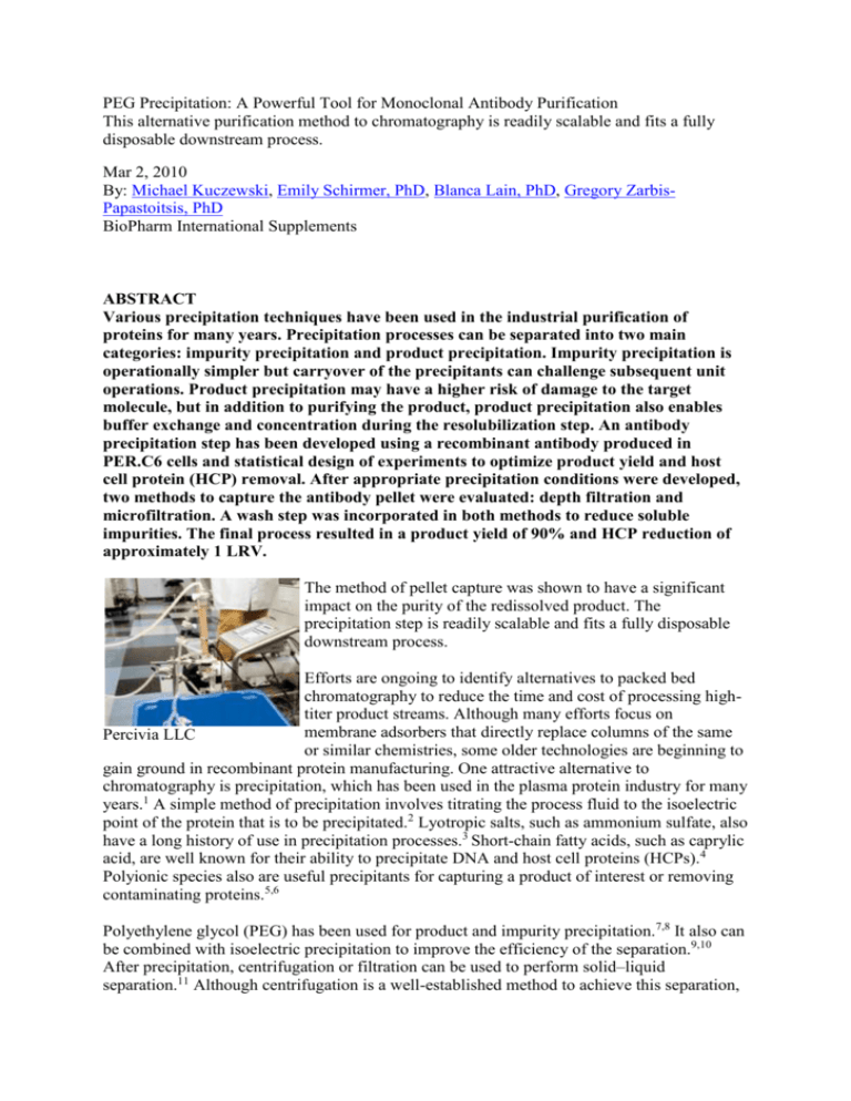
PEG Precipitation: A Powerful Tool for Monoclonal Antibody Purification This alternative purification method to chromatography is readily scalable and fits a fully disposable downstream process. Mar 2, 2010 By: Michael Kuczewski, Emily Schirmer, PhD, Blanca Lain, PhD, Gregory ZarbisPapastoitsis, PhD BioPharm International Supplements ABSTRACT Various precipitation techniques have been used in the industrial purification of proteins for many years. Precipitation processes can be separated into two main categories: impurity precipitation and product precipitation. Impurity precipitation is operationally simpler but carryover of the precipitants can challenge subsequent unit operations. Product precipitation may have a higher risk of damage to the target molecule, but in addition to purifying the product, product precipitation also enables buffer exchange and concentration during the resolubilization step. An antibody precipitation step has been developed using a recombinant antibody produced in PER.C6 cells and statistical design of experiments to optimize product yield and host cell protein (HCP) removal. After appropriate precipitation conditions were developed, two methods to capture the antibody pellet were evaluated: depth filtration and microfiltration. A wash step was incorporated in both methods to reduce soluble impurities. The final process resulted in a product yield of 90% and HCP reduction of approximately 1 LRV. The method of pellet capture was shown to have a significant impact on the purity of the redissolved product. The precipitation step is readily scalable and fits a fully disposable downstream process. Efforts are ongoing to identify alternatives to packed bed chromatography to reduce the time and cost of processing hightiter product streams. Although many efforts focus on membrane adsorbers that directly replace columns of the same Percivia LLC or similar chemistries, some older technologies are beginning to gain ground in recombinant protein manufacturing. One attractive alternative to chromatography is precipitation, which has been used in the plasma protein industry for many years.1 A simple method of precipitation involves titrating the process fluid to the isoelectric point of the protein that is to be precipitated.2 Lyotropic salts, such as ammonium sulfate, also have a long history of use in precipitation processes.3 Short-chain fatty acids, such as caprylic acid, are well known for their ability to precipitate DNA and host cell proteins (HCPs).4 Polyionic species also are useful precipitants for capturing a product of interest or removing contaminating proteins.5,6 Polyethylene glycol (PEG) has been used for product and impurity precipitation.7,8 It also can be combined with isoelectric precipitation to improve the efficiency of the separation.9,10 After precipitation, centrifugation or filtration can be used to perform solid–liquid separation.11 Although centrifugation is a well-established method to achieve this separation, washing the product pellet to remove impurities could be problematic, and it is not suited to a single-use process. Filtration—normal- or tangential-flow—requires more development, but washing the pellet is simpler, and it is readily adaptable to a single-use process. In the present work, a product precipitation step was developed using PEG to recover a monoclonal antibody (MAb) from clarified PER.C6 cell culture media. Appropriate precipitation conditions were identified through the use of full factorial experimental designs. Two filtration steps were evaluated for the capture and washing of the precipitated product, and the superior method was scaled-up 10-fold. The total precipitation process resulted in yields of approximately 90% and HCP reduction of 1 LRV with no significant increase in the aggregate level of the redissolved MAb. Finally, the impact of the precipitation step on the subsequent cation exchange (CEX) capture step was investigated. Materials and Methods Reagents USP grade salts, Tween 20, hydrochloric acid, acetic acid, and sodium hydroxide were purchased from JT Baker (Phillipsburg, NJ). PEG was of reagent grade and purchased from JT Baker or EMD Chemicals (Gibbstown, NJ). All buffers were prepared using MilliQ-grade water (Millipore, Billerica, MA) and were filtered by 0.22-µm filtration before use. Feedstock A human MAb (IgG1, pI = 8.3, 150 kDa) was produced at Percivia, LLC using a PER.C6 cell line. PER.C6 cells are human embryonic retinal cells immortalized by the adenovirus E1 gene, as described in US patent 5,994,128.12 The cells were cultured in a standard fed-batch process or the XD process, both using chemically defined media.13,14 The fed-batch media were clarified by sedimentation and depth filtration, and the XD media were clarified by the enhanced cell settling (ECS) method followed by depth filtration.15 During ECS, Silica-PEI resin was used to enhance cell settling and also reduce DNA and HCP. Precipitation Condition Optimization The conditions used to precipitate the MAb—PEG molecular weight, PEG concentration, and pH—were optimized by full factorial experimental designs using Minitab software (State College, PA). The pH of the clarified XD media was adjusted to the desired level with 2-M Tris in a 15-mL conical tube. The PEG was added as a 40% (w/w) stock solution to the desired final concentration. The tube was then centrifuged at 1,000g and the supernatant decanted. Finally, the pellet was redissolved in phosphate-buffered saline (PBS). Pellet Capture by Depth Filtration or Microfiltration Depth filtration was performed with various grades of filter media. Millistak+HC D0HC, C0HC, and X0HC were purchased from Millipore Corp. (Billerica, MA), and ZetaPlus 60SP02A was purchased from Cuno (Meriden, CT). Precipitation was carried out using a 40% (w/w) stock solution of PEG-3350 and the precipitated media was loaded at a feed flux of 50 L/m2 /h until all of the material was loaded or the transmembrane pressure (TMP) was 15 psid. The filters were then washed with 20–30 L/m2 of 20 mM Tris pH 8.5 + 14.4% (w/w) PEG-3350. After washing, 80 L/m2 of resolubilization buffer was passed through the filters at 100 L/m2 /h, and the permeate was recirculated through the device at 600 L/m2 /h until the A280 of the permeate pool was stable indicating complete MAb dissolution. Finally, any held-up product was recovered with a 20 L/m2 buffer flush and air blowdown of the filter module. In some tests, the filter media was subsequently washed with 85-mM acetate pH 5.3 followed by 1-M NaCl. Pressure and flow data were collected using a custom engineered system from ARC Technology Services (Nashua, NH). Microfiltration was performed with a 0.22-µm hollow fiber membrane from GE Healthcare Life Sciences (Piscataway, NJ). The PEG-3350 was added as a 40% (w/w) stock solution for the small-scale experiment and in powder form for the scale-up work. The feed flux was 710 L/m2 /h and the retentate and permeate were unrestricted. The precipitate was first concentrated 10- to 14-fold and then washed with three diafiltration volumes of 20 mM Tris pH 8.5 plus 14.4% (w/w) PEG-3350. Finally, the precipitate was redissolved in 85 mM sodium acetate pH 5.3 or 20 mM Tris plus 50 mM NaCl pH 7.5. Pressure, flow, and conductivity data were collected using a Slice 200 benchtop system (Sartorius, Gottingen, Germany) for small-scale testing and a SciPro system (SciLog, Middleton, WI) for the scaleup experiment. Cation Exchange Chromatography Toyopearl GigaCap S-650 was procured from Tosoh Bioscience (Montgomeryville, PA) in the Toyoscreen 5-mL format. This resin has been previously demonstrated as a high capacity capture step for MAbs.16 The column was equilibrated with 74-mM sodium acetate pH 5.3 and loaded to 90–95 mg-MAb/mL-resin using either clarified media or clarified and PEGtreated material, each adjusted to the same pH and conductivity as the equilibration buffer. The column was then washed with equilibration buffer and the antibody eluted with 50 mM sodium acetate pH 5.3 plus 90 mM NaCl. Analytical Techniques The MAb concentration in media-containing samples was determined by analytical Protein A HPLC (Applied Biosystems, Foster City, CA). Aggregate levels were measured by sizeexclusion chromatography (SEC) using a TSKgel G3000SWXL column from Tosoh Bioscience (Montgomeryville, PA), with peak detection by UV absorbance at 280 nm. HCP levels were quantified by a PER.C6-specific ELISA from Cygnus Technologies (Southport, NC). SDS-PAGE was performed with NuPAGE 4–12% Bis-Tris gels and staining was done with SimplyBlue SafeStain, both from Invitrogen (Carlsbad, CA). Results and Discussion Precipitation Conditions For both molecular weights of PEG, 3,350 and 6,000 Da, the PEG concentration was the dominant factor in the recovery and purity of the redissolved MAb. Precipitation with PEG-3350 resulted in the highest recovery (Figure 1). However, the higher recovery came at the expense of higher HCP burden in the redissolved MAb. In the case of aggregated MAb, the levels were not significantly different from the starting material, but it should be noted that the particular MAb used in this work is not prone to forming aggregates. The improvement of the HCP reduction with the use of PEG-6000 was offset by the reduction in product yield. The final Figure 1 condition selected was 14% (w/v) PEG-3350 (equivalent to 14.4% w/w) and pH 8.5. Pellet Capture by Depth Filtration In the first experiment, ECS-clarified XD media was precipitated and the pellet was captured by depth filtration with X0HC media. No substantial increase in the transmembrane pressure (TMP) was observed during the loading of 361 g-MAb/m2 (data not shown). After washing, resolubilization of the immobilized pellet was done with 85 Table 1. Yield and impurity mM acetate pH 5.3. Even after recirculation for 2 h, the removal for the PEG precipitation operation using antibody had not completely redissolved, so 0.1% v/v Tween depth filtration with X0HC to 20 was added to the resolubilization buffer. After an additional 30 min of resolubilization, the MAb was fully recover the product dissolved. Mass balance data are summarized in Table 1. The HCP reduction was lower than in the previous experiment (Figure 1), where only 5,000 ppm of HCP was in the final pool as compared to 8,300 ppm in this experiment. Some depth filters, however, are known to have hydrophobic and anion exchange (AEX) adsorptive characteristics.17 The precipitated media was loaded at a relatively high pH (8.5) and low conductivity (<10 mS/cm), which may have induced binding of acidic proteins on an AEX matrix. Furthermore, in the presence of PEG, the protein binding Figure 2 capacity of ion-exchange matrices has been shown to increase.18,19 Therefore, it is probable that some of the HCPs that remained in solution after precipitation bound to the depth filter media and eluted into the product during resolubilization. This hypothesis also is supported by the difference in the HCP content of the precipitation supernatant and the depth filter flow-through (9,900 versus 240 mg) which indicates HCP removal from the solution by the depth filter. Following immobilization of the antibody onto the depth filter, it was redissolved at a significantly lower pH (5.3) resulting in the elution of bound HCP, and reducing the purity of the final product. This phenomenon could be mitigated by using less adsorptive depth filter media, such as Millistak+HC D0HC/C0HC and ZetaPlus SP filters like 60SP02A, or by redissolving the product in a low-salt, higher-pH buffer, like 20-mM Tris pH 7.5. Table 2. Yield and impurity removal for the PEG precipitation operation using depth filtration with various filter media to recover the product These strategies were tested by loading precipitated fed-batch media onto D0HC, C0HC, X0HC, and 60SP02A filters. All filters were washed identically with the PEG/Tris buffer, but the resolubilization was done with 20 mM Tris pH 7.5 plus Tween 20 for the Millipore filters and 85 mM acetate pH 5.3 plus Tween 20 for the Cuno filter. The Millipore filters also were stripped with a low pH buffer (85 mM acetate pH 5.3) and high salt (1 M NaCl) after the product was recovered to determine if any bound HCPs could be eluted. Figure 2 shows that there was substantial fouling of all filters tested. Because the fedbatch media was not clarified by the ECS method, which has been shown to significantly reduce DNA levels in XD harvests, there may be more DNA present, which precipitates in high PEG concentrations.20 The higher DNA content in the precipitate may have reduced filter capacities, but this has not been investigated. Table 2 shows that each of the experiments resulted in better HCP reduction (84–88% versus 46%), and even though the starting HCP burden was higher (49,000 ppm versus 13,000 ppm), the redissolved MAb pools generally had lower HCP contents (6,000–7,800 ppm versus 8,300 ppm). The use of low-adsorptive filters and redissolving in a higher pH buffer appear to solve the problem of HCPs eluting from the depth filter media. SDS-PAGE of the strip fractions revealed that both HCPs and product were bound to the filters—including the less adsorptive D0HC and C0HC filters—and confirmed that X0HC media were more adsorptive than D0HC and C0HC media (Figure 3). Pellet Capture by Microfiltration Although the HCP burden of the redissolved MAb was reduced by using a buffer with a higher pH and less adsorptive depth filter media, the capacities of the depth filters were low for fed-batch media (<400 g-MAb/m2 ). Microfiltration (tangential flow filtration, MF TFF) was tested with the aim of improving capacity with the added benefit of being able to use any buffer for resolubilization because of the low binding characteristic of the hollow fiber. The hydraulic performance of the MF TFF concentration and washing of fed-batch precipitate is shown in Figure 4. The permeate flux was about 100 L/m2 /h and the TMP was between 1.0 and 2.5 psid for the entire operation. The mass loading of 475 gFigure 3 MAb/m2 was better than that achieved in any of the depth filtration operations. The product recovery and HCP reduction were both around 90%, slightly better than that achieved in depth filtration (Table 3). The resolubilization was much simpler and faster than for the depth filtration; rather than recirculating buffer through the device, the buffer was pumped through the inside of the lumens into the retentate vessel with the permeate closed and allowed to mix for about 60 min. This was sufficient to redissolve the antibody, and no excipients were needed. Because of the difficulty in resolubilization with depth filtration, the product was redissolved at or near the concentration in the feed media. The final pool from the MF TFF process was nearly two-fold more concentrated than the starting material. Figure 4 Table 3. Yield and impurity Because the retentate and permeate were open to atmospheric removal for the PEG pressure, minimal instrumentation was required: a crossflow precipitation operation using pump, one pressure sensor, and a transfer pump for the microfiltration to recover the diafiltration. The combination of high capacity, high product at bench scale recovery and HCP clearance, and operational simplicity make MF TFF a preferable option for pellet capture as compared with depth filtration. The precipitation and MF TFF step was scaled-up 10-fold to a 0.12 m2 hollow fiber device, and the performance was comparable to the small scale (Table 4). Cation Exchange Chromatography Table 4. Yield and impurity removal for the PEG precipitation operation using microfiltration to recover the product at pilot scale High capacity cation exchange (CEX) chromatography was evaluated with feeds pretreated with or without PEG precipitation. The precipitation step did not have any significant impact on the step yield or the percentage reduction of HCPs, but the eluate resulting from the precipitated load material had seven-fold less HCP (Table 5). Conclusions Precipitation has long been used in the plasma protein industry to purify proteins at large scales. The technique has been adapted here to the initial MAb purification from clarified fed-batch and XD media in a scalable manner. Two Table 5. Yield and HCP single-use filtration steps have been developed to capture and removal for GigaCap S-650 wash the precipitated product, eliminating the need for loaded with precipitated centrifugation. It was shown that the precipitation operation (+PEG) and nonprecipitated (– did not negatively affect the yield of the CEX capture step, PEG) antibody and it reduced the HCP content of the eluate by a factor of seven. The ability to reduce the impurity burden so far upstream in the purification train is key to truncating the downstream process or replacing traditional chromatography with other singleuse technologies. Lower impurity burdens can improve the loading capacity of flow-through membrane adsorbers and possibly virus filters, which are generally very expensive items in a process. An added benefit is the ability to redissolve the antibody in a buffer that facilitates the subsequent unit operation. For example, cell culture media typically requires extensive titration and dilution or a UF–DF step to prepare for capture chromatography with a cation exchanger. Here, the precipitated antibody can be dissolved in equilibration buffer at high concentration, thus shortening the processing time. This can be important for products that do not tolerate long exposure to low pH/conductivity conditions. In the case of this particular antibody, the clarified media requires a more than two-fold dilution to be loaded onto a CEX column, whereas the redissolved MAb could be loaded directly at nearly two-fold the concentration of the unadjusted media. This is at least a four-fold reduction in the load volume, which can result in substantial time savings for modern, high-capacity CEX resins. Protein purification From Wikipedia, the free encyclopedia Jump to: navigation, search Protein purification is a series of processes intended to isolate a single type of protein from a complex mixture. Protein purification is vital for the characterization of the function, structure and interactions of the protein of interest. The starting material is usually a biological tissue or a microbial culture. The various steps in the purification process may free the protein from a matrix that confines it, separate the protein and non-protein parts of the mixture, and finally separate the desired protein from all other proteins. Separation of one protein from all others is typically the most laborious aspect of protein purification. Separation steps may exploit differences in (for example) protein size, physico-chemical properties, binding affinity and biological activity. Purpose Purification may be preparative or analytical. Preparative purifications aim to produce a relatively large quantity of purified proteins for subsequent use. Examples include the preparation of commercial products such as enzymes (e.g. lactase), nutritional proteins (e.g. soy protein isolate), and certain biopharmaceuticals (e.g. insulin). Analytical purification produces a relatively small amount of a protein for a variety of research or analytical purposes, including identification, quantification, and studies of the protein's structure, posttranslational modifications and function. Pepsin and urease were the first proteins purified to the point that they could be crystallized.[1] Strategies Recombinant bacteria can be grown in a flask containing growth media. Choice of a starting material is key to the design of a purification process. In a plant or animal, a particular protein usually isn't distributed homogeneously throughout the body; different organs or tissues have higher or lower concentrations of the protein. Use of only the tissues or organs with the highest concentration decreases the volumes needed to produce a given amount of purified protein. If the protein is present in low abundance, or if it has a high value, scientists may use recombinant DNA technology to develop cells that will produce large quantities of the desired protein (this is known as an expression system). Recombinant expression allows the protein to be tagged, e.g. by a His-tag, to facilitate purification, which means that the purification can be done in fewer steps. In addition, recombinant expression usually starts with a higher fraction of the desired protein than is present in a natural source. An analytical purification generally utilizes three properties to separate proteins. First, proteins may be purified according to their isoelectric points by running them through a pH graded gel or an ion exchange column. Second, proteins can be separated according to their size or molecular weight via size exclusion chromatography or by SDS-PAGE (sodium dodecyl sulfate-polyacrylamide gel electrophoresis) analysis. Proteins are often purified by using 2D-PAGE and are then analysed by peptide mass fingerprinting to establish the protein identity. This is very useful for scientific purposes and the detection limits for protein are nowadays very low and nanogram amounts of protein are sufficient for their analysis. Thirdly, proteins may be separated by polarity/hydrophobicity via high performance liquid chromatography or reversed-phase chromatography. Evaluating purification yield The most general method to monitor the purification process is by running a SDS-PAGE of the different steps. This method only gives a rough measure of the amounts of different proteins in the mixture, and it is not able to distinguish between proteins with similar apparent molecular weight. If the protein has a distinguishing spectroscopic feature or an enzymatic activity, this property can be used to detect and quantify the specific protein, and thus to select the fractions of the separation, that contains the protein. If antibodies against the protein are available then western blotting and ELISA can specifically detect and quantify the amount of desired protein. Some proteins function as receptors and can be detected during purification steps by a ligand binding assay, often using a radioactive ligand. In order to evaluate the process of multistep purification, the amount of the specific protein has to be compared to the amount of total protein. The latter can be determined by the Bradford total protein assay or by absorbance of light at 280 nm, however some reagents used during the purification process may interfere with the quantification. For example, imidazole (commonly used for purification of polyhistidine-tagged recombinant proteins) is an amino acid analogue and at low concentrations will interfere with the bicinchoninic acid (BCA) assay for total protein quantification. Impurities in low-grade imidazole will also absorb at 280 nm, resulting in an inaccurate reading of protein concentration from UV absorbance. Another method to be considered is Surface Plasmon Resonance (SPR). SPR can detect binding of label free molecules on the surface of a chip. If the desired protein is an antibody, binding can be translated directly to the activity of the protein. One can express the active concentration of the protein as the percent of the total protein. SPR can be a powerful method for quickly determining protein activity and overall yield. It is a powerful technology that requires an instrument to perform. Methods of protein purification The methods used in protein purification can roughly be divided into analytical and preparative methods. The distinction is not exact, but the deciding factor is the amount of protein that can practically be purified with that method. Analytical methods aim to detect and identify a protein in a mixture, whereas preparative methods aim to produce large quantities of the protein for other purposes, such as structural biology or industrial use. In general, the preparative methods can be used in analytical applications, but not the other way around. Extraction Depending on the source, the protein has to be brought into solution by breaking the tissue or cells containing it. There are several methods to achieve this: Repeated freezing and thawing, sonication, homogenization by high pressure, filtration, or permeabilization by organic solvents. The method of choice depends on how fragile the protein is and how sturdy the cells are. After this extraction process soluble proteins will be in the solvent, and can be separated from cell membranes, DNA etc. by centrifugation. The extraction process also extracts proteases, which will start digesting the proteins in the solution. If the protein is sensitive to proteolysis, it is usually desirable to proceed quickly, and keep the extract cooled, to slow down proteolysis. Precipitation and differential solubilization Main article: Ammonium sulfate precipitation In bulk protein purification, a common first step to isolate proteins is precipitation with ammonium sulfate (NH4)2SO4. This is performed by adding increasing amounts of ammonium sulfate and collecting the different fractions of precipitate protein. Ammonium sulphate can be removed by dialysis.The hydrophobic groups on the proteins gets exposed to the atmosphere and it attracts other protein hydrophobic groups and gets aggregated. Protein precipitated will be large enough to be visible. One advantage of this method is that it can be performed inexpensively with very large volumes. The first proteins to be purified are water-soluble proteins. Purification of integral membrane proteins requires disruption of the cell membrane in order to isolate any one particular protein from others that are in the same membrane compartment. Sometimes a particular membrane fraction can be isolated first, such as isolating mitochondria from cells before purifying a protein located in a mitochondrial membrane. A detergent such as sodium dodecyl sulfate (SDS) can be used to dissolve cell membranes and keep membrane proteins in solution during purification; however, because SDS causes denaturation, milder detergents such as Triton X-100 or CHAPS can be used to retain the protein's native conformation during complete purification. Ultracentrifugation Main article: Ultracentrifuge Centrifugation is a process that uses centrifugal force to separate mixtures of particles of varying masses or densities suspended in a liquid. When a vessel (typically a tube or bottle) containing a mixture of proteins or other particulate matter, such as bacterial cells, is rotated at high speeds, the inertia of each particle yields an outward force proportional to its mass. The tendency of a given particle to move through the liquid because of this force is offset by the resistance the liquid exerts on the particle. The net effect of "spinning" the sample in a centrifuge is that massive, small, and dense particles move outward faster than less massive particles or particles with more "drag" in the liquid. When suspensions of particles are "spun" in a centrifuge, a "pellet" may form at the bottom of the vessel that is enriched for the most massive particles with low drag in the liquid. Non-compacted particles remain mostly in the liquid called "supernatant" and can be removed from the vessel thereby separating the supernatant from the pellet. The rate of centrifugation is determined by the angular acceleration applied to the sample, typically measured in comparison to the g. If samples are centrifuged long enough, the particles in the vessel will reach equilibrium wherein the particles accumulate specifically at a point in the vessel where their buoyant density is balanced with centrifugal force. Such an "equilibrium" centrifugation can allow extensive purification of a given particle. Sucrose gradient centrifugation — a linear concentration gradient of sugar (typically sucrose, glycerol, or a silica based density gradient media, like Percoll) is generated in a tube such that the highest concentration is on the bottom and lowest on top. Percoll is a trademark owned by GE Healthcare companies. A protein sample is then layered on top of the gradient and spun at high speeds in an ultracentrifuge. This causes heavy macromolecules to migrate towards the bottom of the tube faster than lighter material. During centrifugation in the absence of sucrose, as particles move farther and farther from the center of rotation, they experience more and more centrifugal force (the further they move, the faster they move). The problem with this is that the useful separation range of within the vessel is restricted to a small observable window. Spinning a sample twice as long doesn't mean the particle of interest will go twice as far, in fact, it will go significantly further. However, when the proteins are moving through a sucrose gradient, they encounter liquid of increasing density and viscosity. A properly designed sucrose gradient will counteract the increasing centrifugal force so the particles move in close proportion to the time they have been in the centrifugal field. Samples separated by these gradients are referred to as "rate zonal" centrifugations. After separating the protein/particles, the gradient is then fractionated and collected. Chromatographic methods Chromatographic equipment. Here set up for a size exclusion chromatography. The buffer is pumped through the column (right) by a computer controlled device. Usually a protein purification protocol contains one or more chromatographic steps. The basic procedure in chromatography is to flow the solution containing the protein through a column packed with various materials. Different proteins interact differently with the column material, and can thus be separated by the time required to pass the column, or the conditions required to elute the protein from the column. Usually proteins are detected as they are coming off the column by their absorbance at 280 nm. Many different chromatographic methods exist: Size exclusion chromatography Main article: Gel permeation chromatography Chromatography can be used to separate protein in solution or denaturing conditions by using porous gels. This technique is known as size exclusion chromatography. The principle is that smaller molecules have to traverse a larger volume in a porous matrix. Consequentially, proteins of a certain range in size will require a variable volume of eluent (solvent) before being collected at the other end of the column of gel. In the context of protein purification, the eluent is usually pooled in different test tubes. All test tubes containing no measurable trace of the protein to purify are discarded. The remaining solution is thus made of the protein to purify and any other similarly-sized proteins. Separation based on charge or hydrophobicity Hydrophobic Interaction Chromatography Resin used in the column are amphiphiles with both hydrophobic and hydrophilic regions. The hydrophobic part of the resin attracts hydrophobic region on the proteins. The greater the hydrophobic region on the protein the stronger the attraction between the gel and that particular protein. Ion exchange chromatography Main article: Ion exchange chromatography Ion exchange chromatography separates compounds according to the nature and degree of their ionic charge. The column to be used is selected according to its type and strength of charge. Anion exchange resins have a positive charge and are used to retain and separate negatively charged compounds, while cation exchange resins have a negative charge and are used to separate positively charged molecules. Before the separation begins a buffer is pumped through the column to equilibrate the opposing charged ions. Upon injection of the sample, solute molecules will exchange with the buffer ions as each competes for the binding sites on the resin. The length of retention for each solute depends upon the strength of its charge. The most weakly charged compounds will elute first, followed by those with successively stronger charges. Because of the nature of the separating mechanism, pH, buffer type, buffer concentration, and temperature all play important roles in controlling the separation. Ion exchange chromatography is a very powerful tool for use in protein purification and is frequently used in both analytical and preparative separations. Nickel-affinity column. The resin is blue since it has bound nickel. Affinity chromatography Main article: Affinity chromatography Affinity Chromatography is a separation technique based upon molecular conformation, which frequently utilizes application specific resins. These resins have ligands attached to their surfaces which are specific for the compounds to be separated. Most frequently, these ligands function in a fashion similar to that of antibody-antigen interactions. This "lock and key" fit between the ligand and its target compound makes it highly specific, frequently generating a single peak, while all else in the sample is unretained. Many membrane proteins are glycoproteins and can be purified by lectin affinity chromatography. Detergent-solubilized proteins can be allowed to bind to a chromatography resin that has been modified to have a covalently attached lectin. Proteins that do not bind to the lectin are washed away and then specifically bound glycoproteins can be eluted by adding a high concentration of a sugar that competes with the bound glycoproteins at the lectin binding site. Some lectins have high affinity binding to oligosaccharides of glycoproteins that is hard to compete with sugars, and bound glycoproteins need to be released by denaturing the lectin. Metal binding Main article: Polyhistidine-tag A common technique involves engineering a sequence of 6 to 8 histidines into the N- or Cterminal of the protein. The polyhistidine binds strongly to divalent metal ions such as nickel and cobalt. The protein can be passed through a column containing immobilized nickel ions, which binds the polyhistidine tag. All untagged proteins pass through the column. The protein can be eluted with imidazole, which competes with the polyhistidine tag for binding to the column, or by a decrease in pH (typically to 4.5), which decreases the affinity of the tag for the resin. While this procedure is generally used for the purification of recombinant proteins with an engineered affinity tag (such as a 6xHis tag or Clontech's HAT tag), it can also be used for natural proteins with an inherent affinity for divalent cations. Immunoaffinity chromatography A HPLC. From left to right: A pumping device generating a gradient of two different solvents, a steel enforced column and an apparatus for measuring the absorbance. Main article: Immunoaffinity chromatography Immunoaffinity chromatography uses the specific binding of an antibody to the target protein to selectively purify the protein. The procedure involves immobilizing an antibody to a column material, which then selectively binds the protein, while everything else flows through. The protein can be eluted by changing the pH or the salinity. Because this method does not involve engineering in a tag, it can be used for proteins from natural sources.[2] Purification of a tagged protein Another way to tag proteins is to engineer an antigen peptide tag onto the protein, and then purify the protein on a column or by incubating with a loose resin that is coated with an immobilized antibody. This particular procedure is known as immunoprecipitation. Immunoprecipitation is quite capable of generating an extremely specific interaction which usually results in binding only the desired protein. The purified tagged proteins can then easily be separated from the other proteins in solution and later eluted back into clean solution. When the tags are not needed anymore, they can be cleaved off by a protease. This often involves engineering a protease cleavage site between the tag and the protein. HPLC Main article: High performance liquid chromatography High performance liquid chromatography or high pressure liquid chromatography is a form of chromatography applying high pressure to drive the solutes through the column faster. This means that the diffusion is limited and the resolution is improved. The most common form is "reversed phase" hplc, where the column material is hydrophobic. The proteins are eluted by a gradient of increasing amounts of an organic solvent, such as acetonitrile. The proteins elute according to their hydrophobicity. After purification by HPLC the protein is in a solution that only contains volatile compounds, and can easily be lyophilized.[3] HPLC purification frequently results in denaturation of the purified proteins and is thus not applicable to proteins that do not spontaneously refold. Concentration of the purified protein A selectively permeable membrane can be mounted in a centrifuge tube. The buffer is forced through the membrane by centrifugation, leaving the protein in the upper chamber. At the end of a protein purification, the protein often has to be concentrated. Different methods exist. Lyophilization If the solution doesn't contain any other soluble component than the protein in question the protein can be lyophilized (dried). This is commonly done after an HPLC run. This simply removes all volatile components, leaving the proteins behind. Ultrafiltration Ultrafiltration concentrates a protein solution using selective permeable membranes. The function of the membrane is to let the water and small molecules pass through while retaining the protein. The solution is forced against the membrane by mechanical pump, gas pressure, or centrifugation. Analytical Denaturing-Condition Electrophoresis Gel electrophoresis is a common laboratory technique that can be used both as preparative and analytical method. The principle of electrophoresis relies on the movement of a charged ion in an electric field. In practice, the proteins are denatured in a solution containing a detergent (SDS). In these conditions, the proteins are unfolded and coated with negatively charged detergent molecules. The proteins in SDS-PAGE are separated on the sole basis of their size. In analytical methods, the protein migrate as bands based on size. Each band can be detected using stains such as Coomassie blue dye or silver stain. Preparative methods to purify large amounts of protein, require the extraction of the protein from the electrophoretic gel. This extraction may involve excision of the gel containing a band, or eluting the band directly off the gel as it runs off the end of the gel. In the context of a purification strategy, denaturing condition electrophoresis provides an improved resolution over size exclusion chromatography, but does not scale to large quantity of proteins in a sample as well as the late chromatography columns. Non-Denaturing-Condition Electrophoresis An important non-denaturing electrophoretic procedure for isolating bioactive metalloproteins in complex protein mixtures is termed 'quantitative native continuous polyacrylamide gel electrophoresis (QPNC-PAGE).
