Supplemental Methods and Data
Supplemental information
Acute zone and insect vision
Insect eyes are composed of large numbers of individual facets, so there are tradeoffs between the number and size of the ommatidia over the surface of the eye [1–3].
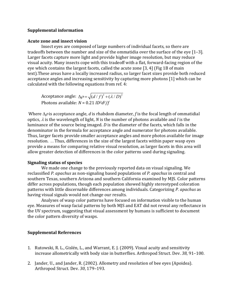
Larger facets capture more light and provide higher image resolution, but may reduce visual acuity. Many insects cope with this tradeoff with a flat, forward-facing region of the eye which contains the largest facets, called the acute zone [3, 4] (Fig 1B of main text).These areas have a locally increased radius, so larger facet sizes provide both reduced acceptance angles and increasing sensitivity by capturing more photons [1] which can be calculated with the following equations from ref. 4:
Acceptance angle: D r =
( d / f )
2 +
( l
/ D )
2
Photons available: N = 0.21 ID 2 d 2 /f
Where is acceptance angle, d is rhabdom diameter, f is the focal length of ommatidial optics, is the wavelength of light, N is the number of photons available and I is the luminance of the source being imaged. D is the diameter of the facets, which falls in the denominator in the formula for acceptance angle and numerator for photons available.
Thus, larger facets provide smaller acceptance angles and more photos available for image resolution. . . Thus, differences in the size of the largest facets within paper wasp eyes provide a means for comparing relative visual resolution, as larger facets in this area will allow greater detection of differences in the color patterns used during signaling.
Signaling status of species
We made one change to the previously reported data on visual signaling. We reclassified P. apachus as non-signaling based populations of P. apachus in central and southern Texas, southern Arizona and southern California examined by MJS. Color patterns differ across populations, though each population showed highly stereotyped coloration patterns with little discernable differences among individuals. Categorizing P. apachus as having visual signals would not change our results.
Analyses of wasp color patterns have focused on information visible to the human eye. Measures of wasp facial patterns by both MJS and EAT did not reveal any reflectance in the UV spectrum, suggesting that visual assessment by humans is sufficient to document the color pattern diversity of wasps.
Supplemental References
1. Rutowski, R. L., Gislén, L., and Warrant, E. J. (2009). Visual acuity and sensitivity increase allometrically with body size in butterflies. Arthropod Struct. Dev. 38, 91–100.
2. Jander, U., and Jander, R. (2002). Allometry and resolution of bee eyes (Apoidea).
Arthropod Struct. Dev. 30, 179–193.
3. Land, M. F. (1997). Visual acuity in insects. Annu. Rev. Entomol. 42, 147–177.
4. Land, M. F., and Nilsson, D.-E. (2012). Animal eyes (Oxford University Press).
Data used in analaysis y y n y n n n n y y y n n n y n n signal y n n species actaeon buyssoni carnifex carnifex comanchus navajoe consubrinus crinitus americanus jokahamae pacificus snelleni stigma bernadii anularis apachus bahamensis bellicosus dominulus dorsalis exclamans fuscatus major metricus surface area maximum facet diameter
0.196520262 1.418335944
0.280942525
0.273985319
1.454871407
1.461196196
0.309878104
0.242912948
0.19381238
0.309007097
1.427479323
1.451317422
1.402256388
1.474591868
0.226091398
0.204440611
0.17444675
0.36845136
0.307426527
0.290063648
0.310479154
0.233834768
0.224035811
0.249036881
0.286347988
0.293121784
0.307496237
1.379706542
1.40970343
1.368063246
1.531839957
1.459882038
1.479291711
1.4597893
1.420830236
1.413287067
1.452997192
1.465675379
1.456734394
1.47906219
NOTE:
1. Values for surface area are transformed as followed: sqrt(log(surface area mm2))
2. Values for maximum facet diameter are the log transformed values (measured in um)


