outline
advertisement
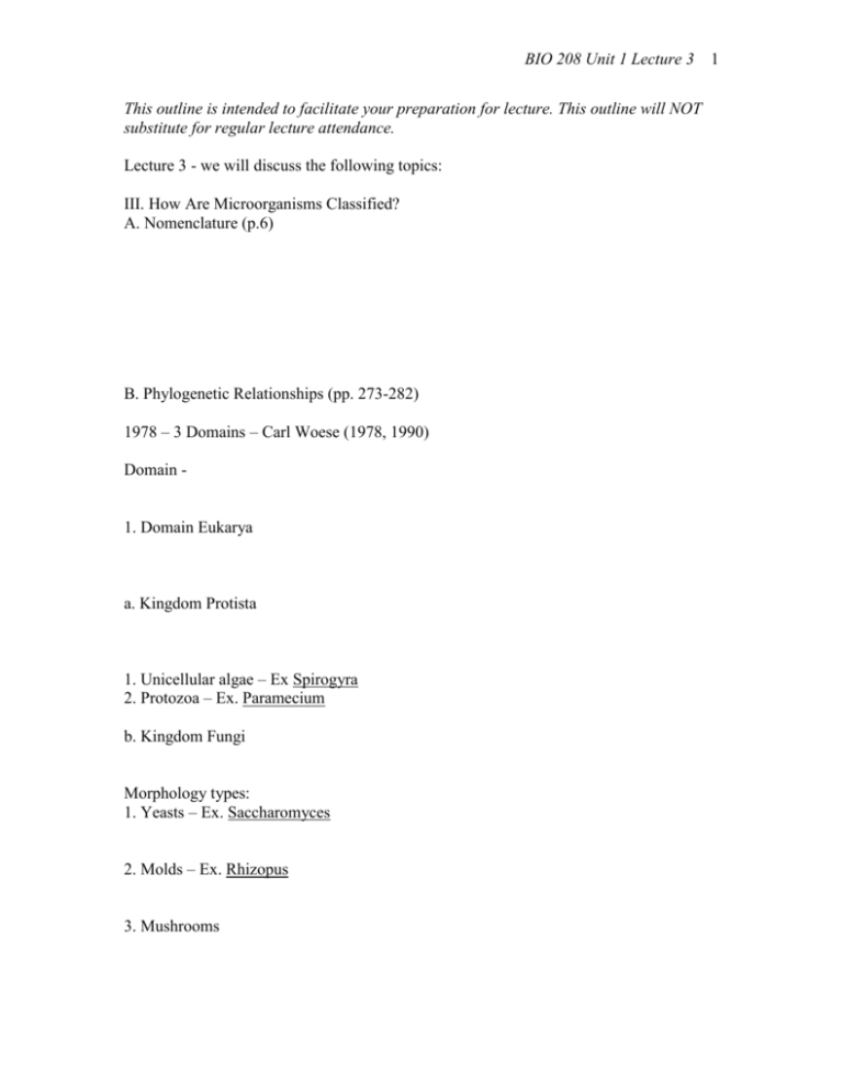
BIO 208 Unit 1 Lecture 3 This outline is intended to facilitate your preparation for lecture. This outline will NOT substitute for regular lecture attendance. Lecture 3 - we will discuss the following topics: III. How Are Microorganisms Classified? A. Nomenclature (p.6) B. Phylogenetic Relationships (pp. 273-282) 1978 – 3 Domains – Carl Woese (1978, 1990) Domain - 1. Domain Eukarya a. Kingdom Protista 1. Unicellular algae – Ex Spirogyra 2. Protozoa – Ex. Paramecium b. Kingdom Fungi Morphology types: 1. Yeasts – Ex. Saccharomyces 2. Molds – Ex. Rhizopus 3. Mushrooms 1 BIO 208 Unit 1 Lecture 3 Domains Bacteria and Archaea What do they have in common? What are the differences? 2. Domain Bacteria Ex. Escherichia coli 3. Domain Archaea Ex. Halobacterium salinarium Comparing general characteristics of the 3 Domains Domain has a nucleus? has histones? arrangement of membrane Eukarya yes yes bilayer type of phospholipids has peptidoglycan? FA no Archaea no yes bilayer or monolayer No FA no C. What do we do with: 1. Viruses - 2. Prions – BSE (bovine spongiform encephalopathy = mad cow disease) Creutzfeldt-Jacob Disease (CJD and nvCJD) Kuru Bacteria no no bilayer FA yes 2 BIO 208 Unit 1 Lecture 3 3 IV. Observing Microbes With Microscopes CH 3 p 55 Units of Measurement and pp 55-65 The Instruments A. What problems need to be overcome to see a microorganism? B. Units of measurement (you should be very comfortable with these; and converting back and forth) 1m 1 mm (millimeter) = 0.001 or 10-3 m 1 m (micrometer) = 0.000001m or 10-6 m 1 nm (nanometer) = 0.000000001 m or 10-9 m C. Properties of Light in Relation to Microscopy 1. Light - waves (wavelength = ) 2. Resolution – ability to see 2 things that are close together as 2 separate & discrete things visible light = 550 nm resolution maximum = 200 nm (0.2 m) ultraviolet light = 100-400 nm resolution max. = 100 nm electrons = 0.005 nm resolution max. = 0.2 nm 3. Refraction – bending of light as it passes from one medium to another of a different density. BIO 208 Unit 1 Lecture 3 D. Microscopy 1. Light Microscopy (LM) magnification resolving power a. Bright field b. Dark field Ex. c. Phase-contrast d. Fluorescence Ex. 4 BIO 208 Unit 1 Lecture 3 2. Electron Microscopy (EM) a. Transmission Electron Microscopy (TEM) r.p. = 0.2nm mg = 10,000-100,000x b. Scanning Electron Microscopy (SEM) r.p. = 20nm mag. = 1,000-10,000x Comparison of LM and EM Light Microscopy Electron Microscopy Illumination Light Electrons Objective Lens Glass Electromagnetic Ocular Lens Glass Electromagnetic Projector Visualize Through eye piece On view screen Assignment to complete before next class CH 10: Multiple Choice 3, 4 CH 3: Review 1, 4, 6, 7, 8, 9 5
