 Fatima Adam
Functional Histology of the Kidney (Physiology)
Functional histology of the kidney - Dr Daniel Berwick Session summary This session explains basic terminology and structural information that will enable students to understand physiological and pathological descriptions of the urinary tract, and related matters such as the control of blood pressure. Histopathologists study specimens of the urinary tract taken at biopsy, operation or autopsy. In order to diagnose diseases they must be able to recognize normal tissues and cells and identify abnormalities. Learning objectives Nephrons and their overall organization The layout of a glomerulus and its associated arterioles and Bowman’s capsule, in relation to function The three layers of the glomerular membrane and their roles in ultrafiltration of plasma The cellular organization of each of the segments of a renal tubule in relation to function, namely the proximal convoluted tubule, thin and ascending thick limbs of loop of Henle, distal convoluted tubule and collecting tubule The relation of the structure of a collecting duct to its function The juxtaglomerular apparatus and its function Transitional epithelium and its function The cellular structure of the normal walls of the ureter and bladder, in relation to the retention of urine and protection against its toxic components Session resources Functional Histology of the Kidney 2023-24 Berwick.pptxDownload Functional Histology of the Kidney 2023-24 Berwick.pptx Session activities Introduction This session continues a series of lectures on the kidney. The material has been adapted from a previous lecture written by Prof Dot Bennett. Before we get going, a quick reminder of basics: Kidney Function Kidneys are required for blood homeostasis. In particular, maintenance of: Plasma composition, via regulated secretion of water, ions and organic waste into urine; Blood pressure, via secretion of renin; Red blood cell number, via secretion of erythropoietin. In consequence, the kidney is served by very large blood vessels and receives around 25% of cardiac blood output. Basic Kidney Structure The following animation summarises the most fundamental anatomical features within a kidney:
-
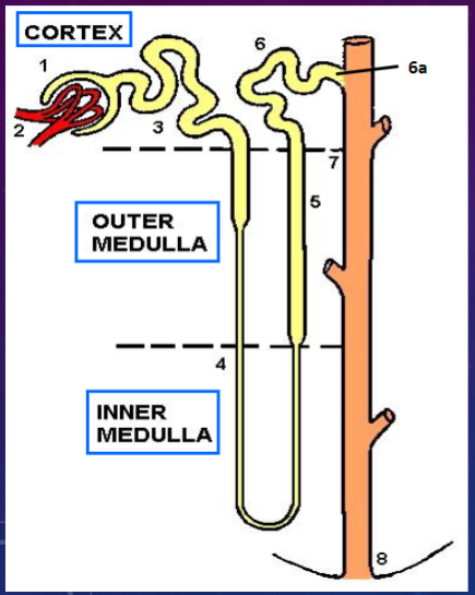
What are the parts and locations of the nephron? (8)

𖹭 Bowman’s capsule
𖹭 Glomerulus (blood capillaries)
𖹭 Proximal convoluted tubule
𖹭 Loop of Henle - thin arms (descending & ascending)
𖹭 Loop of Henle - thick arm (ascending)
𖹭 Distal convoluted tubule
𖹭 Collecting tubule (straighter, epithelium is like the collecting duct)
𖹭 Collecting duct
𖹭 Papilla
-
What are the components of the renal corpuscle? (2)
𖹭 Bowman’s capsule
𖹭 Glomerulus
-
What are the components of the renal tubule? (4)

𖹭 Proximal convoluted tubule
𖹭 Loop of Henle - thin arms (descending & ascending)
𖹭 Loop of Henle - thick arm (ascending)
𖹭 Distal convoluted tubule
𖹭 Collecting tubule
-
What does the nephron consist of? (6)
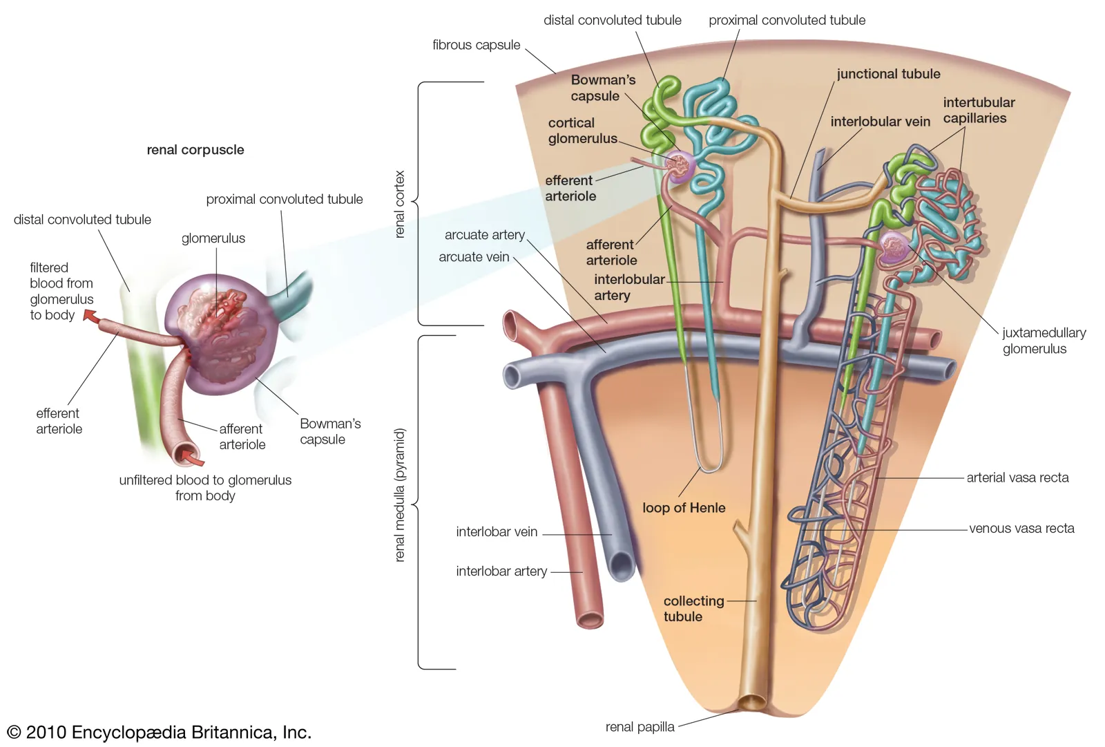
𖹭 Bowman’s capsule
𖹭 Glomerulus
𖹭 Proximal convoluted tubule
𖹭 Loop of Henle - thin arms (descending & ascending)
𖹭 Loop of Henle - thick arm (ascending)
𖹭 Distal convoluted tubule
𖹭 Collecting tubule
-
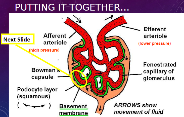
What is the structure of the glomerulus?
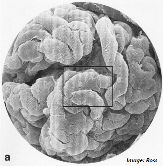
𖹭 The glomerulus is a knot of capillaries.
-
What forms part of the filter in the glomerulus and Bowman’s capsule?
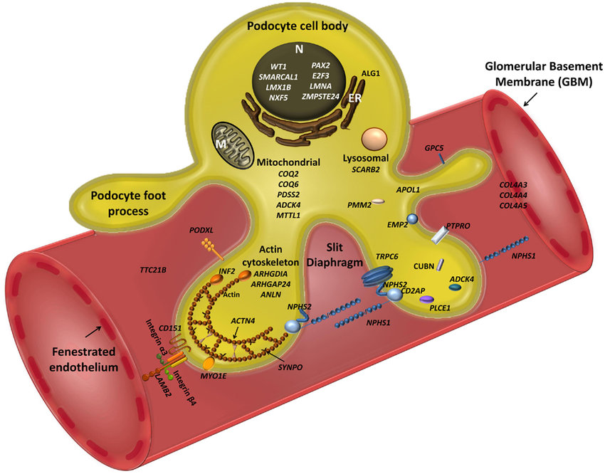
𖹭 The filter is formed by specialized epithelial cells coating the capillaries, known as podocytes.
-
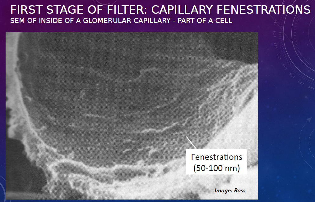
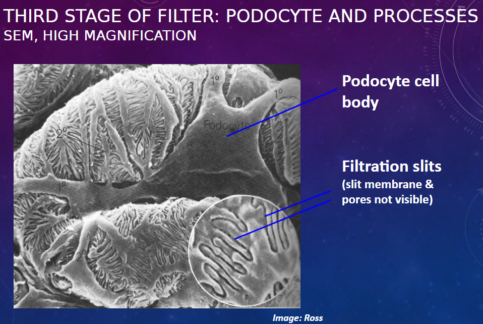
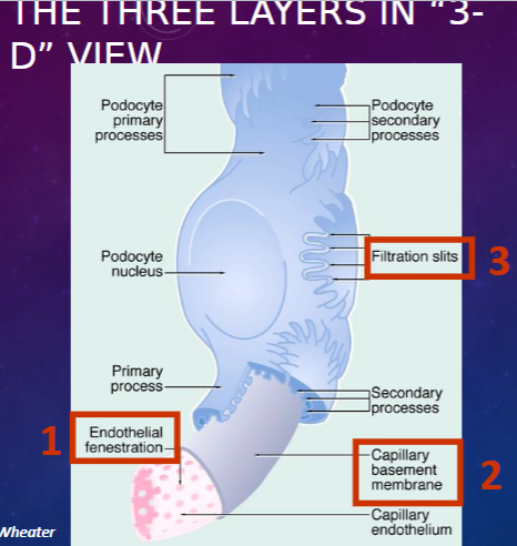
What are the three components of the filtration barrier in the glomerulus? (3)
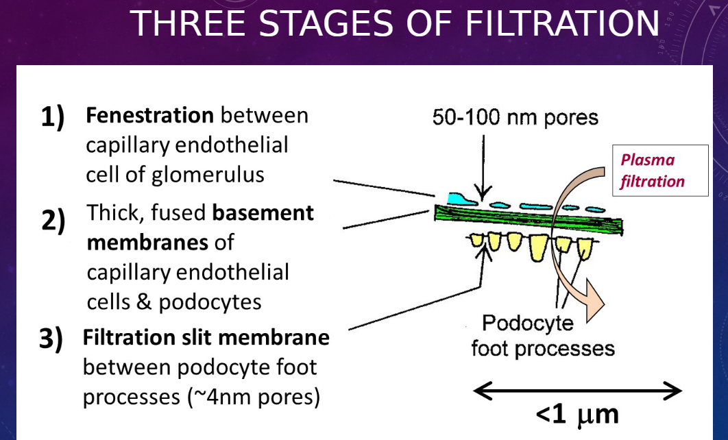
𖹭 Fenestration between capillary endothelial cells of the glomerulus
𖹭 Thick, fused basement membranes of capillary endothelial cells and podocytes
𖹭 Filtration slit membrane between podocyte foot processes (~4nm pores)
-
Picture demonstrating FIRST STAGE OF FILTER: CAPILLARY FENESTRATIONS:
-
Picture THIRD STAGE OF FILTER: PODOCYTE AND PROCESSESSEM, HIGH MAGNIFICATION:
-
Picture demonstrating the glomerulus:
-
Picture demonstrating THE THREE LAYERS IN “3-D” VIEW:
-
What is the function of the proximal convoluted tubule?
𖹭 Reabsorption from ultrafiltrate
-
What are the mechanisms of reabsorption in the proximal convoluted tubule and what molecules do they transport? (3)
𖹭 Active transport: Small molecules against a concentration gradient (e.g., glucose, amino acids, Na+ ions)
𖹭 Pinocytosis: Macromolecules, especially proteins (proteins are degraded within lysosomes and amino acids are recycled to blood)
𖹭 Passive flux: Small molecules with a concentration gradient (e.g., H2O, Cl- ions)
-
What are the features of a proximal convoluted tubule (PCT) epithelial cell?
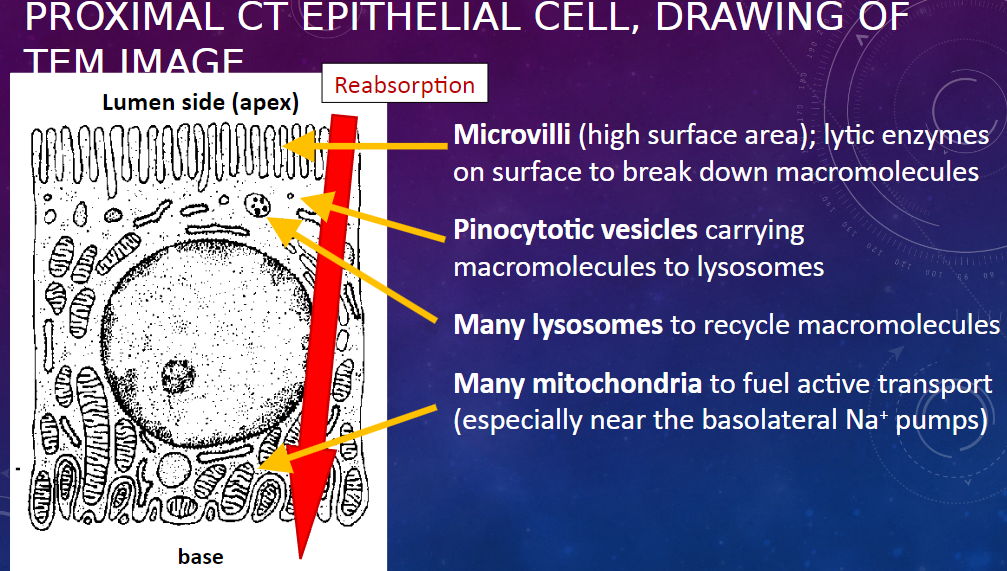
𖹭 Microvilli: High surface area for absorption
𖹭 Lytic enzymes on surface: Break down macromolecules
𖹭 Pinocytotic vesicles: Carry macromolecules to lysosomes
𖹭 Many lysosomes: Recycle macromolecules
𖹭 Many mitochondria: Fuel active transport, especially near the basolateral Na+ pumps
-
What is the function of the thin arms of the Loop of Henle?
𖹭 Reabsorption from ultrafiltrate
-
What is the mechanism of reabsorption in the thin arms of the Loop of Henle?
𖹭 By passive flux only (i.e., osmosis)
-
What are the features of the epithelial cell in the thin Loop of Henle?
𖹭 Thin, squamous epithelium: Allows passive fluxes
𖹭 A minimum of organelles
-
What is the function of the thick ascending Loop of Henle and distal convoluted tubule?
𖹭 Blood homeostasis
-
What are the mechanisms involved in their function?
𖹭 Regulated active transport and ion exchange, including Na+/K+ and H+/HCO3- exchange.
-
What are the features of the epithelial cell in the distal convoluted tubule (DCT)?
𖹭 Cuboidal epithelium: Thicker than squamous, to reduce passive fluxes and accommodate organelles
𖹭 Many mitochondria: To fuel active transport, can show as a pale or striped basal area in H&E-stained sections.
𖹭 Few, short microvilli: Unlike proximal convoluted tubule (PCT)
-
What are the functions of the collecting duct and collecting tubule?
𖹭 Transport of urine to ureter
𖹭 Water homeostasis: Passive reabsorption of water, regulated through epithelial permeability
-
What are the features of the epithelial cell in the collecting duct?
𖹭 Cuboidal to columnar epithelium: To prevent unregulated passive flux of water (and urea)
𖹭 Dense membranes at cell contacts: Function unclear, probably also helping to prevent passive flux.
-
What are the components visible in the light microscopy (H&E stain) of the nephron?
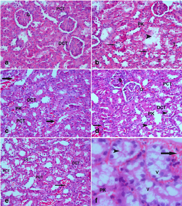
𖹭 Renal corpuscle (glomerulus + Bowman’s capsule)
𖹭 Proximal convoluted tubules
𖹭 Distal convoluted tubules (paler)
𖹭 Collecting ducts, in medullary ray
-
What are the components visible in the histology of a normal glomerulus (longitudinal view)?
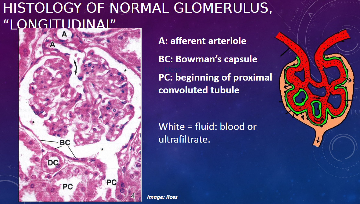
𖹭 A: Afferent arteriole
𖹭 BC: Bowman’s capsule
𖹭 PC: Beginning of proximal convoluted tubule
𖹭 White areas represent fluid, which can be either blood or ultrafiltrate.
-
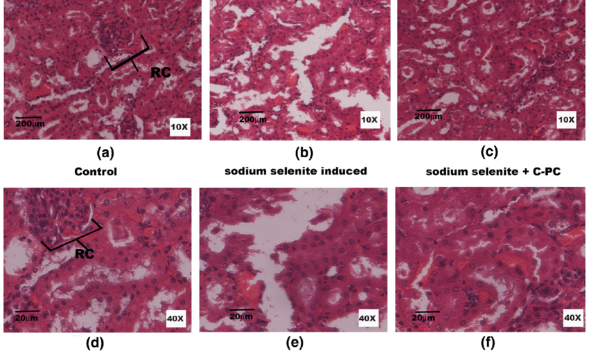
What is an example of kidney disease involving glomerular damage?
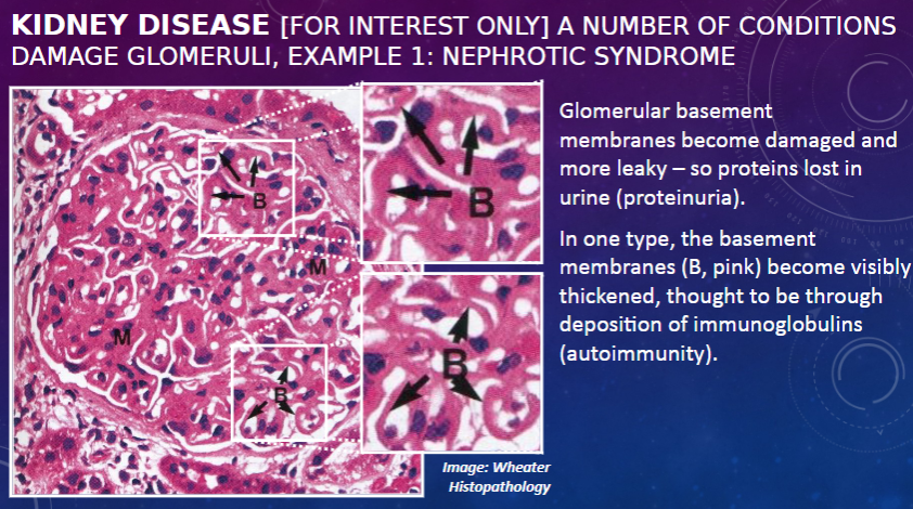
𖹭 Nephrotic syndrome: Glomerular basement membranes become damaged and more leaky, leading to protein loss in urine (proteinuria).
-
What is a characteristic feature of one type of nephrotic syndrome?

𖹭 In one type, the basement membranes become visibly thickened, thought to be through deposition of immunoglobulins, indicating autoimmunity.
-
What is an example of kidney disease associated with high blood pressure?
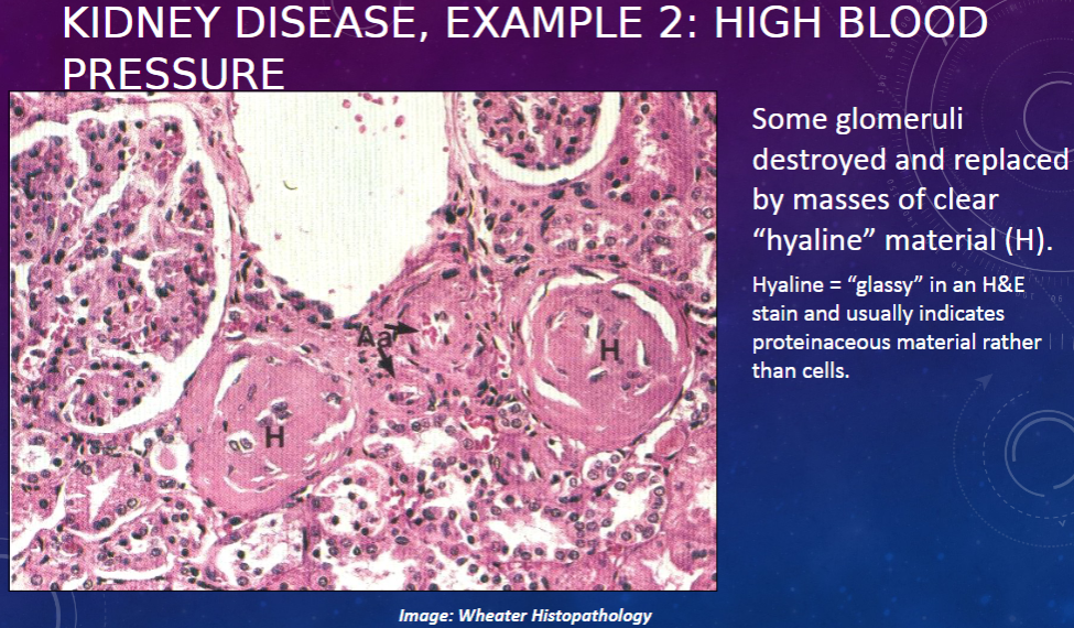
𖹭 High blood pressure: Some glomeruli are destroyed and replaced by masses of clear "hyaline" material (H). Hyaline appears "glassy" in an H&E stain and usually indicates proteinaceous material rather than cells.
-
What are the distinguishing features between proximal convoluted tubules (PCT) and distal convoluted tubules (DCT) in the cortex?
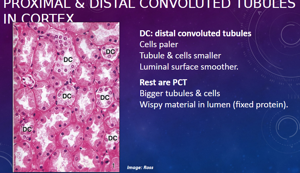
𖹭 DCT (distal convoluted tubules):
Cells appear paler
Tubules and cells are smaller
Luminal surface is smoother
𖹭 PCT (proximal convoluted tubules):
Bigger tubules and cells
Wispy material present in the lumen (fixed protein)
-
What are the components visible in the medulla section of various tubules?
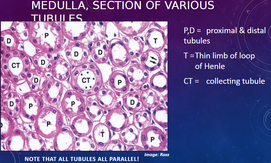
𖹭 P,D = Proximal and distal tubules
𖹭 T = Thin limb of the loop of Henle
𖹭 CT = Collecting tubule
Note: All tubules are parallel in the image.
-
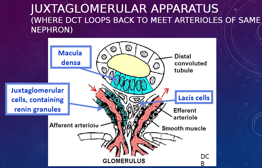
Picture demonstrating the juxtaglomerular apparatus:
-
What are the functions of the cells in the juxtaglomerular apparatus?
𖹭 Macula densa: Senses [Na+] in the DCT fluid. Appears to signal to the juxtaglomerular cells to release renin when the [Na+] is low, thereby helping to regulate blood pressure and glomerular filtration rate
𖹭 Juxtaglomerular cells: Release renin, more so in response to lower [Na+] in DCT. Renin indirectly increases vascular tone and sodium resorption.
𖹭 Lacis cells: Function unknown. (Signaling between the other cells)
-
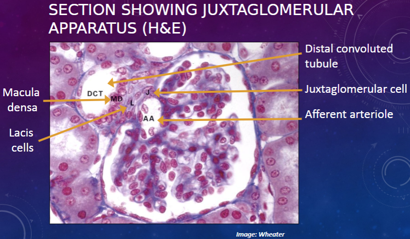
Picture demonstrating SECTION SHOWING JUXTAGLOMERULARAPPARATUS (H&E):
-
What are the components visible in the transverse section of the ureter?
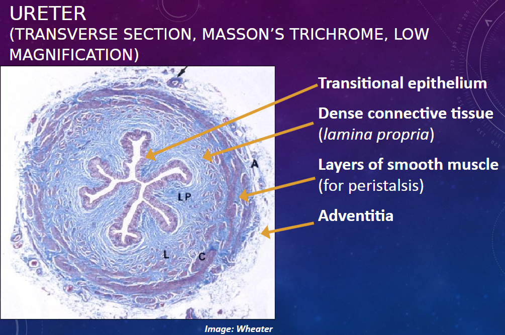
𖹭 Transitional epithelium
𖹭 Dense connective tissue (lamina propria)
𖹭 Layers of smooth muscle (for peristalsis)
𖹭 Adventitia
-
What are the characteristics of transitional epithelium?
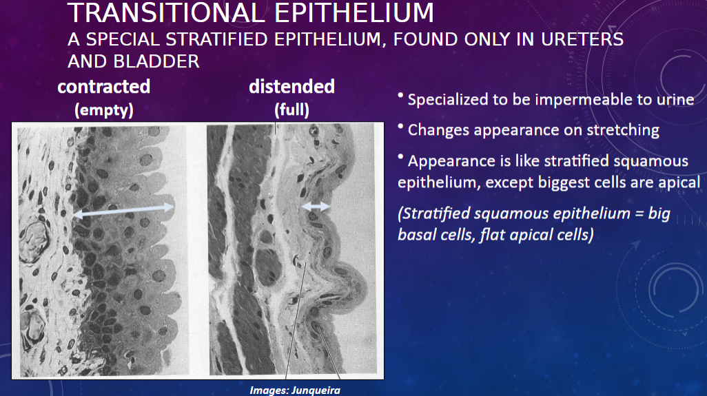
𖹭 Specialized stratified epithelium, found only in ureters and bladder.
𖹭 Impermeable to urine.
𖹭 Changes appearance on stretching.
𖹭 Appearance is like stratified squamous epithelium, except the biggest cells are apical. (Stratified squamous epithelium = big basal cells, flat apical cells).
-
Here's the flashcard based on the provided information:
What is an oddity in transitional epithelium?

𖹭 Plaques of specialized (urine-resistant) plasma membrane in apical cells: These impermeable, rigid membrane patches protect apical cells from toxic urine.
-
How do these plaques behave in different bladder states?
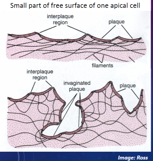
𖹭 Distended (bladder full): The plaques remain rigid, protecting apical cells.
𖹭 Contracted (bladder empty): The rigid plaques are invaginated, forming pits, allowing bladder volume to decrease.

