What are the parts and locations of the nephron? (8)
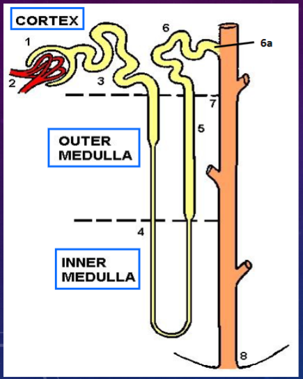
𖹭 Bowman’s capsule
𖹭 Glomerulus (blood capillaries)
𖹭 Proximal convoluted tubule
𖹭 Loop of Henle - thin arms (descending & ascending)
𖹭 Loop of Henle - thick arm (ascending)
𖹭 Distal convoluted tubule
𖹭 Collecting tubule (straighter, epithelium is like the collecting duct)
𖹭 Collecting duct
𖹭 Papilla
What are the components of the renal corpuscle? (2)
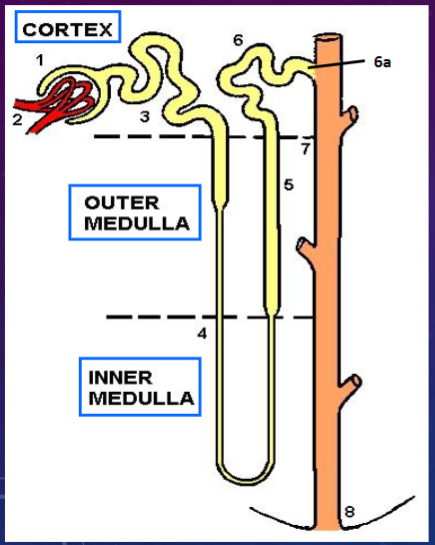
𖹭 Bowman’s capsule
𖹭 Glomerulus
What are the components of the renal tubule? (4)
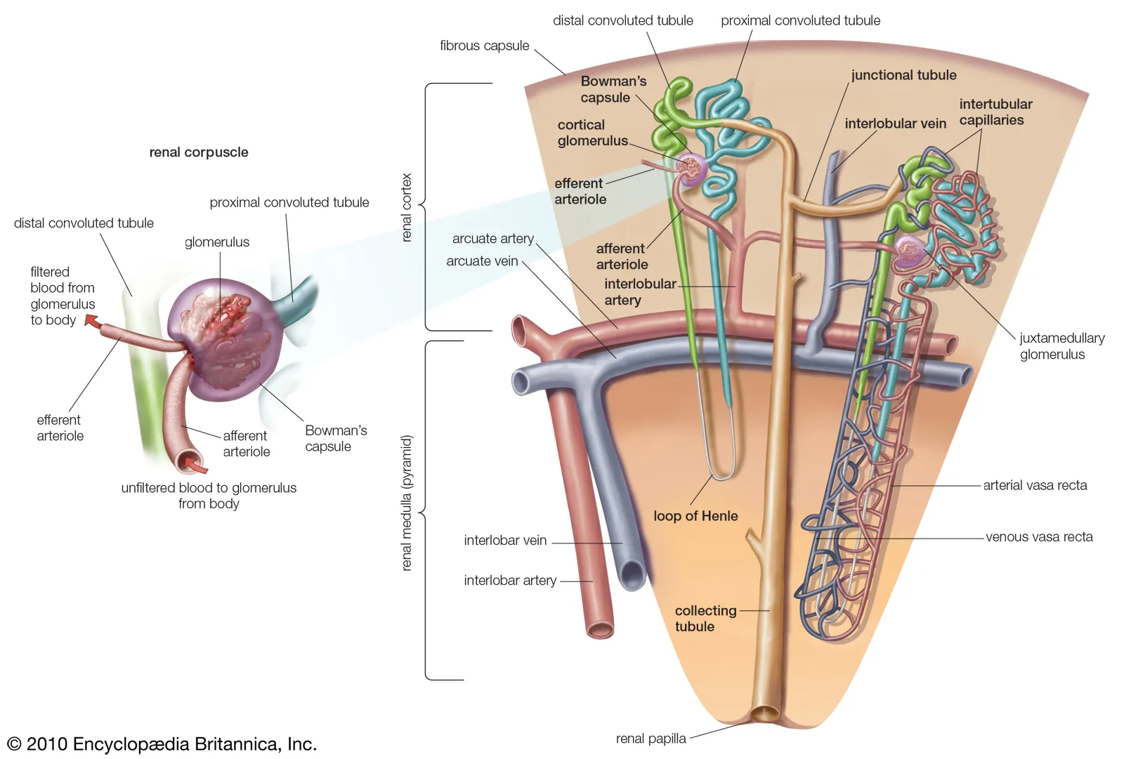
𖹭 Proximal convoluted tubule
𖹭 Loop of Henle - thin arms (descending & ascending)
𖹭 Loop of Henle - thick arm (ascending)
𖹭 Distal convoluted tubule
𖹭 Collecting tubule
What does the nephron consist of? (6)
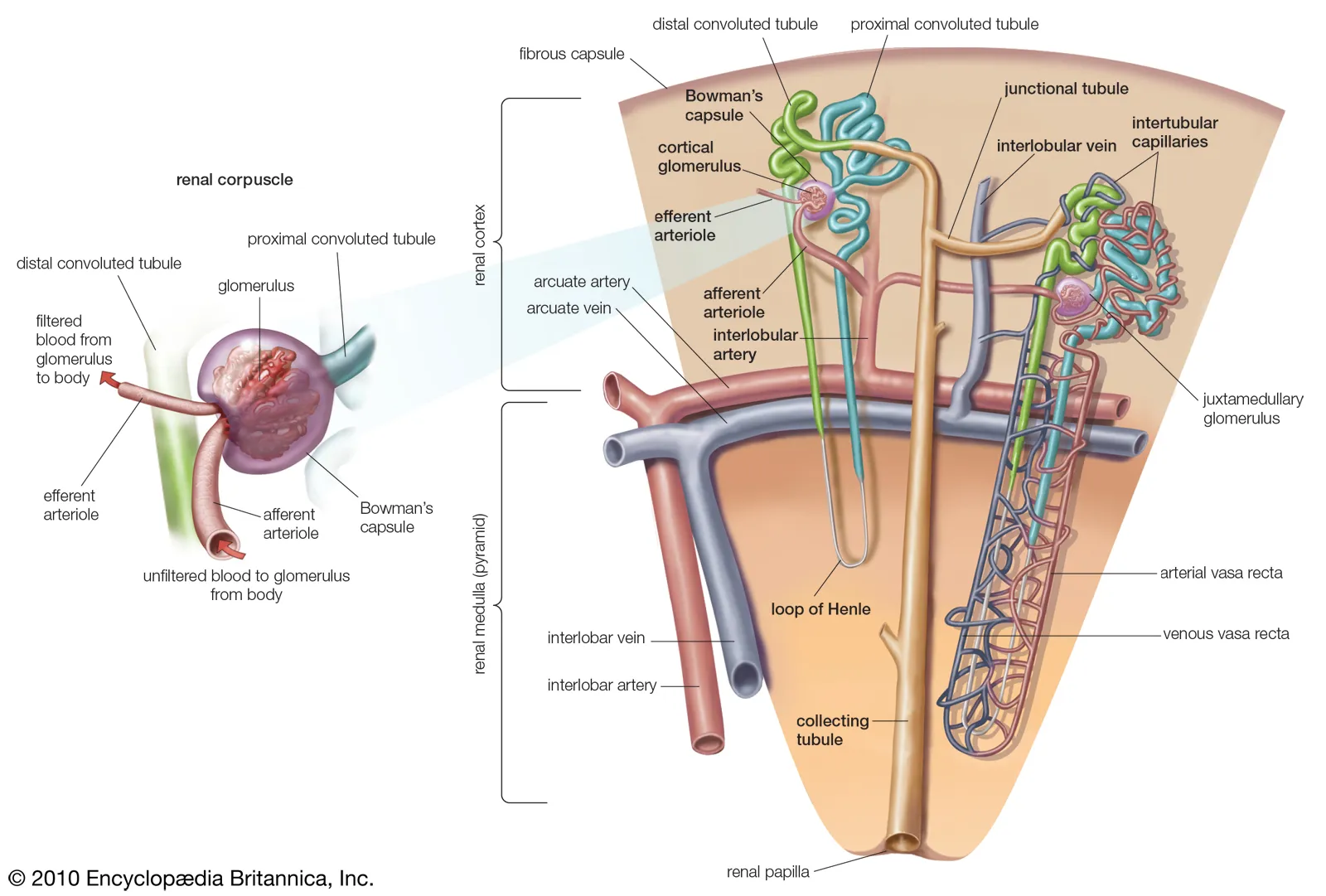
𖹭 Bowman’s capsule
𖹭 Glomerulus
𖹭 Proximal convoluted tubule
𖹭 Loop of Henle - thin arms (descending & ascending)
𖹭 Loop of Henle - thick arm (ascending)
𖹭 Distal convoluted tubule
𖹭 Collecting tubule
What is the structure of the glomerulus?
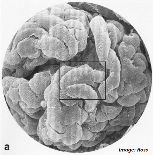
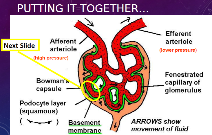
𖹭 The glomerulus is a knot of capillaries.
What forms part of the filter in the glomerulus and Bowman’s capsule?
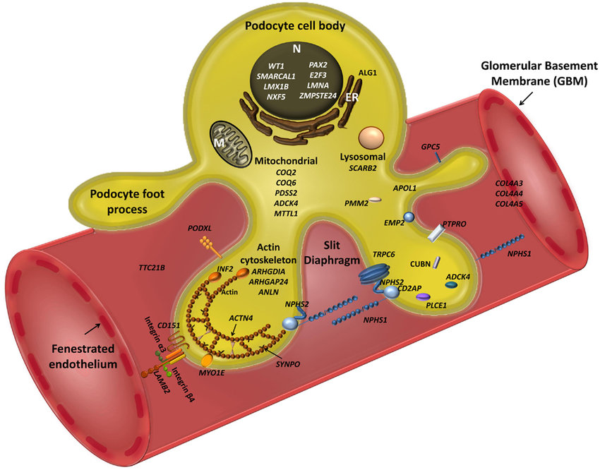
𖹭 The filter is formed by specialized epithelial cells coating the capillaries, known as podocytes.
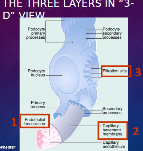
What are the three components of the filtration barrier in the glomerulus? (3)
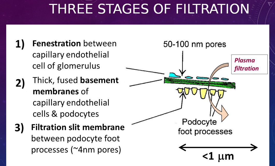
𖹭 Fenestration between capillary endothelial cells of the glomerulus
𖹭 Thick, fused basement membranes of capillary endothelial cells and podocytes
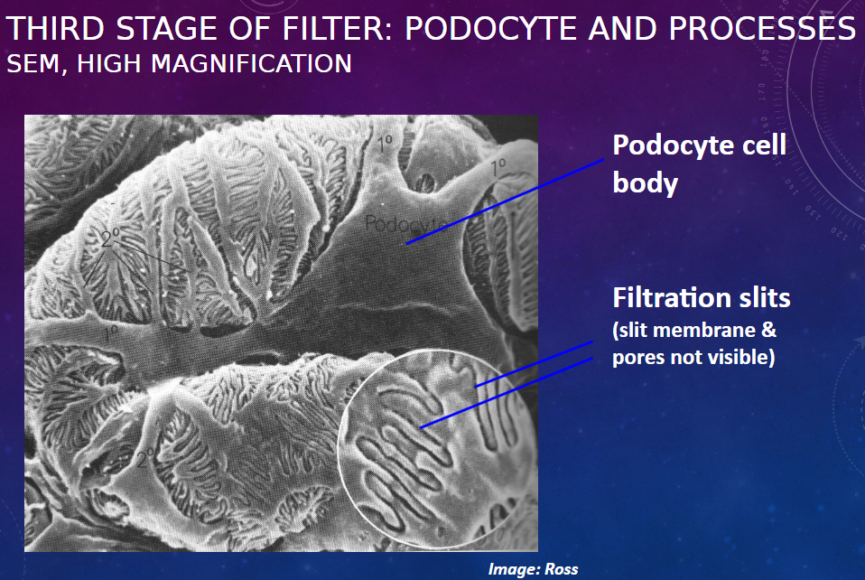
𖹭 Filtration slit membrane between podocyte foot processes (~4nm pores)
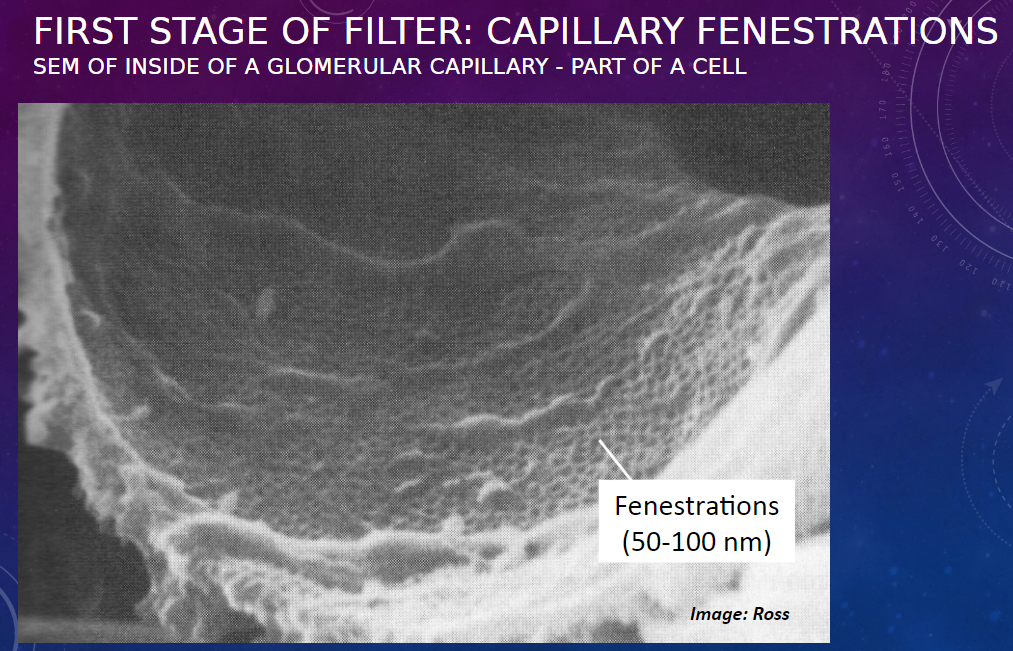
Picture demonstrating FIRST STAGE OF FILTER: CAPILLARY FENESTRATIONS:
Picture THIRD STAGE OF FILTER: PODOCYTE AND PROCESSESSEM, HIGH MAGNIFICATION:
Picture demonstrating the glomerulus:
Picture demonstrating THE THREE LAYERS IN “3-D” VIEW:
What is the function of the proximal convoluted tubule?
𖹭 Reabsorption from ultrafiltrate
What are the mechanisms of reabsorption in the proximal convoluted tubule and what molecules do they transport? (3)
𖹭 Active transport: Small molecules against a concentration gradient (e.g., glucose, amino acids, Na+ ions)
𖹭 Pinocytosis: Macromolecules, especially proteins (proteins are degraded within lysosomes and amino acids are recycled to blood)
𖹭 Passive flux: Small molecules with a concentration gradient (e.g., H2O, Cl- ions)
What are the features of a proximal convoluted tubule (PCT) epithelial cell?
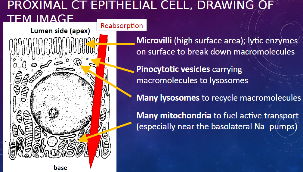
𖹭 Microvilli: High surface area for absorption
𖹭 Lytic enzymes on surface: Break down macromolecules
𖹭 Pinocytotic vesicles: Carry macromolecules to lysosomes
𖹭 Many lysosomes: Recycle macromolecules
𖹭 Many mitochondria: Fuel active transport, especially near the basolateral Na+ pumps
What is the function of the thin arms of the Loop of Henle?
𖹭 Reabsorption from ultrafiltrate
What is the mechanism of reabsorption in the thin arms of the Loop of Henle?
𖹭 By passive flux only (i.e., osmosis)
What are the features of the epithelial cell in the thin Loop of Henle?
𖹭 Thin, squamous epithelium: Allows passive fluxes
𖹭 A minimum of organelles
What is the function of the thick ascending Loop of Henle and distal convoluted tubule?
𖹭 Blood homeostasis
What are the mechanisms involved in their function?
𖹭 Regulated active transport and ion exchange, including Na+/K+ and H+/HCO3- exchange.
What are the features of the epithelial cell in the distal convoluted tubule (DCT)?
𖹭 Cuboidal epithelium: Thicker than squamous, to reduce passive fluxes and accommodate organelles
𖹭 Many mitochondria: To fuel active transport, can show as a pale or striped basal area in H&E-stained sections.
𖹭 Few, short microvilli: Unlike proximal convoluted tubule (PCT)
What are the functions of the collecting duct and collecting tubule?
𖹭 Transport of urine to ureter
𖹭 Water homeostasis: Passive reabsorption of water, regulated through epithelial permeability
What are the features of the epithelial cell in the collecting duct?
𖹭 Cuboidal to columnar epithelium: To prevent unregulated passive flux of water (and urea)
𖹭 Dense membranes at cell contacts: Function unclear, probably also helping to prevent passive flux.
What are the components visible in the light microscopy (H&E stain) of the nephron?
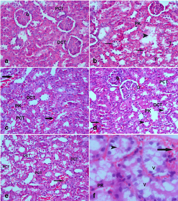
𖹭 Renal corpuscle (glomerulus + Bowman’s capsule)
𖹭 Proximal convoluted tubules
𖹭 Distal convoluted tubules (paler)
𖹭 Collecting ducts, in medullary ray
What are the components visible in the histology of a normal glomerulus (longitudinal view)?
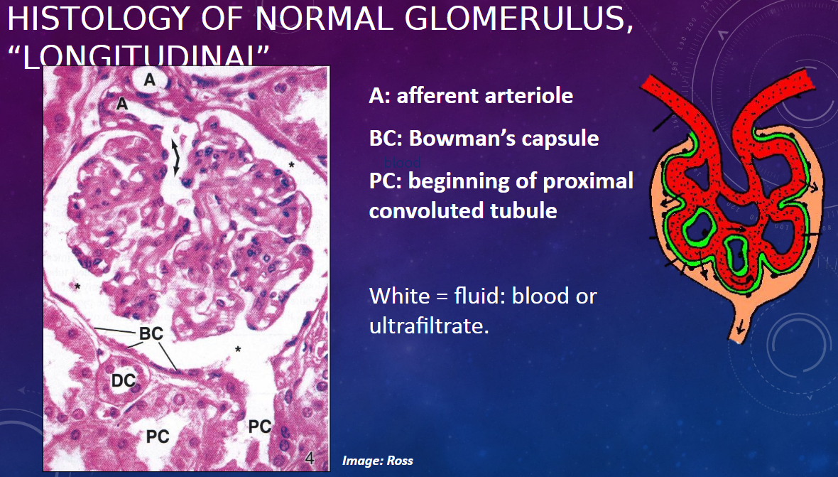
𖹭 A: Afferent arteriole
𖹭 BC: Bowman’s capsule
𖹭 PC: Beginning of proximal convoluted tubule
𖹭 White areas represent fluid, which can be either blood or ultrafiltrate.
What is an example of kidney disease involving glomerular damage?
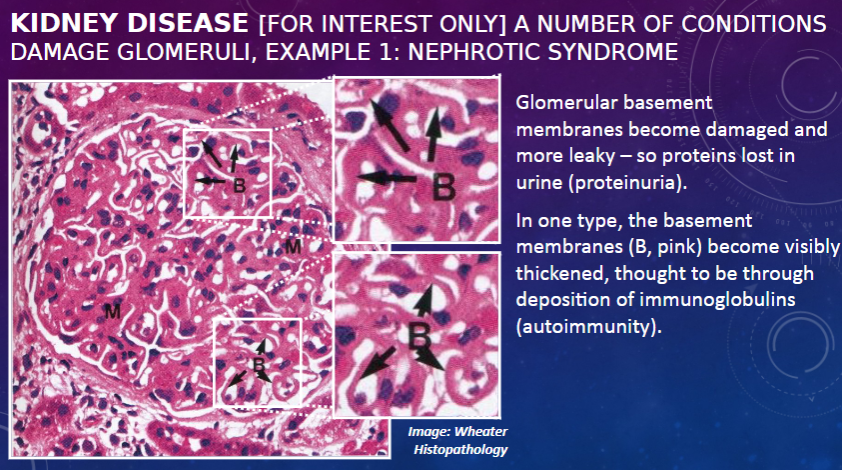
𖹭 Nephrotic syndrome: Glomerular basement membranes become damaged and more leaky, leading to protein loss in urine (proteinuria).
What is a characteristic feature of one type of nephrotic syndrome?
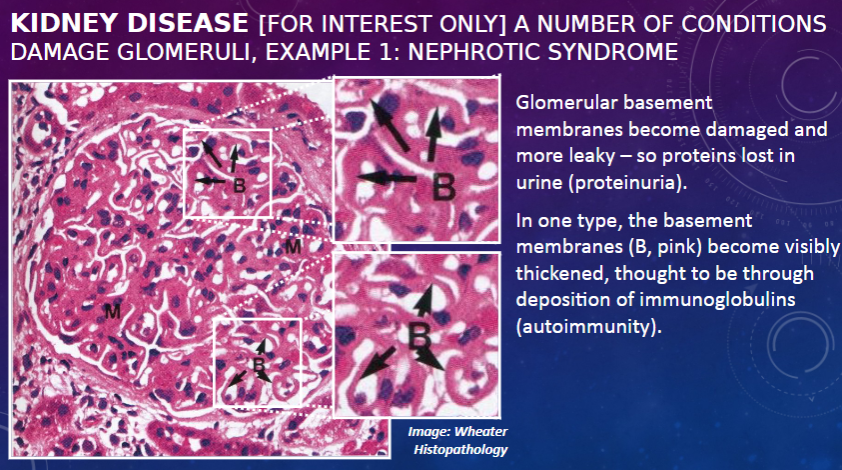
𖹭 In one type, the basement membranes become visibly thickened, thought to be through deposition of immunoglobulins, indicating autoimmunity.
What is an example of kidney disease associated with high blood pressure?
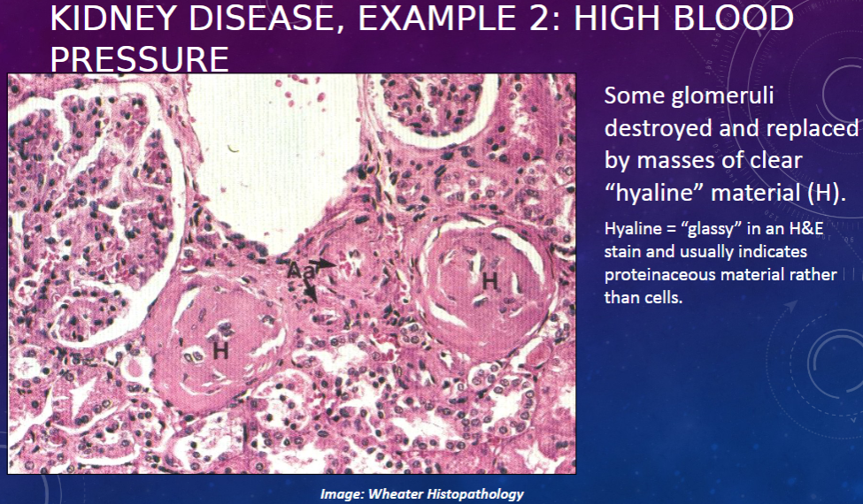
𖹭 High blood pressure: Some glomeruli are destroyed and replaced by masses of clear "hyaline" material (H). Hyaline appears "glassy" in an H&E stain and usually indicates proteinaceous material rather than cells.
What are the distinguishing features between proximal convoluted tubules (PCT) and distal convoluted tubules (DCT) in the cortex?
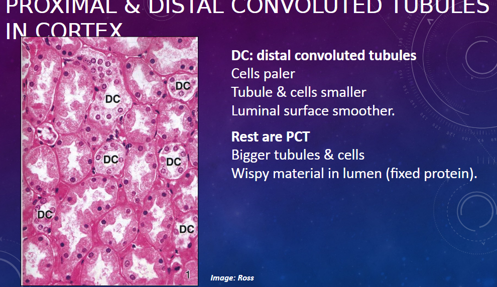
𖹭 DCT (distal convoluted tubules):
Cells appear paler
Tubules and cells are smaller
Luminal surface is smoother
𖹭 PCT (proximal convoluted tubules):
Bigger tubules and cells
Wispy material present in the lumen (fixed protein)
What are the components visible in the medulla section of various tubules?
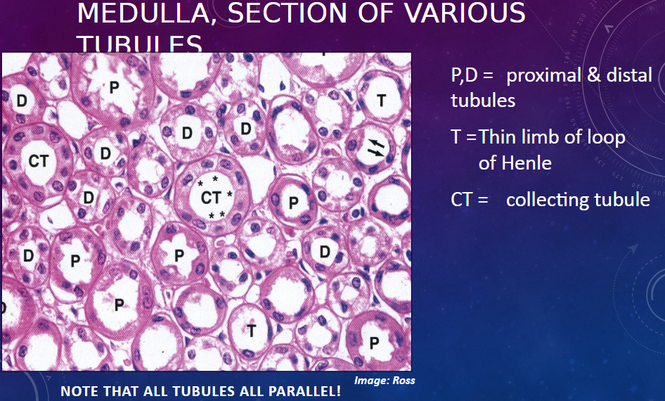
𖹭 P,D = Proximal and distal tubules
𖹭 T = Thin limb of the loop of Henle
𖹭 CT = Collecting tubule
Note: All tubules are parallel in the image.
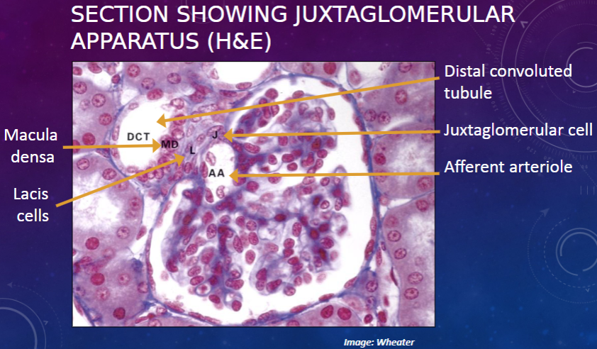
Picture demonstrating the juxtaglomerular apparatus:
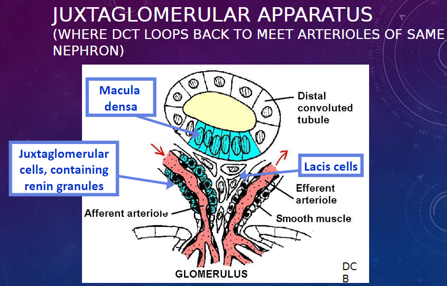
What are the functions of the cells in the juxtaglomerular apparatus?
𖹭 Macula densa: Senses [Na+] in the DCT fluid. Appears to signal to the juxtaglomerular cells to release renin when the [Na+] is low, thereby helping to regulate blood pressure and glomerular filtration rate
𖹭 Juxtaglomerular cells: Release renin, more so in response to lower [Na+] in DCT. Renin indirectly increases vascular tone and sodium resorption.
𖹭 Lacis cells: Function unknown. (Signaling between the other cells)
Picture demonstrating SECTION SHOWING JUXTAGLOMERULARAPPARATUS (H&E):
What are the components visible in the transverse section of the ureter?
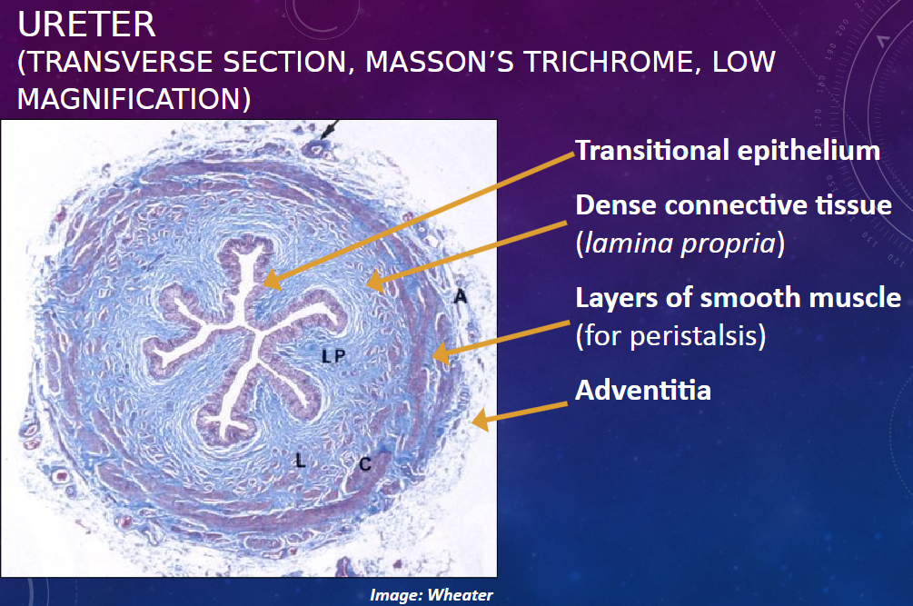
𖹭 Transitional epithelium
𖹭 Dense connective tissue (lamina propria)
𖹭 Layers of smooth muscle (for peristalsis)
𖹭 Adventitia
What are the characteristics of transitional epithelium?
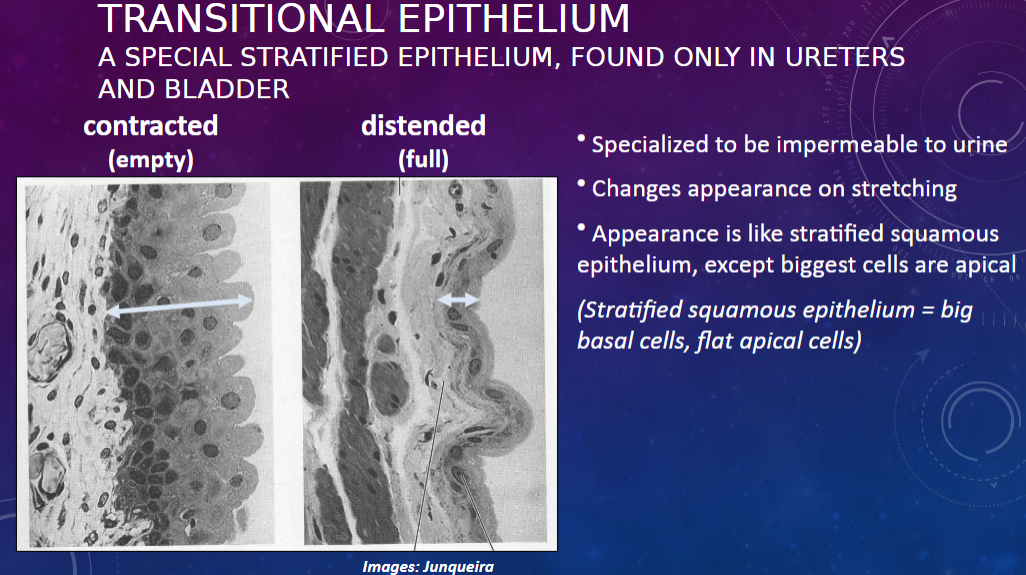
𖹭 Specialized stratified epithelium, found only in ureters and bladder.
𖹭 Impermeable to urine.
𖹭 Changes appearance on stretching.
𖹭 Appearance is like stratified squamous epithelium, except the biggest cells are apical. (Stratified squamous epithelium = big basal cells, flat apical cells).
Here's the flashcard based on the provided information:
What is an oddity in transitional epithelium?
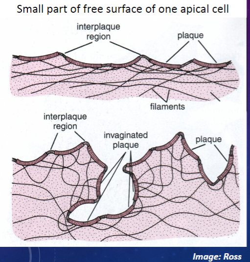
𖹭 Plaques of specialized (urine-resistant) plasma membrane in apical cells: These impermeable, rigid membrane patches protect apical cells from toxic urine.
How do these plaques behave in different bladder states?
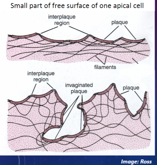
𖹭 Distended (bladder full): The plaques remain rigid, protecting apical cells.
𖹭 Contracted (bladder empty): The rigid plaques are invaginated, forming pits, allowing bladder volume to decrease.