 Faith_LaBry
-
Respiratory surfaces in mammals

Large network of alveoli in lung tissue
-
Respiratory surfaces in insects
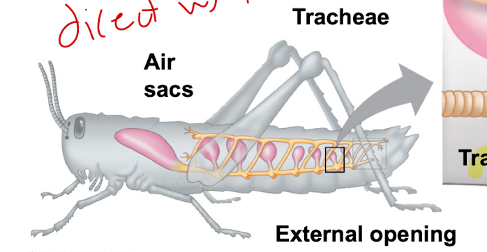
Extensive tracheal system
-
Tracheal Systems in Insects consist of...

Tiny branching tubes that penetrate the body
-
Respiratory system order in mammals
1. Nasal cavity and pharynx
2. Trachea
3. Bronchi
4. Bronchioles
5. Alveoli (site of gas exchange)
-
Tidal Volume
The volume of the air inhaled and exhaled during a normal breath
-
Vital Capacity
The maximum volume of air that can be inhaled and exchaled
-
Residual Volume
air remaining after full exhalation (incomplete removal)
-
Negative Pressure Breathing
Pulls air into the lungs
-
Positive Pressure Breathing
Forces air down the trachea
-
Who performs negative pressure breathing?
Mammals, birds, reptiles
MY BLOODY RALENTINE
-
Who performs positive pressure breathing?
Amphibians, fish
-
Is breathing associated with a negative or positive feedback loop?
Negative feedback loop
-
What happens if breathing is ineffective?
PCO2 increases --> pH of blood and cerebrospinal fluid decreases --> ventilation increases by blowing off CO2
-
What controls O2/CO2/breathing concentration in humans?
Chemosensors in the aorta and carotid arteries (which signals the breathing control centers)
-
How many air sacs does a bird have?
8-9
-
How do the air sacs help a bird breathe?
By functioning as bellows that keep the air flowing through the lungs; have parabronchi (air tubes) instead of bronchi
-Air passes in one direction only
-Fresh air does not mix
-
Partial Pressure in the alveoli

Favors diffusion of O2 into the blood and CO2 into the air
AI
-
Partial Pressure in the capillaries

Favors diffusion of O2 out of the blood and CO2 into the blood
CO
-
How many subunits does a single hemoglobin molecule have?
4 (2 alpha and 2 beta)
-
How many molecules of O2 can each Hemoglobin (Hb) carry?
4
-
What facilitates the binding and offloading of O2 among the 4 subunits of Hb?
Allosteric cooperation
-
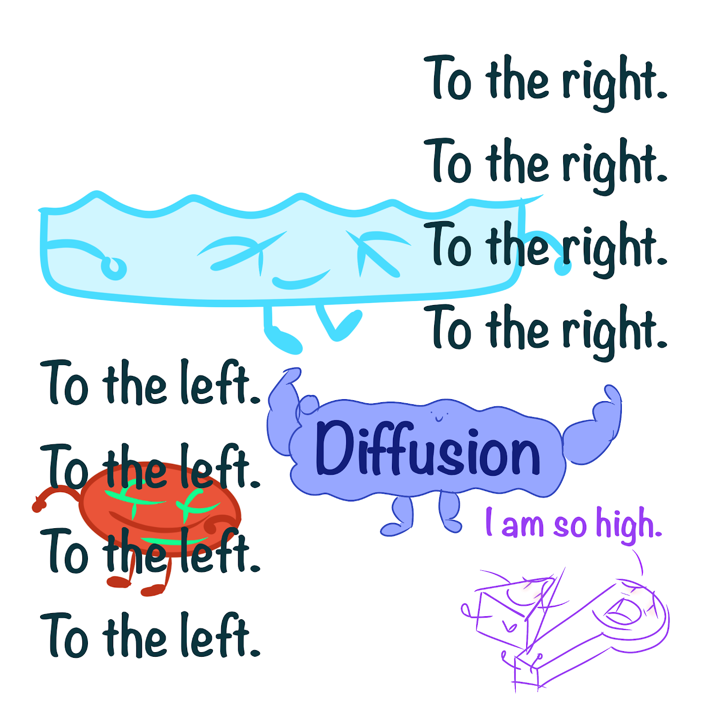
What causes the Bohr shift?
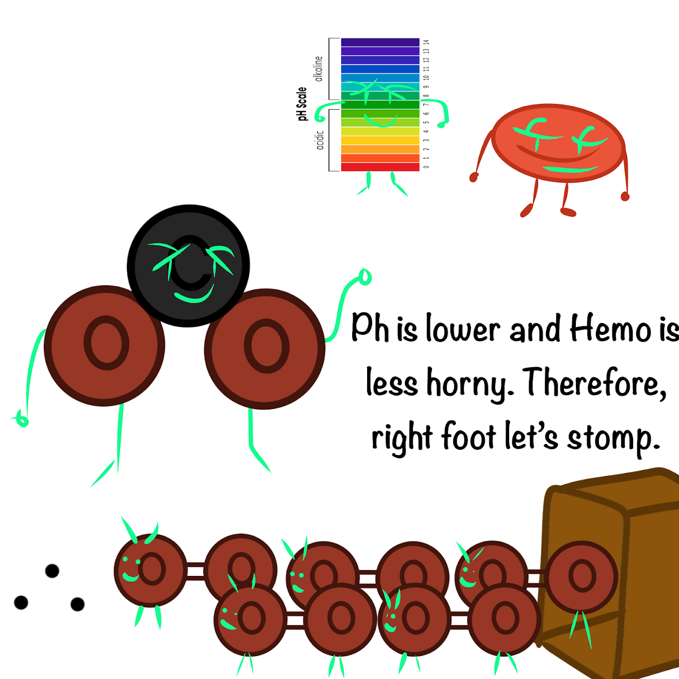
CO2 produced during cellular respiration lowers blood pH and decreases the affinity of hemoglobin for O2, causing a rightward shift and facilitating the offloading of oxygen
-
CO2 is transported into the lungs as
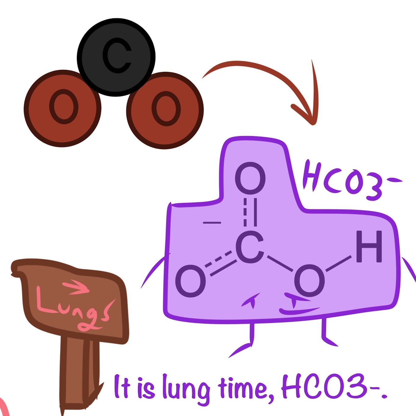
HCO3- ions in the plasma
-
Why is O2 more challenging in the water than the air to get?
There is less O2 available in water than in air
-
Why are gills extremely thin?
Offsets low O2 content in water by minimizing diffusive distance
-
_____, in which blood flows in the opposite direction to water, maintains high △p
Countercurrent exchange
-
A skeletal muscle consists of a bundle of ____, each a single cell, running parallel to the length of the muscle
long fibers
-
Each muscle fiber itself is a bundle of smaller _____ arranged longitudinally
myofibrils
-
myofibrils are composed of 2 kinds of myofilaments:
Thick filaments and thin filaments
-
Thick filaments
Staggered arrays of myosin molecules
-
Thin filaments
Consist of 2 strands of actin and 2 strands of a regulatory protein
-
Functional unit of a muscle
Sarcomere
-
Actin filaments are anchored by
Z lines
-
Myosin filaments are anchored by
M lines
-
First step of the Sliding-Filament model of muscle contraction
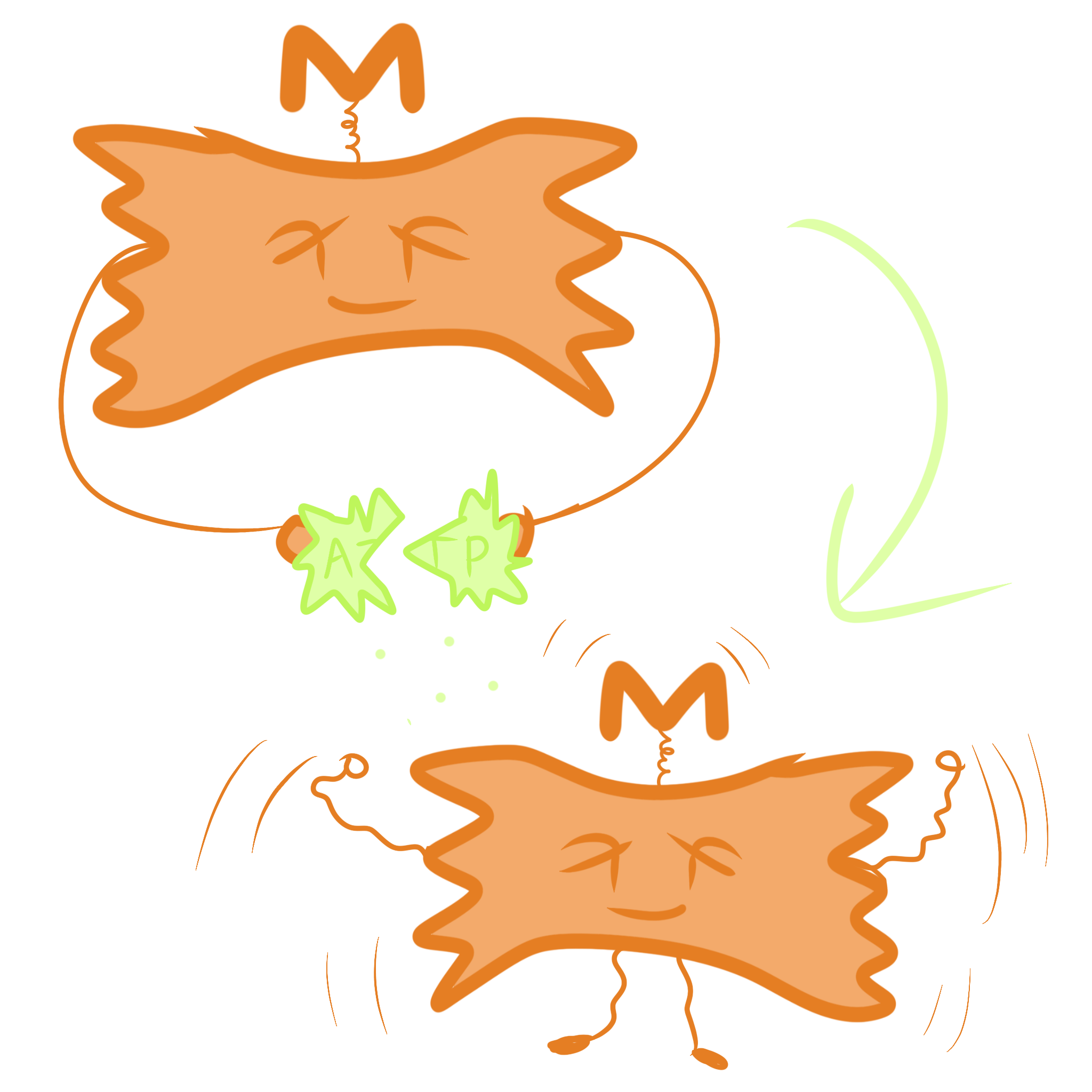
Myosin heads split ATP and become reoriented/energized
-
Second step of the Sliding-Filament model of muscle contraction

Ca2+ binds to troponin so that the binding site is uncovered; Head of the myosin molecule binds to an actin filament, forming a cross-bridge
-
Third step of the Sliding-Filament model of muscle contraction
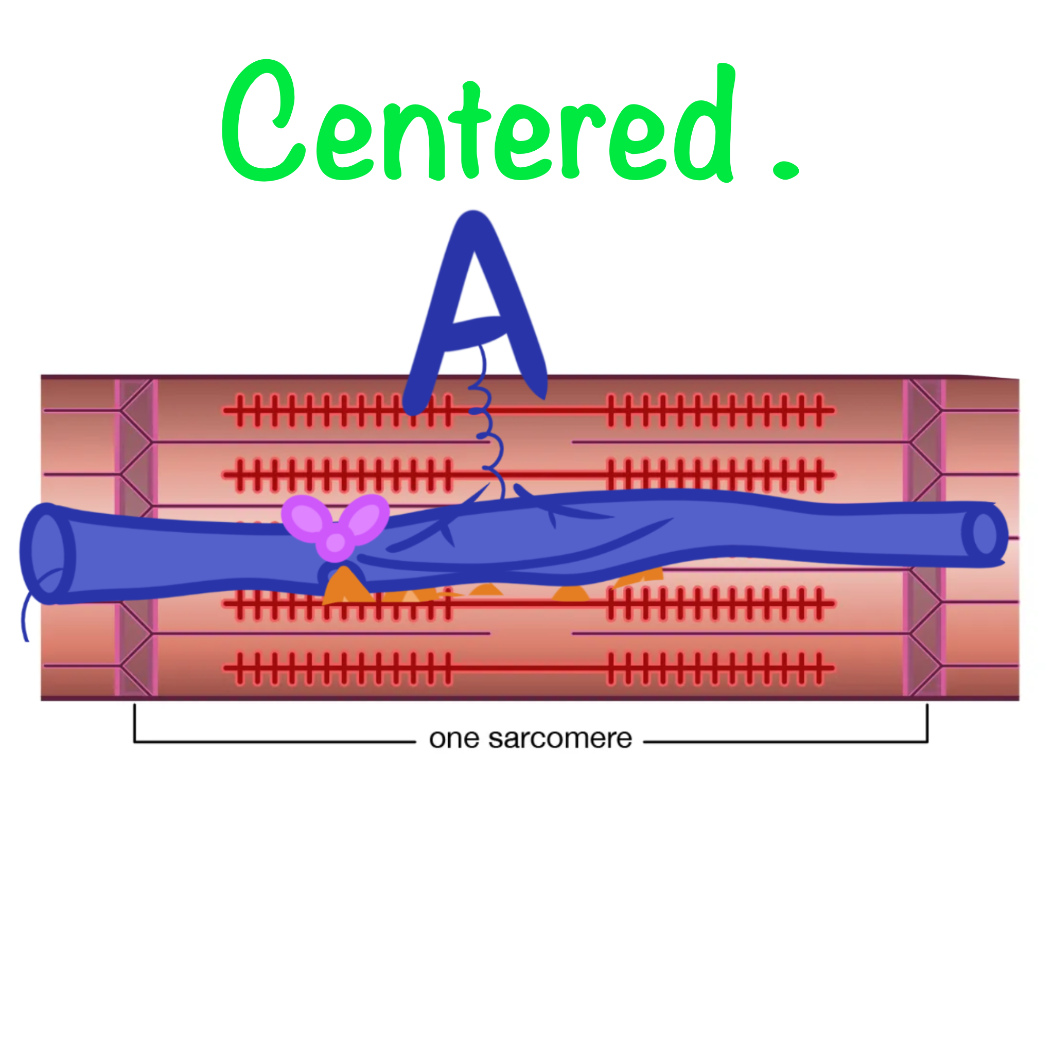
Myosin head rotates towards the center of the sacromere (Power stroke); thin filament moves toward the center of the sarcomere.
-
Tropomyosin/Troponin complex
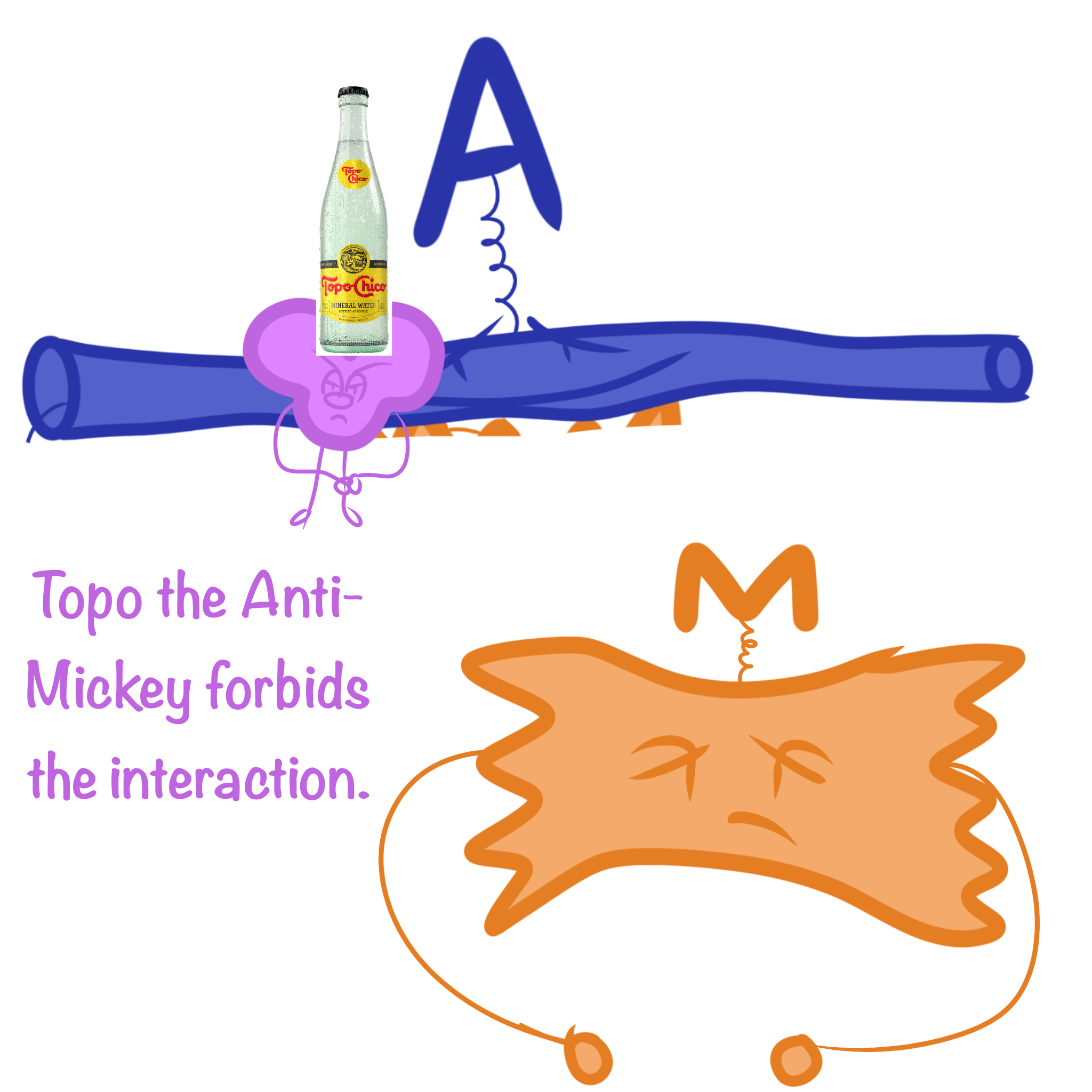
binds to actin strands on thin filaments, preventing actin and myosin from interacting
-
What concentration ions are involved in muscle contraction?
Calcium ions (Ca2+)
-
An action potential in a motor neuron that makes a synapse with the muscle fiber is the
stimulus leading to contraction of a muscle fiber
-
First step of muscle contraction
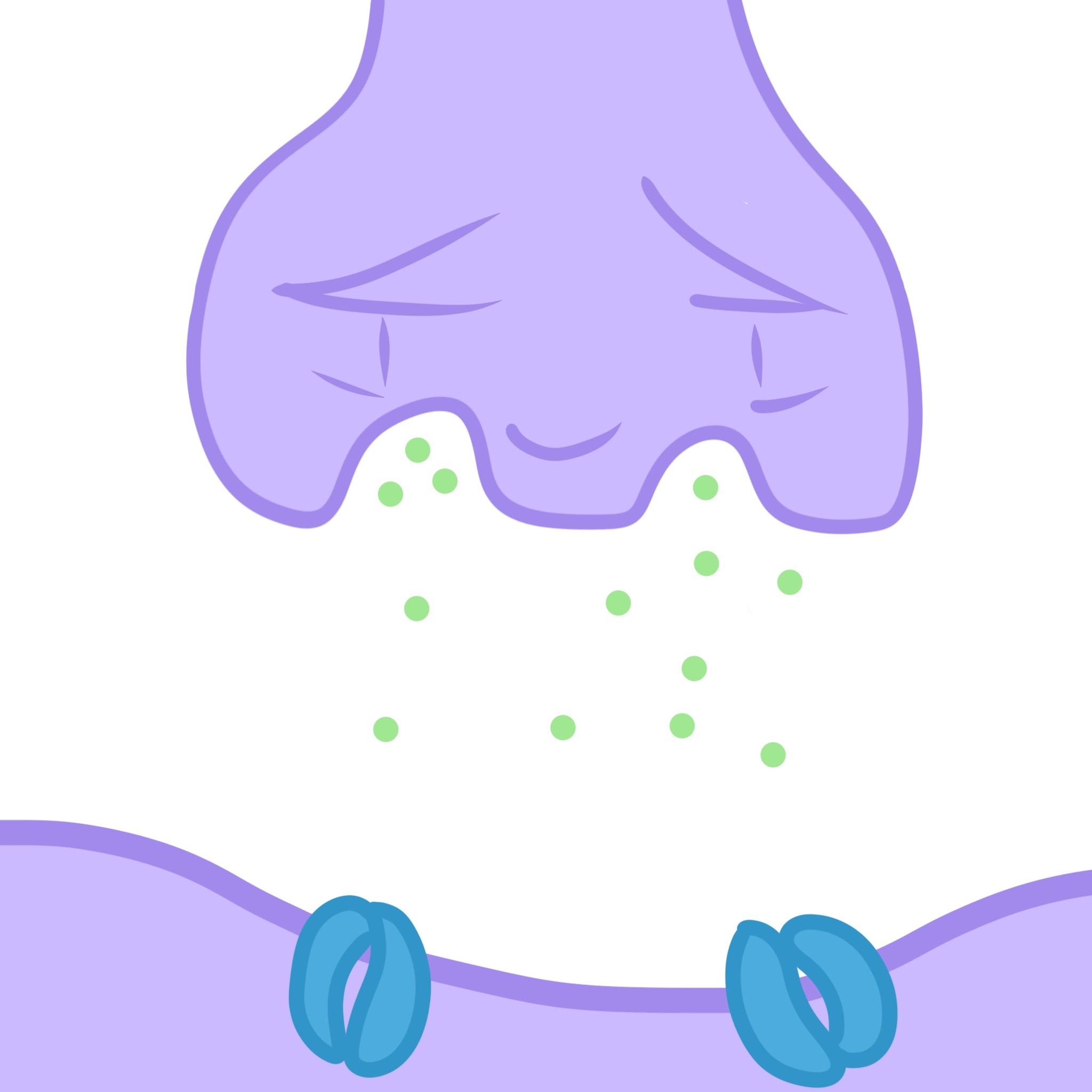
The synaptic terminal of the motor neuron releases the neurotransmitter acetylcholine (Ach)
-
Second step of muscle contraction
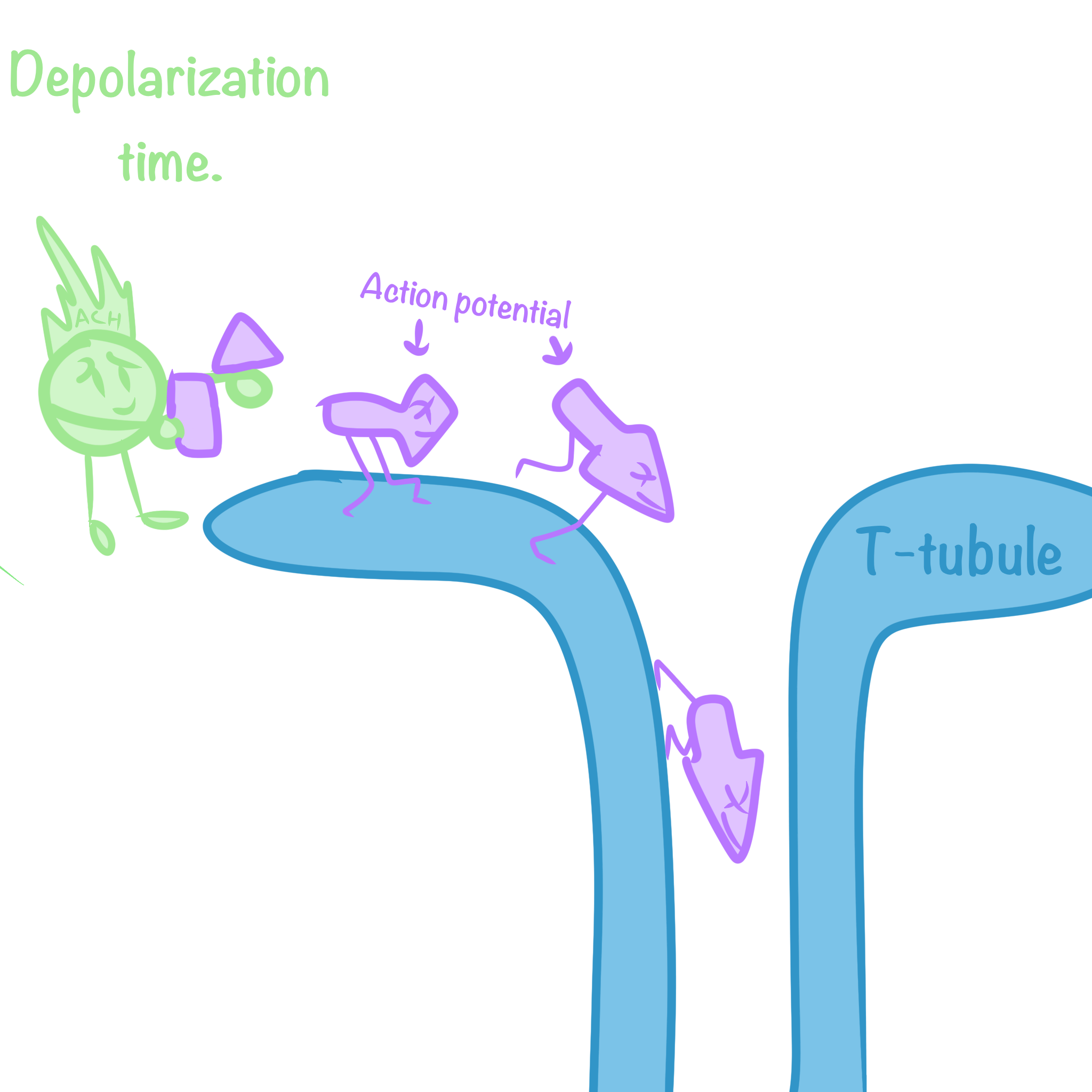
Acetylcholine depolarizes the muscle, causing it to produce an action potential, which travels to the interior of the muscle fiber along transverse (T) tubules
-
Third step of muscle contraction
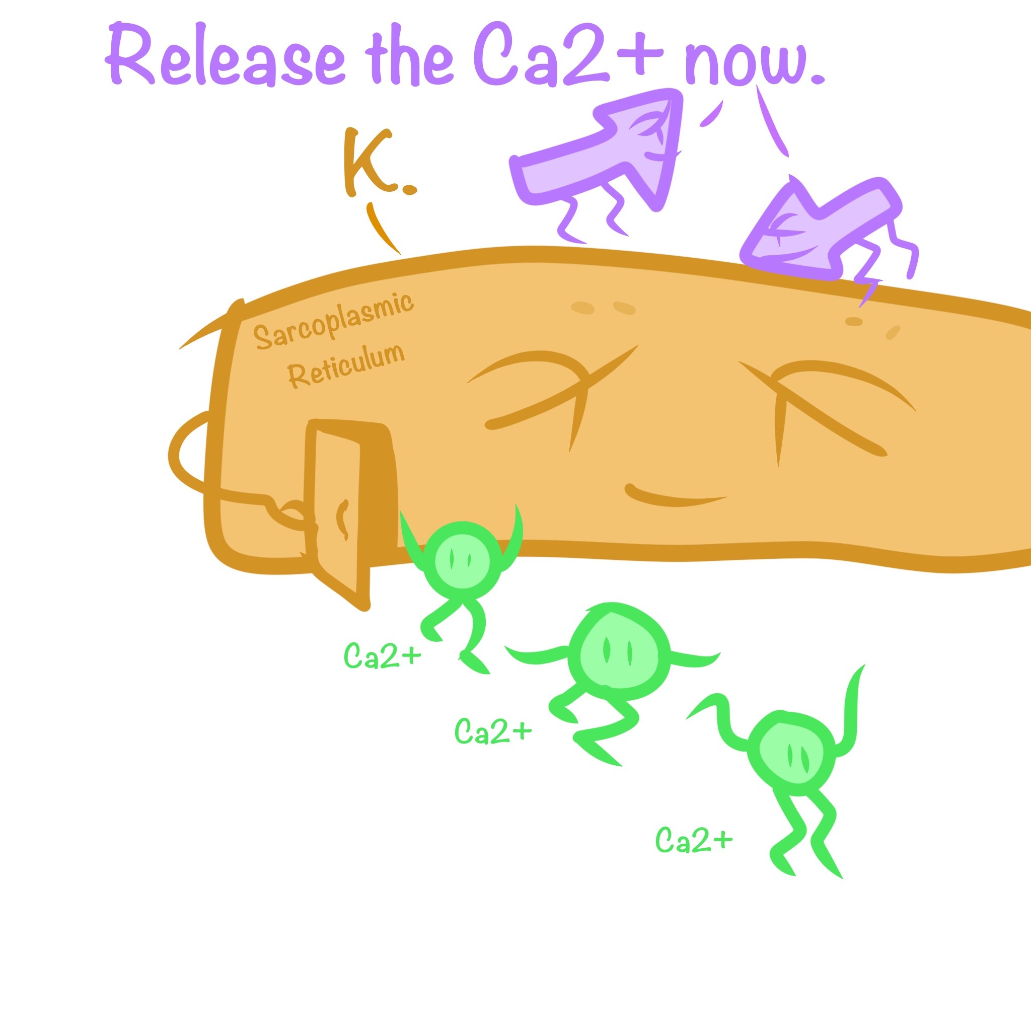
The action potential causes the sarcoplasmic reticulum (SR) to release Ca2+
-
Fourth step of muscle contraction
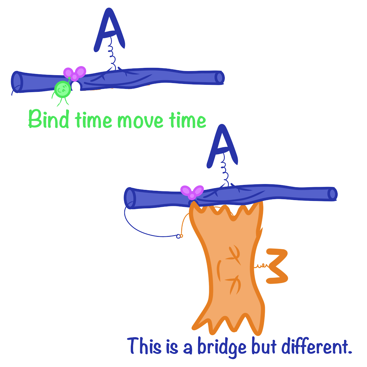
Ca2+ binds to the troponin complex on the thin filaments, exposing myosin-binding sites (allowing the cross-bridge cycle to proceed)
-
Fifth step of muscle contraction
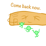
When motor neuron input stops, the muscle cell relaxes; Transport proteins in the SR pump Ca2+ out of the cytosol and regulatory proteins shift back to the myosin-binding sites
-
Sixth step of muscle contraction
Conformational change in troponin
-
2 basic mechanisms by which the nervous system produced graded contractions
Varying the number and varying the rate
-
Tetanus

A state of smooth, sustained contraction when the rate is so high the muscle fiber cannot relax between stimuli
-
Oxidative Fibers
-Rely mostly on aerobic respiration to generate ATP
-Many mitochondria
-Rich blood supply
-Large amount of myoglobin
-Dark meat
-
Glycolytic Fibers
-Uses glycolysis (anaerobic) as their primary source of ATP
-Less myoglobin; tires more easily
-Light meat
-
Slow-twitch fibers
-Contracts slowly
-Sustains longer contractions
-ALL oxidative
-
Fast-twitch fibers
-Contracts rapidly
-Sustains shorter contractions
-Glycolytic or oxidative
-
Slow Oxidative Muscles
-Slow concentration speed
-Aerobic respiration
-Slow rate of fatigue
-Many mitochondria
-High myoglobin content
-
Fast Oxidative Muscles
-Fast concentration speed
-Aerobic respiration
-Intermediate rate of fatigue
-Many mitochondria
-High myoglobin content
-
Fast Glycolytic Muscles
-Fast concentration speed
-Glycolysis
-Fast rate of fatigue
-Few mitochondria
-Low myoglobin content
-
Molarity
Concentration of a dissolved substance
-
Osmolarity
Concentration of all solutes that contribute to the osmotic pressure of a solution (depends on the number of dissolved particles); determines the movement of water across a selectively permeable membrane
-
Isosmotic
Same concentration of solutes
-
Hyposmotic
Lower relative concentration of solutes
-
Hyperosmotic
Higher relative concentration of solutes
-
Osmoconformers
Isosmotic with their surroundings and do not regulate their osmolarity; consists only of some marine animals
-
Osmoregulators
Expends energy to control water uptake and loss in a hyper/hyposmotic environment
-
Stenohaline
Animals that cannot tolerate substantial changes in external osmolarity (most animals)
-
Euryhaline
Animals that can survive large fluctuations in external osmolarity (common in estuaries and tidal pools)
-
Why is life hard for freshwater fish?
They constantly lose salts and gain water by diffusion from their hyposmotic environment
-
How do freshwater fish maintain osmotic balance?
By excreting large amounts of dilute urine and replacing salts by active uptake across the gills and diet
-
Why is life hard for saltwater fish?
They constantly lose water by osmosis and gain salt by diffusion
-
How do saltwater fish maintain osmotic balance?
By drinking seawater and excreting salts across gills, excreting salts and retaining water by kidneys, and diet
-
Hypertonic
A solution that contains more solute than the cell which is placed in it (withered cells)
-
Isotonic
A solution in which the same amount of solute and solution is available inside of the cell and outside of the cell
-
Hypotonic
A solution that contains less solute than the cell which is placed in it (fat cells)
-
3 forms of nitrogenous wastes
Ammonia (NH3), Urea, Uric Acid
-
Ammonia
-Most Toxic
-Freely diffuses across the whole body surface or through gills
-Need access to lots of water
-Fish/Aquatic animals
-
Urea
-Less toxic, requires energy
-NH3 converted to urea in liver, then excreted by kidneys
-Requires less water
-Amphibians, sharks, mammals
-
Uric Acid
-Least toxic, most expensive
-Does not dissolve readily in water
-Can be secreted as a paste with little water loss
-Insects, birds, reptiles
-
4 stages of excretion by the kidney
Filtration, Reabsorption, Secretion, Excretion
(Farting Rats Scream Excitedly.)
-
Stage 1 of excretion
Filtration: blood pressure forces water and small solutes through capillary bed into excretory tubule; cells and large molecules remain in the blood
-
Stage 2 of excretion
Reabsorption: Valuable solutes actively reclaimed back into the blood (glucose, amino acids, etc.); wastes and non-essential solutes remain
-
Stage 3 of excretion
Secretion: Nonessential solutes and wastes are added to the filtrate (toxins, excess ions)
-
Stage 4 of excretion
Excretion: Processed filtrate containing nitrogenous wastes is released
-
Kidney structure
Located on either side of the vertebreal column; connected to a ureter; consists of a renal vein/artery, medulla, cortex, and renal pelvis
-
Nephron position within the kidney
In the renal pyramid among the cortex and medulla
-
What is the nephron organized for?
Stepwise processing of blood filtrate
-
What does the blood filtrate produced in Bowman's capsule contain?
Salts, glucose, amino acids, vitamins, nitrogenous wastes, and other small molecules
-
What are the two primary solutes affecting osmolarity?
NaCl and Urea
-
Proximal tubule
Salts and nutrients are actively reabsorbed; water follows passively by osmosis; as the filtrate passes through the proximal tubule, materials to be excreted become concentrated as water is being reabsorbed in the capillaries
-
Descending limb of loop of Henle
Freely permeable to water through channels formed by aquaporin proteins, but very low permeability for NaCl and other small solutes. Water moves out freely by osmosis; filtrate becomes increasingly concentrated
-
Thin segment of ascending limb
Ascending loop of Henle is impermeable to H2O, but permeable to NaCl so NaCal initially passes into interstitial fluid freely by diffusion
-
Thick segment of ascending limb
Gradient becomes unfavorable for passive diffusion of NaCl --> active transport required; movement of NaCl along ascending limb helps maintain osmolarity of medulla; filtrate becomes increasingly dilute as NaCl is reabsorbed
-
Distal Tububle
Reabsorption of NaCl and H2O
-
How do the proximal and distal tubules regulate pH?
Secretion of h+ and reabsorption of HCO3- ions
-
Collecting Duct (When conserving water)
Aquaporin channels allowed water to be reabsorbed; impermeable to salts; solutes become increasingly concentrated; in the inner medulla, high concentration of urea drives passive reabsorption that helps to maintain high osmolarity of interstitial fluid in this region
-
Collecting Duct (When eliminating water)
Aquaporin channels are absent; impermeable to water; salts actively reabsorbed; hormones control absorption of water in collecting ducts by regulating aquaporin channels
-
Anti-diuretic hormone (ADH) Function
Prevents water loss by regulating water retention by making the collecting duct epithelium more permeable to water
-
When osmolarity rises above its set point, ADH release
increases
-
When osmolarity drops below its set point, ADH release
decreases
-
Function of Aquaporins
Helps reabsorb water and some solutes across the plasma membrane
-
Herbivore
Primarily eats plants and algae
-
Carnivore
Primarily eats other animals
-
Omnivore
Regularly consume animals as well as plants or algae

