 Fatima Adam
Auditory and Vestibular Physiology (Physiology 2)
Auditory and Vestibular Physiology - Dr Penny Murphy Lecture Outline The sensory apparatus of both auditory and vestibular systems is found in the inner ear, and uses the same basic type of receptor. The auditory system plays a vital role in human communication, as well as being a source of great pleasure for many. Loss of hearing, especially late in life, can restrict the ability to socialise with others, to gain information via normal communication routes, and to enjoy some forms of entertainment. The resulting isolation can have detrimental effects on both physical and psychological health, daily activity and economic well-being. The vestibular system is best known as the system responsible for our sense of equilibrium and the ability to maintain upright posture. However, it is also vital for the accurate control of eye movements. The vestibular system provides the afferent drive for a set of reflexes that compensate for self-movement, allowing us to achieve stable visual fixation on a stationary target and to accurately follow a moving target with our eyes. Damage to the vestibular system can lead to dizziness and a sense of disequilibrium, persistent oscillatory eye movements (nystagmus) and an associated sensation that the world is spinning around the head or that the head itself is spinning persistently (vertigo). A mismatch between vestibular and other sensory signals is thought to mimic the effects of poisoning, leading to nausea and vomiting. This lecture will cover the structure and function of the hair cell receptors that serve both systems, and the tunnels and membranes of the inner ear that channel the stimuli in such a way that the different groups of hair cells respond to diverse stimuli (sound waves of different frequencies, rotational movements of the head, linear movements of the head, the direction of gravity). It will cover the basics of auditory system function and stimulus encoding, as well as some vulnerabilities in the system that lead to loss of hearing. Likewise it will cover how the vestibular system responds to and hence encodes different types of head movement and gravity, and how it integrates this information with visual input in order to ensure accurate eye movements. Desired learning outcomes Describe the structure of a generic hair cell receptor, and explain how it converts stimulation of the stereocilia into a receptor potential. Describe how the structure of the ear channels sound waves of different frequencies to activate specific populations of auditory receptor cells (hair cells) Explain how the auditory system captures and encodes information about the loudness, frequency, origin and timing of a sound, and how this is passed on to and used by the cerebral cortex Explain the distinction between inner and outer hair cells and their respective roles in hearing Explain the vulnerabilities in the system that can lead to conductive and sensorineuronal deafness. Describe how the structure of the inner ear creates fluid movement in response to different types of head movement and changes in the direction of gravity, such as to generate responses in different populations of vestibular hair cells. Describe the neural pathways for vestibulo-ocular reflexes, including the role of the cerebellum for short- and long-term modulation of these responses Session description This is an on-line, self-learning session sub-divided into three sections. 1. This lecture connects with a number of previous sessions, which you should identify and review before starting some sections. Don't move on without ensuring that you are confident in the relevant background knowledge. 2. Each section has a powerpoint and Panopto presentation, which include outlines of the contents and a check-list of things you should be able to do before moving on to the next section To see the slides properly, use the download icon and run in powerpoint presentation view. Session Checklist The three sections will take approximately 1 hour to listen to, but studying them properly will take 2-3 times as long: Part a - 15 mins Part b - 25 mins Part c - 20 mins Session Recording and Powerpoints Part a: Hair Cells This segment introduces the hair cell receptors that serve both systems, and gives an overview of the auditory and vestibular sensory apparatus. Preparation - before going further: review your previous teaching on membrane potentials, and ensure that you understand the changes that will occur if cation (ie +vely charged ion) channels are opened in a nerve cell membrane. Once you've done that, go ahead with the lecture. Part a powerpoint slidesDownload Part a powerpoint slides Part b: Auditory System This segment goes back through the structures of the outer, middle and inner ears, adding functional information, and then concentrates on the cochlea of the inner ear and the neural apparatus needed to transduce and encode sound. The cochlea is made up of three chambers, separated by flexible membranes that channel and vibrate in time with the sound waves. One of these is the basilar membrane, which supports the spiral organ which in turn houses the auditory receptors. The auditory receptors are hair cells - so called because they have a tuft of sterocilia sprouting from their apical surface. Thanks to the arrangement of chambers, the body of each hair cell is surrounded by a normal extracellular fluid while the stereocilia stick out into a compartment filled with potassium rich endolymph. Endolymph has an excess of positive charge compared with the surrounding tissue, which creates an electrical gradient that drives K+ into the hair cell when sound waves open the mechanically-gated channels in the stereocilia. This depolarises the cell and causes it to release glutamate from the synapse. The structure of the cochlea, the location and function of the inner hair cells, and further processing in the superior olivary nuclei capture and encode key characteristics of the sound. Their signal is transmitted to the auditory cortex, where these characteristics are used to reconstruct and understand the sound. The outer hair cells act as an amplification system (and famously dance to Elvis). Preparation - before going further: Make sure that you understand how hair cells function and that you've grasped the overall anatomy of the auditory part of the system. Once you've done that, go ahead with the lecture. Part b powerpoint slidesDownload Part b powerpoint slides Part c: Vestibular System This segment will cover the two systems that are found within the vestibular labyrinth. One picks up and encodes linear movements of the head and changes in the direction of gravity. The other deals with rotational movements. In both cases it is changes in the speed of movement that matter - ie acceleration or deceleration - because tilting of the hair cell stereocilia depends on the effect of inertia on the structures that are associated with them. Both systems feed into the motor system, providing compensation for externally generated movements of the body. The semi-circular canals are particularly important for the control of eye movements, and this will be covered in a little more detail. Preparation - before going further: Make sure that you've grasped the overall anatomy of the vestibular part of the system. Note that vestibular hair cells function in much the same way as those in the auditory system, so if anything isn't clear review parts a and b. Note that I assume you have some understanding of the role of extraocular eye muscles and the importance of foveal vision. Part c powerpoint slidesDownload Part c powerpoint slides Well done, you've reached the end of the session If you want to test test yourself, either now or during exam revision, this link will take you to a quiz that uses exam-style questions and provides feedback on incorrect answers. Auditory & Vestibular quiz Follow up activities This is where the real learning takes place. Discuss the material in this lecture with others You need to practice using the information, and to do so in different contexts. Answering quiz questions is one way to do that, but any quiz will cover only a small part of the material. Active discussion is more wide-ranging and very effective. Each PowerPoint presentation includes a check-list of things you should be able to do before moving on to the next part. If you are struggling with any of the concepts and the reading material hasn't helped, then post questions on the "Discussion Forum". Use the forum to discuss your questions with other students and to offer suggestions to others who have posted questions of their own. I will monitor the forum and add explanations where needed. Some suggested reading material: You need to read around to consolidate your knowledge and clear up any difficulties in understanding the lecture material. NB - use the reading to back up and better understand the information given in the lecture, but don't get bogged down in the extra information given or the additional fine detail: "Neuroscience: Exploring the Brain" Bear, Connors and Paradiso, 3rd ed or higher; Chapter 11. The Cochlear Amplifier - Ashmore & Gail (2004) Current Biology 14:R403-4.Links to an external site. Short review article, for interest. J. Ashmore dancing hair cell , including a brief explanation of the role of outer hair cells in the "cochlear amplifier". These video resources are relatively simplistic in their content but the animations may clarify the mechanics of the systems. If you can't access them, then ask the library for help (we have a subscription, but it depends on how you're logged onto the University intranet): JoVE - Overview of hearing JoVE - The cochlea JoVE - Hair cells JoVE Vestibular system Some important terms for sensory neuroscience: Receptor - has multiple meanings, especially in sensory neuroscience: a) protein complex that binds to and is activated by a signalling molecule b) nerve cell, often highly specialised, that is adapted to detect and respond to an external (ie external to the receptor) event, converting the energy in that event into a change in membrane potential (the receptor potential / generator potential) Primary afferent - first cell in the chain that carries a sensory signal from the sensory apparatus to the cerebral cortex. In some systems the receptive apparatus is part of the primary afferent, but in the "special senses" the receptors are separate and highly specialised nerve cells. Threshold - has multiple meanings, especially in sensory neuroscience: a) action potential threshold, which is the level of depolarisation necessary to allow voltage-gated sodium channels to open and hence trigger an action potential. Roughly -50mV. b) minimum sensory input necessary to activate a sensory receptor, and hence potentially generate a sensation. Different types of receptor have varying sensitivity and hence activation threshold (NB action potential threshold does not vary). c) perceptual threshold, which is the minimum sensory input needed to generate conscious perception of the stimulus. Depends not only on the strength of activation of the receptors, but also the attention of the subject (see modulatory systems lecture). Sensitivity - the relative responsiveness of a sensory system (or component thereof) to stimulation. Higher sensitivity correlates with lower response thresholds, larger response magnitude to a given stimulus, and a tendency to saturate at lower stimulus strengths. Specificity - a measure of how finely a system (or component thereof) differentiates between similar stimuli. Receptor adaptation - in sensory physiology this refers to a common feature of receptor cells, which respond strongly to changes in stimulus strength but reset their membrane potentials back to rest if a stimulus remains constant. Some adapt very rapidly (eg Pacinian corpuscles in the skin, which only respond repeatedly to rapid vibration), others very slowly (eg cone photoreceptors adapt over several seconds). C fibre nociceptors are probably the only ones that don't obviously adapt. Adaptation damps down neural responses to homogeneous stimuli while allowing small differences in stimulus strength to be signaled strongly. This extends dynamic range and as well as saving energy. Lateral inhibition - a key mechanism in sensory neurophysiology, typified by inhibition of one part of a sensory pathway by excitation of a neighbouring part. It is seen for example in the somatosensory system, where the cells carrying information from neighbouring patches of skin inhibit on another, and in the visual system where cells carrying information from neighbouring patches of retina inhibit one another. Less obviously, cells carrying information about similar qualities rather than location (eg sound frequencies) may inhibit one another. In each case the inhibition damps down neural responses to homogeneous stimuli while allowing small differences in stimulus strength to be signaled strongly. This inhibition extends dynamic range as well as saving energy. Dynamic range - the range of stimulus strengths over which a sensory system can respond. A single receptor type without the ability to adapt would either have to have very poor sensitivity (it could only afford to respond weakly, even to big jumps in stimulus strength) or a very limited response range (if it responded strongly to even small changes in stimulus strength, it would rapidly reach its maximum response magnitude and be unable to signal changes in stimulus strength beyond that point). Sensory systems overcome this by 1) having receptors that vary in sensitivity and 2) through adaptation of those receptors to extend their individual dynamic ranges. Saturation - point at which a sensory system (or component thereof) has reached its maximum response magnitude, beyond which it cannot signal increases in stimulus strength or differentiate between stimuli that differ in strength. Synaptic plasticity - the changes in the strength and effectiveness of synapses in response to patterns of use. The best understood mechanism is long-term potentiation (LTP), which increases the strength of fast excitatory (ionotropic) synapse by increasing transmitter availability, receptor numbers and receptor effectiveness. The trigger for LTP is that the synapse releases glutamate and immediately afterwards the postsynaptic cell strongly depolarises. The strong depolarisation indicates that many other synapses activated simultaneously, suggesting that they are all involved in a useful circuit. Specific circuit / pathway - neural machinery performing "computations" linked to sensory, motor and cognitive functions. Modulatory pathway - neural inputs that adjust how a cell responds to its specific inputs, which control the sleep-wake cycle, attention and concentration, mood and some emotional states. Related inputs are essential to the function of some motor and cognitive pathways.
-
What are hair cells and what do they detect? (7)
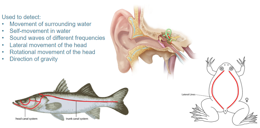
Hair cells are motion-detecting mechanoreceptors that are responsible for detecting:
Movement of surrounding water
Self-movement in water
Sound waves of different frequencies
Lateral movement of the head
Rotational movement of the head
Direction of gravity
-
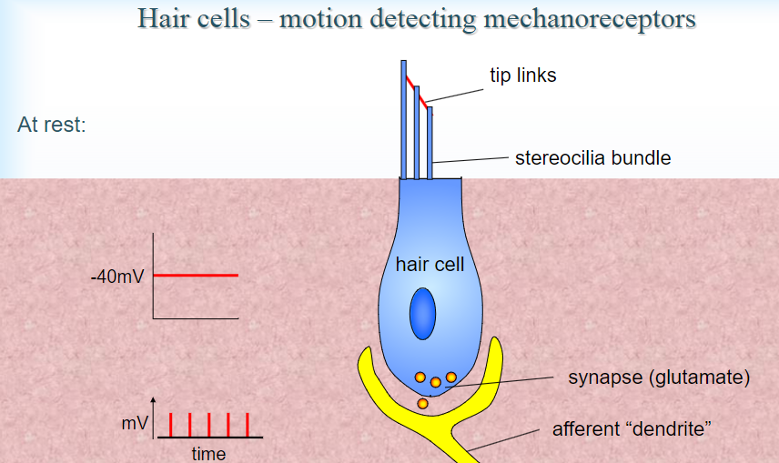
Picture demonstrating Hair cells – motion detecting mechanoreceptors:
-
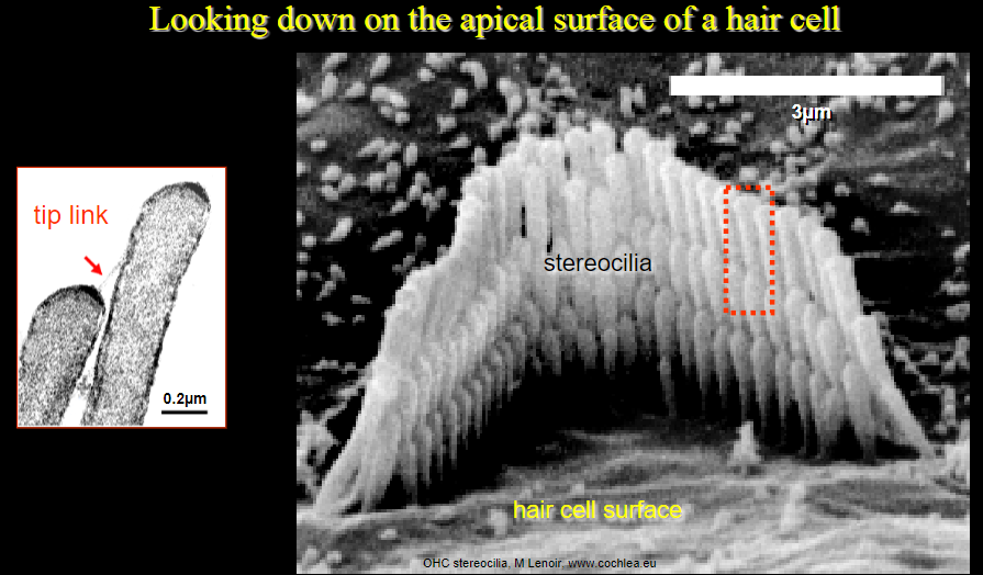
Picture demonstrating Looking down on the apical surface of a hair cell:
-
How do hair cells function as motion-detecting mechanoreceptors at rest? (4)
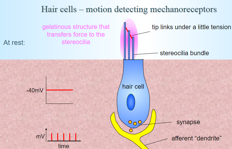
Tip links: Under a slight tension, these connect the stereocilia bundles.
Gelatinous structure: Transfers force to the stereocilia.
Resting membrane potential: The hair cell has a resting potential of around -40mV.
Synapse activity: The synapse connects to an afferent "dendrite," which is involved in transmitting signals to the brain.
-
Picture demonstrating the anatomy of the ear:
-
Picture demonstrating the anatomy of the inner ear:
-
Picture demonstrating the anatomy of the inner ear (auditory picture 1):
-
Picture demonstrating the anatomy of the inner ear (vestibular picture 1):
-
Picture demonstrating the anatomy of the inner ear (vestibular picture 2):
-
Picture demonstrating the anatomy of the inner ear (vestibular picture 3):
-
Picture demonstrating the anatomy of the inner ear (vestibular picture 4):
-
Picture demonstrating the anatomy of the inner ear (vestibular picture 5):
-
Picture demonstrating the anatomy of the inner ear (vestibular picture 6):
-
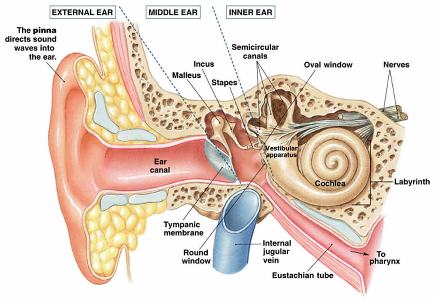
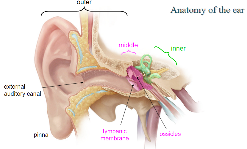
What are the components of the auditory system and their roles? (5)
Pinna: Collects sound waves and directs them into the ear canal.
Tympanic membrane (eardrum): Vibrates in response to sound waves.
Oval window: Vibrations from the tympanic membrane transfer to the inner ear.
Ossicles (malleus, incus, stapes): Amplify sound vibrations.
Cochlea: Converts sound vibrations into neural signals.
-
What is conductive hearing loss? (2)
Definition: A blockage or damage that prevents sound waves from reaching the oval window.
Common causes: Blockage in the ear canal, fluid in the middle ear, otitis media, or damage to ossicles.
-
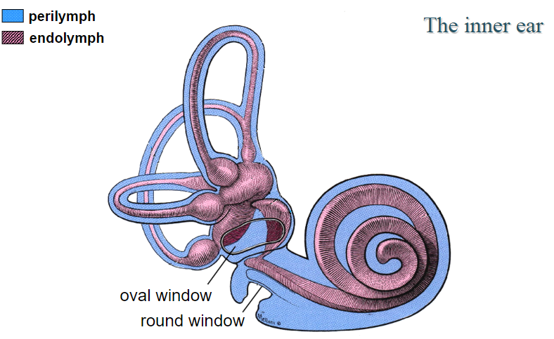
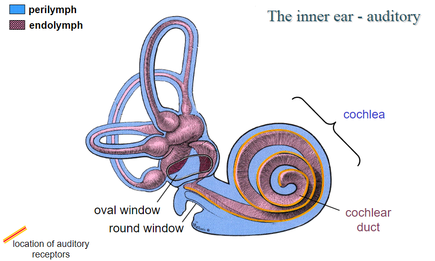
What structures are located in the inner ear and their functions? (6)
Cochlea: Contains auditory receptors and is responsible for converting sound waves into neural signals.
Perilymph: Fluid surrounding the cochlear duct.
Endolymph: Fluid inside the cochlear duct.
Cochlear duct: Location of the auditory receptors.
Round window: Helps dissipate sound pressure.
Cochlear branch of the vestibulocochlear nerve: Transmits auditory information from the cochlea to the brain.
-
What is the structure and function of the spiral organ (organ of Corti)? (7)
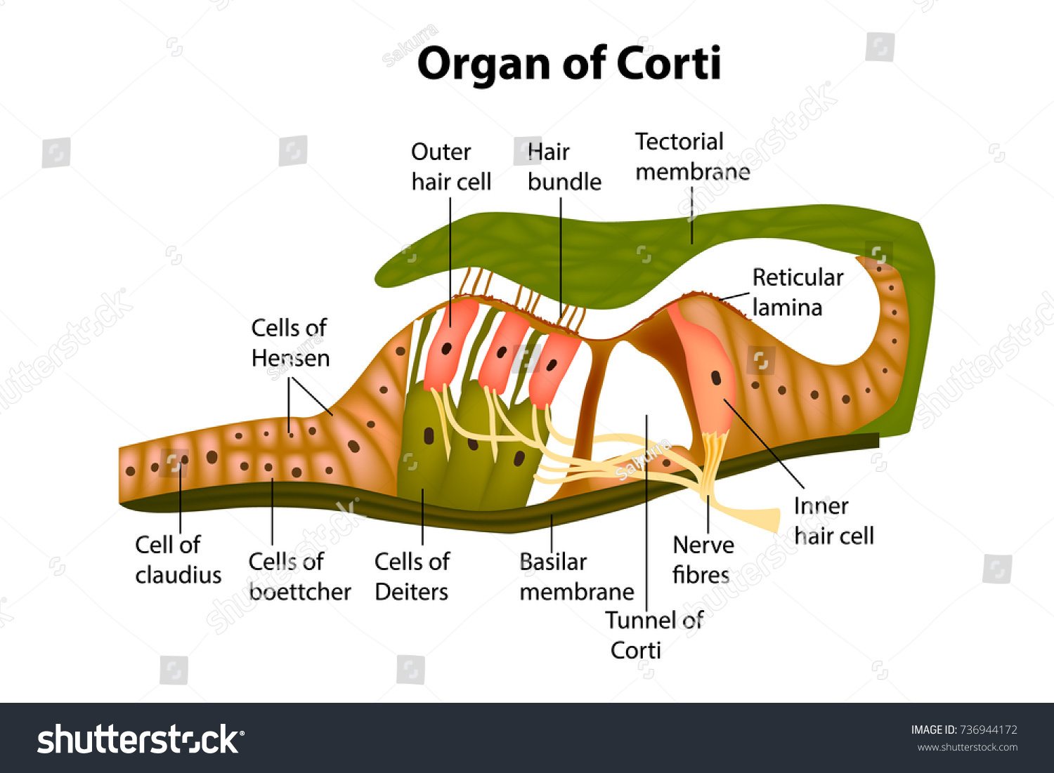
Basilar membrane: Supports hair cells and vibrates in response to sound.
Tectorial membrane: Provides support for stereocilia.
Stereocilia: Hair-like projections that detect sound vibrations.
Inner hair cells: Convert sound vibrations into electrical signals.
Outer hair cells: Amplify sound vibrations through mechanical feedback.
Support cells: Help maintain the structure of the organ.
Vestibular membrane: Separates the cochlear duct from the scala vestibuli.
-
What is the place code in the cochlea? (3)
Frequency encoding: Different parts of the basilar membrane vibrate at different frequencies.
Base of cochlea: Narrow and stiff, responds to high frequencies (~20kHz).
Apex of cochlea: Broad and flexible, responds to low frequencies (~20Hz).
-
How does the auditory system encode different qualities of sound? (4)
Frequency: Encoded by the place code (location of hair cells on the basilar membrane).
Loudness: Encoded by the firing rate of action potentials (more firing = louder sound).
Timing: Preserved by fast axons and synapses, essential for detecting the timing of sounds.
Origin: Encoded by the superior olivary nuclei which compare sound input from both ears.
-
How do the medial and lateral superior olivary nuclei contribute to sound localization? (2)
Medial superior olivary nuclei: Compare the timing of sounds reaching both ears (important for localizing low-frequency sounds).
Lateral superior olivary nuclei: Compare the loudness of sounds in both ears (important for localizing high-frequency sounds).
-
What is the pathway for sound localization? (5)
Cochlear nuclei: Receive input from the cochlear nerve.
Superior olivary nuclei: Process sound information for localization.
Inferior colliculus: Integrates auditory information and assists in sound localization.
Medial geniculate nucleus: Relays sound information to the auditory cortex.
Primary auditory cortex: Processes sound information in the temporal lobe.
-
What is the role of outer hair cells in the cochlea? (3)
Amplify sound: Outer hair cells amplify sound vibrations through mechanical feedback.
Electromotility: Outer hair cells change their length in response to sound, enhancing the movement of the basilar membrane.
Improve sensitivity: Outer hair cells increase the cochlear sensitivity to quiet sounds.
-
What are some conditions that can damage hair cells? (3)
Loud sounds: Can cause mechanical damage to hair cells.
Genetic conditions: Some genetic disorders can lead to the loss of hair cells.
Age-related hearing loss: Over time, hair cells naturally degrade.
-
What are cochlear implants and when are they used? (3)
Cochlear implants: Devices that bypass damaged hair cells and directly stimulate the auditory nerve.
Used for: Severe sensorineural hearing loss, particularly when hair cells in the cochlea are non-functional.
Benefit: Restores hearing by converting sound into electrical signals that stimulate the auditory nerve.
-
How do vibrations in the cochlea affect the hair cells? (2)
Vibration of the basilar membrane: Causes tilting of the stereocilia on hair cells.
Depolarization: When the stereocilia bend, ion channels open, leading to depolarization of the hair cells and triggering action potentials.
-
What is the role of afferent fibers in the auditory system? (2)
Transmit signals: Afferent fibers carry electrical signals from the inner hair cells to the brain.
Essential for hearing: The brain interprets these signals to perceive sound.
-
What is the tonotopic organization of the auditory cortex? (3)
Tonotopic mapping: Different frequencies are processed in different regions of the auditory cortex.
High frequencies: Processed in the anterior part of the cortex.
Low frequencies: Processed in the posterior part of the cortex.
-
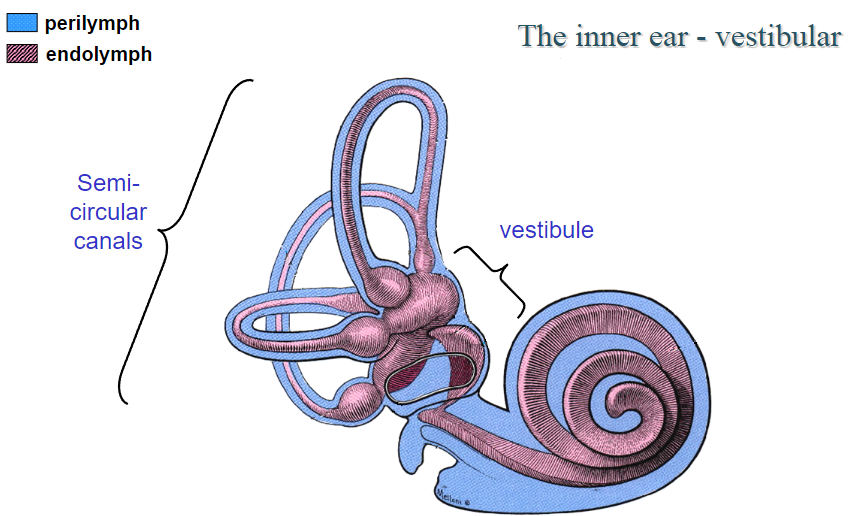
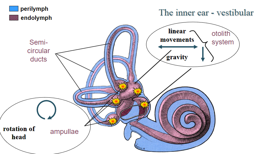
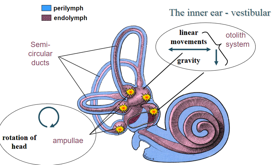
What is the vestibular system and its primary components? (2)
The vestibular system is responsible for detecting changes in head position and movement to help maintain balance and coordinate eye movements.
Primary components: Perilymph, endolymph, utricle, saccule, otolith system, semi-circular canals, ampullae, and hair cells.
-
What are the functions of the perilymph and endolymph in the vestibular system? (2)
Perilymph: Found in the space between the bony labyrinth and membranous labyrinth; provides structural support.
Endolymph: Found within the membranous labyrinth; aids in the transduction of mechanical movements into neural signals.
-
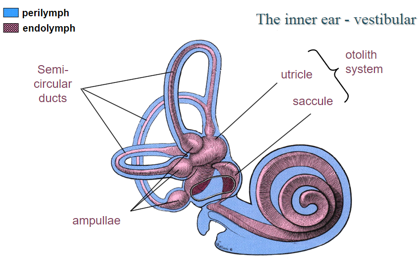
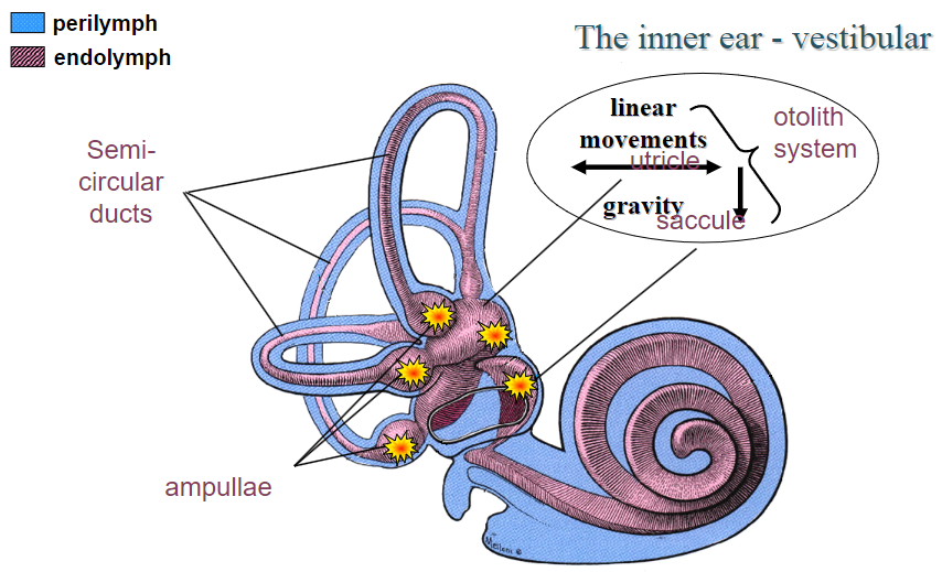
What are otolith organs and their role in the vestibular system? (4)
Otolith organs (utricle and saccule) detect linear acceleration and the direction of gravity.
They contain hair cells embedded in a gelatinous membrane (otolithic membrane) with otoconia (crystals) that provide mass and inertia.
The macula of the utricle and saccule detect changes in the head's linear movements.
The otolith system helps maintain upright posture and compensates for externally induced movements.
-
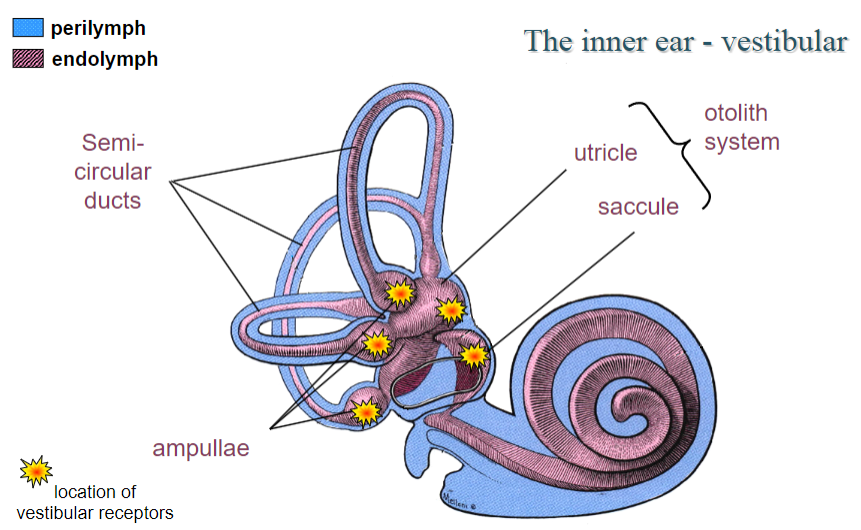
Describe the macula of the otolith system. (3)
The macula contains hair cells that are oriented in different directions to detect changes in head position.
Each hair cell is activated by linear movements, either in the utricle or saccule.
Hair cells project afferent signals to the vestibular nuclei and via the vestibulospinal tract to motor neurons.
-
How do otolith organs detect linear acceleration and gravity? (3)
During linear acceleration, the otoliths lag behind movement due to inertia.
As the head decelerates, the otoliths continue to move due to inertia.
The direction of gravity is detected when the otoliths shift in response to the head tilting.
-
What is the role of the vestibulospinal tract in the vestibular system? (2)
The vestibulospinal tract carries afferent signals from the otolith organs to motor neurons controlling the trunk and legs.
It helps maintain postural stability, particularly in response to changes in body position.
-
What is the function of the semi-circular canal system? (3)
The semi-circular canals detect angular acceleration of the head in three dimensions (anterior, posterior, and horizontal canals).
They consist of hair cells located in the ampulla, surrounded by endolymph and perilymph.
The canals help detect rotational movements by measuring the delay of endolymph fluid movement in response to head turns.
-
Describe the structure of the semi-circular canal ampulla. (3)
The ampulla is a swelling at the end of each semi-circular canal.
It contains the crista ampullaris, a structure with hair cells embedded in a gelatinous membrane called the cupula.
The movement of the endolymph during head rotation causes deflection of the hair cells, leading to neural activation.
-
How do movements of the head activate the hair cells in the semi-circular canal? (3)
When the head rotates, the endolymph fluid inside the canals lags behind.
This causes the cupula (gelatinous membrane) to bend, which deflects the hair cells.
The bending of hair cells alters the firing rate of afferent nerves, signaling the direction and velocity of head movement.
-
How is the direction of movement encoded in the semi-circular canal system? (2)
Each semi-circular canal detects rotation along a specific axis (anterior, posterior, or horizontal).
The arrangement of the canals allows for the detection of all possible rotational movements of the head.
-
What are the main outputs of the vestibular system in terms of motor control? (4)
The vestibular system projects afferent signals via the vestibulospinal tracts to control posture and movement.
It regulates eye movements through the vestibulo-ocular reflex (VOR) to stabilize the gaze.
It coordinates the movements of neck and shoulder muscles to maintain head stability.
It helps control the movement of limbs by adjusting posture in response to changes in head position.
-
What is the vestibulo-ocular reflex (VOR)? (3)
The VOR helps maintain stable vision during head movements by coordinating eye movements in the opposite direction of head rotation.
The VOR involves the oculomotor (CN III), abducens (CN VI), and vestibular nuclei.
It ensures that the gaze remains steady and compensates for head movements, contributing to visual stability.
-
What are conjugate eye movements and their connection to the vestibular system? (3)
Conjugate eye movements, such as smooth pursuit, are coordinated movements of both eyes in the same direction.
The vestibular system helps guide smooth pursuit movements through signals sent from the vestibular nuclei to the extraocular muscles.
These eye movements are critical for maintaining visual stability during head or body movements.
-
How does the vestibular system contribute to eye movement control? (4)
The vestibular system ensures the stability of gaze during head movements through the vestibulo-ocular reflex.
It controls the extraocular muscles via the vestibular nuclei and medial longitudinal fasciculus.
It coordinates movements of both eyes to maintain visual focus.
It helps adjust the speed and direction of eye movements, such as during smooth pursuit or saccades.
-
What is the role of the medial longitudinal fasciculus in the vestibular system? (2)
The medial longitudinal fasciculus is a neural pathway that connects the vestibular nuclei with the oculomotor and abducens nuclei, which control eye movements.
It ensures the coordination of eye movements with head movements for stable vision, and its dysfunction can lead to issues like nystagmus or double vision.
-
What is the relationship between the vestibular system and multiple sclerosis? (2)
The pathways involved in the vestibular system require rapid conduction and are heavily myelinated.
Damage to the myelinated pathways, such as in multiple sclerosis, can impair vestibular function and affect balance, coordination, and eye movement stability.
-
What are the effects of head stability and visually-guided movements on the vestibular system? (3)
The vestibular system helps maintain head stability by compensating for head and body movements, ensuring that the visual field remains steady.
It adjusts the activity of eye muscles to keep the gaze focused during head movements.
It plays a key role in coordinating visually-guided movements, allowing smooth and accurate tracking of objects in space.
-
What is a receptor and its significance? (2)
A protein complex that binds to and is activated by a signaling molecule.
A nerve cell that detects external stimuli and converts it into a receptor potential.
-
What is a primary afferent? (1)
The first cell in the sensory pathway that transmits sensory signals from the sensory apparatus to the cerebral cortex.
-
What is threshold and its different types? (3)
Action potential threshold: The depolarization level necessary to trigger an action potential (approximately -50mV).
Minimum sensory input threshold: The least sensory input needed to activate a receptor.
Perceptual threshold: The minimum sensory input required for conscious perception, dependent on both receptor activation and attention.
-
What is sensitivity and how does it relate to sensory systems? (2)
The relative responsiveness of a sensory system to stimuli.
High sensitivity correlates with lower response thresholds and larger responses to stimuli.
-
What is specificity in sensory systems? (1)
The ability of a system to differentiate between similar stimuli.
-
What is receptor adaptation and why is it important? (3)
The process where receptors adjust their responses to constant stimuli.
Some adapt quickly (e.g., Pacinian corpuscles), others slowly (e.g., cone photoreceptors).
Helps extend dynamic range and save energy.
-
What is lateral inhibition and its purpose in sensory systems? (2)
A mechanism where excitation of one part of a pathway inhibits its neighbors.
Enhances contrast and allows small differences in stimuli to be detected.
-
What is dynamic range? (1)
The range of stimulus strengths a sensory system can respond to, extended through receptor sensitivity and adaptation.
-
What is saturation in sensory systems? (1)
The point at which a sensory system reaches its maximum response, beyond which it can't distinguish stronger stimuli.
-
What is synaptic plasticity and how does it function? (2)
Changes in synaptic strength due to activity patterns.
Long-term potentiation (LTP) increases synapse strength by enhancing transmitter availability and receptor effectiveness.
-
What is a specific circuit/pathway? (1)
A neural circuit involved in processing sensory, motor, or cognitive information.
-
What is a modulatory pathway? (2)
Neural inputs that adjust a cell's response to sensory or motor input.
Crucial for regulating sleep, attention, mood, and emotions, impacting motor and cognitive functions.

