Analysis of Proteins in Different Animal Organs
advertisement

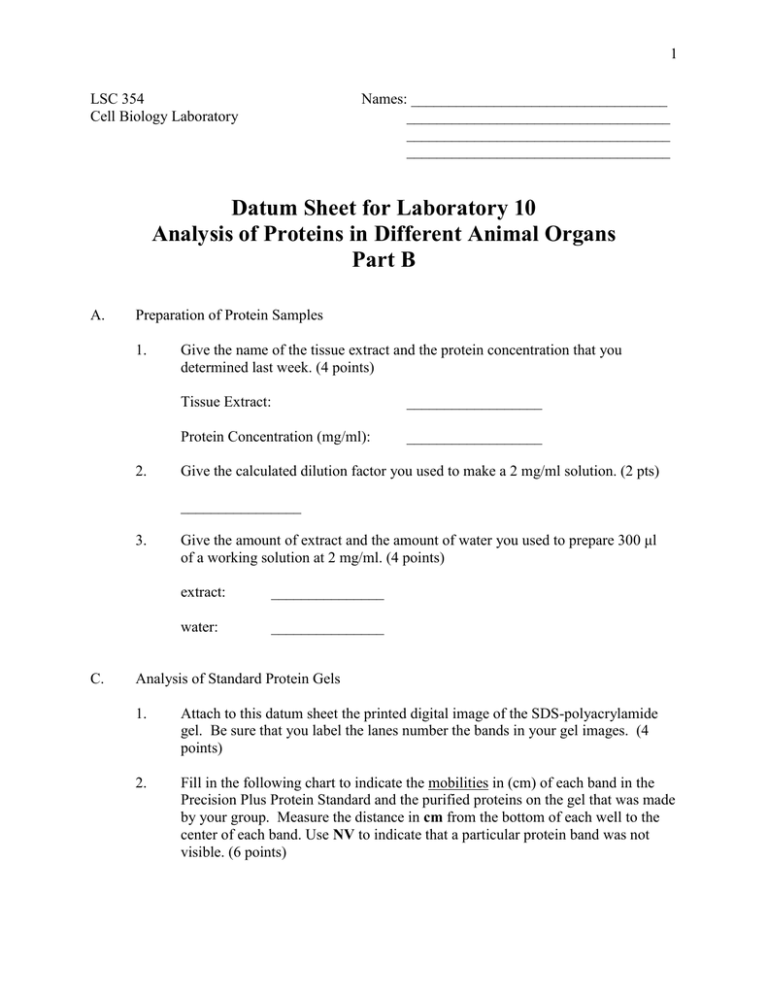
1 LSC 354 Cell Biology Laboratory Names: __________________________________ ___________________________________ ___________________________________ ___________________________________ Datum Sheet for Laboratory 10 Analysis of Proteins in Different Animal Organs Part B A. Preparation of Protein Samples 1. 2. Give the name of the tissue extract and the protein concentration that you determined last week. (4 points) Tissue Extract: __________________ Protein Concentration (mg/ml): __________________ Give the calculated dilution factor you used to make a 2 mg/ml solution. (2 pts) ________________ 3. C. Give the amount of extract and the amount of water you used to prepare 300 μl of a working solution at 2 mg/ml. (4 points) extract: _______________ water: _______________ Analysis of Standard Protein Gels 1. Attach to this datum sheet the printed digital image of the SDS-polyacrylamide gel. Be sure that you label the lanes number the bands in your gel images. (4 points) 2. Fill in the following chart to indicate the mobilities in (cm) of each band in the Precision Plus Protein Standard and the purified proteins on the gel that was made by your group. Measure the distance in cm from the bottom of each well to the center of each band. Use NV to indicate that a particular protein band was not visible. (6 points) 2 purified proteins: protein molecular mass mobility (cm) gel A gel B rabbit glyceraldehyde-3-phosphate dehydrogenase 36,000 ___________ bovine albumin 66,000 ___________ phosphorylase b 97,000 ___________ Band Color molecular mass mobility (cm) gel A gel B Blue 10,000 ___________ Blue 15,000 ___________ Blue 20,000 ___________ Pink 25,000 ___________ Blue 37,000 ___________ Blue 50,000 ___________ Pink 75,000 ___________ Blue 100,000 ___________ Blue 150,000 ___________ Blue 250,000 ___________ Precision Plus Protein Standard: 3. Attach to this datum sheet the standard curve for the proteins in the Protein standard for the 4-15% polyacrylamide gels. Plot mobility (in cm) on the X axis and log molecular weight on the Y axis. Draw the best fit line you can through the datum points. (4 points) 3 4. You are provided a protein that has a quaternary structure composed of three subunits. The molecule mass of each of the three subunits is as follows: Subunit A 17,000 daltons Subunit B 25,000 daltons Subunit C 30,000 daltons If this protein was fractionated on a SDS-PAGE gel like you did in class, what would you expect in the lane that contained this protein? Be specific, include number and size of bands and also the expected distance migrated based on your standard curve from #3. (6 points)


